The fetal period and prenatal diagnosis
Fetal period development from week nine to birth, covering growth, differentiation, and viability.
The developing human is termed a fetus from the ninth week of gestation until birth[1]. This period spans approximately thirty weeks and is characterised by continued cell growth and differentiation[1]. These processes fully develop the structures and functions of the immature organ systems formed during the embryonic period[1]. The completion of fetal development results in a newborn capable of survival outside the womb[1].
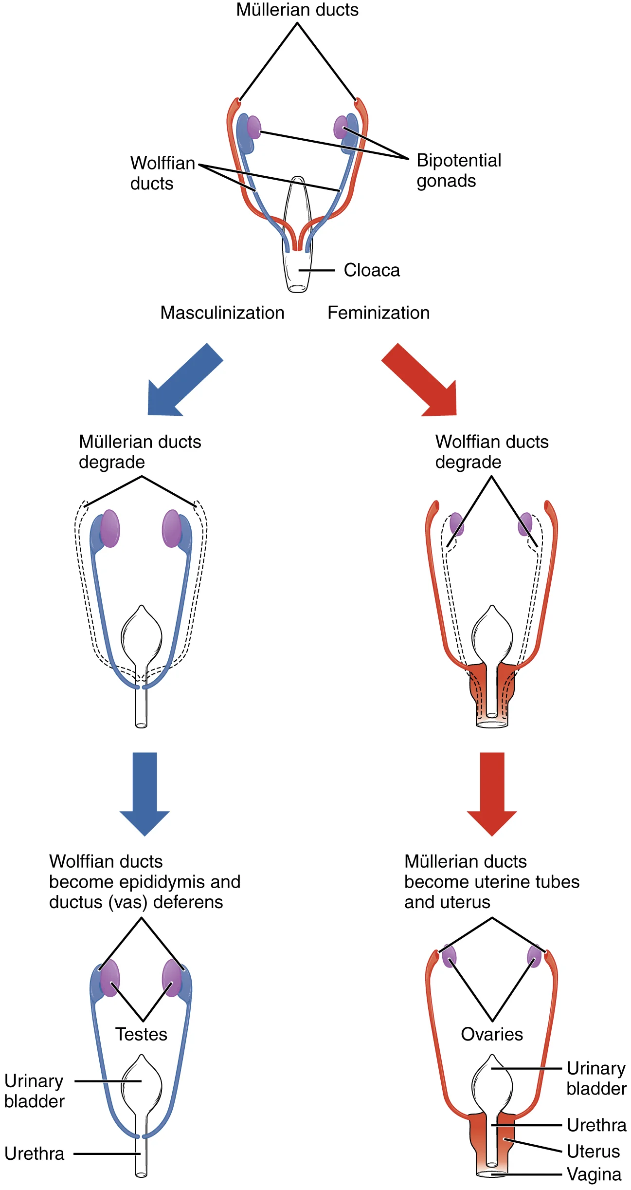
Sexual differentiation begins during weeks nine to twelve[1]. Embryonic males and females are morphologically identical despite genetic differences[1]. Bipotential gonads connect to the cloaca via Müllerian and Wolffian ducts[1]. The fetal circulatory system becomes specialised, including three shunts: the ductus venosus, foramen ovale, and ductus arteriosus[1]. These shunts bypass the semifunctional liver and pulmonary circuit until after childbirth[1].
During weeks nine to twelve, the brain expands and the body elongates[1]. Ossification continues, and fetal movements become frequent but jerky[1]. The bone marrow begins erythrocyte production, taking over from the liver[1]. The liver secretes bile, and the fetus circulates amniotic fluid by swallowing and urinating[1]. Eyes are well-developed with fused eyelids, and nails begin on fingers and toes[1].
Between weeks sixteen and twenty, the pregnant person may feel quickening[1]. Limb movements are powerful but restricted by space, forcing the fetal position[1]. Sebaceous glands coat the skin with vernix caseosa for protection and lubrication[1]. Lanugo covers the skin from weeks seventeen to twenty before being shed[1].
Developmental weeks twenty-one to thirty are marked by rapid weight gain[1]. This weight gain is crucial for maintaining stable body temperature after birth[1]. Spinal cord axons begin myelination, which is necessary for efficient nervous system functioning[1]. Eyelids open and close, and the lungs begin producing surfactant to reduce surface tension[1].
From week thirty-one until birth, the fetus lays down subcutaneous fat[1]. The skin transitions from red and wrinkled to soft and pink[1]. Lanugo is shed, and nails grow to the tips of fingers and toes[1]. Immediately before birth, the average crown-to-rump length is thirty-five point five to forty point five centimetres[1]. The fetus weighs approximately two point five to four kilograms[1]. At birth, the average length is approximately fifty-one centimetres[1].
DefinitionFetal Viability
Viability is the capacity of the fetus to survive outside the womb, achieved through organ maturation such as lung surfactant production and thermoregulation via subcutaneous fat.
| Parameter | Value |
|---|---|
| Start Length (Week 9) | 3.3 cm |
| End Length (Birth) | 51 cm |
| End Weight (Birth) | 3.4 kg |
| Crown-to-Rump (Pre-birth) | 35.5–40.5 cm |
Birth defects in outline arise from disruptions in these differentiation processes. The fetal period is critical for the refinement of organ systems rather than their initial formation.
Figure 12915 Sexual Differentation-02
Long description
Differentiation of the male and female reproductive systems does not occur until the fetal period of development.

Figure 1: Differentiation of the male and female reproductive systems does not occur until the fetal period of development.
The fetal period begins after the embryonic stage, marked by significant growth and functional maturation of organs established previously. This figure illustrates the transition from early organogenesis to the refined anatomical structures seen in the late second trimester. The view is a sagittal section of the fetal trunk and head, oriented with the anterior aspect to the left. In the cranial region, the eyes are shown moving closer together, with blinking motions beginning although the eyes remain sealed shut. The lips exhibit sucking motions, indicating neuromuscular development. The ears have moved upward and lie flatter against the head. The scalp is depicted with growing hair. In the thoracic and abdominal regions, the heart is shown as a four-chambered structure, having folded and differentiated from the initial two parallel tubes. The fetal circulatory system is integrated with the placenta via the umbilical cord, allowing the fetus to receive oxygen and nutrients. Shunts are visible as anatomical diversions that allow blood flow to bypass immature organs such as the lungs and liver. In the abdominal cavity, the kidneys are well-formed, and the intestines contain meconium, or fetal feces. Meconium consists of ingested amniotic fluid, cellular debris, mucus, and bile. The excretory system is actively developing. [1]
Sexual differentiation proceeds along distinct pathways determined by genetic sex. During male fetal development, the bipotential gonads become the testes and associated epididymis. The Müllerian ducts degenerate. This process ensures the formation of male internal genitalia. [1]
DefinitionSexual Differentiation
The divergence of male and female internal genitalia from bipotential precursors driven by specific ductal regression and development.
In contrast, during female fetal development, the bipotential gonads develop into ovaries. The Wolffian ducts degenerate. This pathway establishes the female reproductive tract. [1]
| Feature | Male Development | Female Development |
|---|---|---|
| Gonads | Testes and epididymis | Ovaries |
| Müllerian Ducts | Degenerate | Uterine tubes and uterus |
| Wolffian Ducts | Vas deferens | Degenerate |
| Cloaca | Urethra and rectum | Vagina, urethra, and rectum |
Meconium consists of ingested amniotic fluid, cellular debris, mucus, and bile. [1]
The fetal cardiovascular system differs from the adult system by including circulatory shortcuts, or shunts. A shunt is an anatomical (or sometimes surgical) diversion that allows blood flow to bypass immature organs such as the lungs and liver until childbirth. When the heart first forms in the embryo, it exists as two parallel tubes derived from mesoderm and lined with endothelium, which then fuse together. As the embryo develops into a fetus, the tube-shaped heart folds and further differentiates into the four chambers present in a mature heart. However, unlike a mature cardiovascular system, the fetal cardiovascular system retains these shunts to optimize oxygen delivery to the brain and vital organs while the lungs are non-functional. [1]
NoteFetal Circulation
Shunts bypass the lungs and liver, directing oxygenated blood from the placenta to the brain and heart.
Prenatal diagnosis allows for the identification of structural and genetic anomalies. Although the number of CPSS patients diagnosed prenatally is increasing, studies on the prenatal diagnosis of CPSS are scarce, warranting further studies to expand our knowledge to improve perinatal management and counselling since early diagnosis and appropriate intervention are important to avoid deterioration of the disease. Some patients may have prenatal comorbidities, such as IUGR, and these comorbidities will attract attention of obstetricians, which can be helpful for the detection of CPSS. Early prenatal diagnosis may significantly influence the clinical outcomes because it is helpful to monitor the progression of the disease so that essential treatment can be provided in time. [2]
Prenatal Assessment
- Initial ScreeningUltrasound and maternal history
- Question: Anomaly Detected?
Yes
- Detailed ImagingAssess shunts and organ structure
- CounsellingDiscuss perinatal management
No
- Routine CareMonitor growth and development
The integration of the fetal circulatory system with the placenta via the umbilical cord ensures that the fetus receives both oxygen and nutrients from the placenta. However, after childbirth, the umbilical cord is severed, and the newborn’s circulatory system must be reconfigured. This transition marks the end of the fetal period and the beginning of neonatal adaptation.
Molecular regulation during the fetal period is governed by intricate signalling cascades that coordinate organogenesis and physiological adaptation. A primary example is the role of microRNAs in neural development. MiR-9 is critical for early neural tube patterning, telencephalic neurogenesis, and early differentiation. It functions as a direct negative regulator of Foxg1, a gene that normally prevents neural stem cell differentiation and promotes proliferation. This regulatory balance ensures proper cortical plate thickness; disruption leads to increased neural stem cell proliferation and subsequent microencephaly. [3]
Epigenetic mechanisms further modulate these pathways. Ethanol exposure alters methylation patterns in differentiating neural stem cells, thereby regulating miRNA expression through epigenetic means. For instance, ethanol depletes miR-9 expression while promoting neural stem cell proliferation and inducing the expression of ELAVL2 mRNA. ELAVL2 is an RNA-binding protein that stabilizes Foxg1 mRNA transcripts. This compensatory mechanism becomes refractory to miR-9 depletion in the later half of the second trimester-equivalent period, specifically after gestational day 16.5. This suggests that miR-9 plays a critical role in early second trimester ethanol teratology, coordinating with other ethanol-sensitive miRNAs to maintain developmental stability. [3]
The genomic architecture of these regulators is also significant. The mammalian-specific miR-335 is coded as an intronic miRNA within the MEST/Peg1 imprinted gene locus. This locus contains GC-rich regions known as CpG islands and is part of a broader network of imprinted genes that controls fetal growth. MiR-21 and miR-335 serve as functional antagonists, with miR-21 regulated by GABAAR-dependent mechanisms, whereas miR-335 regulation involves distinct chromosomal structures. [3]
Clinical pearlDevelopmental Timing
Disruption of miR-9/Foxg1 balance in the early second trimester-equivalent period is particularly detrimental, leading to reduced cortical plate thickness and microencephaly.
| Molecule | Target/Role | Effect of Disruption |
|---|---|---|
| MiR-9 | Negative regulator of Foxg1 | Increased proliferation, microencephaly |
| Foxg1 | Prevents NSC differentiation | Reduced cortical plate thickness |
| ELAVL2 | Stabilizes Foxg1 mRNA | Compensatory refractory period |
| MiR-335 | Part of MEST/Peg1 locus | Altered fetal growth control |
DefinitionFetal Circulation
The fetal circulatory system utilises specific shunts to bypass the non-functional liver and lungs, ensuring oxygenated blood from the placenta reaches the fetal heart and brain efficiently.
The transition from embryonic to fetal structures involves the maturation of primitive organ systems. During this period, embryonic organ structures that were previously primitive and nonfunctional develop to the point that the newborn can survive in the outside world. For instance, the respiratory system undergoes significant structural changes, although it remains immature until birth. Inadequate surfactant production in premature newborns may result in respiratory distress syndrome, a condition requiring immediate intervention such as surfactant replacement therapy, supplemental oxygen, or maintenance in a continuous positive airway pressure (CPAP) chamber. [1]
| Embryonic/Fetal Structure | Adult Derivative/Function |
|---|---|
| Umbilical Vein | Ligamentum teres hepatis (Round ligament of liver) |
| Ductus Venosus | Ligamentum venosum |
| Ductus Arteriosus | Ligamentum arteriosum |
| Foramen Ovale | Fossa ovalis |
| Testes (in males) | Descend into scrotum near end of period |
Growth during the fetal period is rapid and measurable. This measurement provides a concrete benchmark for assessing fetal growth and development during the third trimester. [1]
NoteClinical Correlation
In male fetuses, the testes descend into the scrotum near the end of this period. Failure of this descent is a common congenital anomaly. [1]
Congenital portosystemic shunt (CPSS) is a rare vascular malformation which results in anomalous communication between the portal venous system and the systemic vein. This condition disrupts the normal derivative pathway where the umbilical vein and ductus venosus should obliterate. Prenatal ultrasonic characteristics are critical for diagnosis, allowing for perinatal monitoring and assessment of postnatal outcomes. However, hemodynamic changes secondary to cardiac defects might affect the persistence of vitelline veins during embryonic development of the portal system, and contributed to the development or maintenance of CPSS. Thus, fetal echocardiography should be conducted when CPSS is diagnosed. Close monitoring is important if the fetuses have cardiovascular malformations. More attention should be given if the type of the shunt is USS, DVSS or EHPSS. [2]
Exam trapShunt Types
Be aware of specific shunt types such as USS, DVSS, or EHPSS, as they require heightened clinical attention during prenatal diagnosis and monitoring.
The derivatives of the fetal period are not merely structural remnants but functional adaptations that ensure survival in the intrauterine environment. Understanding these pathways is essential for diagnosing anomalies such as CPSS and managing the transition to extrauterine life.
Congenital anomalies are structural or functional abnormalities present at birth that result from errors in embryonic or fetal development. These anomalies can affect any organ system and may be caused by genetic factors, environmental influences, or a combination of both.
Congenital portosystemic shunt (CPSS) is a rare vascular malformation with an incidence of approximately one case per 30,000–50,000 children. This anomaly results from the false evolution or involution of vitelline veins during embryogenesis, leading to anomalous communication between the portal venous system and systemic veins. CPSS often occurs with other comorbidities such as cardiomegaly, intrauterine growth restriction (IUGR), chromosomal aberrations, and encephalopathy. [2]
CPSS can be classified into two main subtypes: extrahepatic porto-systemic shunt (EHPSS) and intrahepatic porto-systemic shunt (IHPSS). Intrahepatic portosystemic shunt (IHPSS) is defined as a shunt between the intrahepatic portal venous system and hepatic veins, and IHPSS patients tend to have a normal intrahepatic portal venous system (IHPVS). This normal anatomy provides better hemodynamic tolerance and better outcomes for IHPSS patients. [2]
The prognosis of CPSS depends on the type and associated comorbidities. IHPSS patients have a lower incidence of severe comorbidities such as cardiovascular malformations and chromosomal aberrations, making them more likely to have better outcomes. In contrast, other groups, particularly those with umbilical-systemic shunt (USS), have higher rates of severe comorbidities and lower rates of good outcomes. The integrity of the intrahepatic portal venous system (IHPVS) plays a pivotal role as a prenatal prognostic factor and in optimizing postnatal care. [2]
Prenatal diagnosis of congenital portosystemic shunts (CPSS) is primarily achieved through ultrasound examinations. This imaging modality allows for the identification of abnormal vascular connections before birth. The in-utero classification system, proposed in 2016, relies on the anatomical origin of the shunt within the intact umbilical vein-portal vein-ductus venosus (UV-PV-DV) structure. This classification divides umbilical portosystemic venous shunts (UPSVS) into three main types: Type I, umbilical–systemic shunt (USS); Type II, ductus venosus–systemic shunt (DVSS); and Type III, portal–systemic shunt. Type III is further subdivided into Type IIIa, intrahepatic portal-systemic shunt (IHPSS), and Type IIIb, extrahepatic portal-systemic shunt (EHPSS). Each type exhibits distinct postnatal clinical characteristics. [2]
Invasive procedures are not routinely required for diagnosis but may be considered in complex cases. The clinical management strategy depends heavily on the specific type of shunt and the presence of clinical manifestations. Patients with IHPSS often have good outcomes and may benefit from a “wait-and-see” approach. These patients might experience spontaneous closure in the first few months of life and remain asymptomatic. In contrast, patients with other groups deserve closer monitoring due to the risk of severe complications. [2]
Postnatal clinical symptoms are crucial factors in the choice of treatment. Neonates diagnosed with EHPSS had the highest incidence of postnatal clinical symptoms. These patients are more likely to need interventional or surgical treatment to close the shunts. It has been proposed that if the shunt does not close spontaneously and remains patent in the second year of life, surgical or endovascular intervention is required. [2]
Clinical pearlSpontaneous Closure
Shunts could close spontaneously before two years old particularly in children with IHPSS.
The diversion of metabolites and vasoactive mediators, such as ammonia and bile acid, from the splanchnic venous system directly into the systemic circulation leads to adverse effects on metabolism. These substances accumulate within the body and lead to corresponding symptoms. In neonates, these metabolites and the vasoactive mediators could also cause damage to their growing brains. Therefore, the volume of the shunt, which could be influenced by CPSS type, was another factor affecting the clinical manifestations and outcomes. [2]
| Type | Shunt Description | Clinical Note |
|---|---|---|
| Type I | Umbilical–systemic shunt (USS) | 9% of cases [2] |
| Type II | Ductus venosus–systemic shunt (DVSS) | 50% of cases; often asymptomatic [2] |
| Type IIIa | Intrahepatic portal-systemic shunt (IHPSS) | 30% of cases; spontaneous closure common [2] |
| Type IIIb | Extrahepatic portal-systemic shunt (EHPSS) | 6% of cases; high symptom incidence [2] |
Each patient should receive personalized treatment, since some patients could have severe clinical manifestations and should be treated. Personalized treatment is essential as CPSS patients can be asymptomatic or have severe complications. Neonates with EHPSS tend to have postnatal clinical symptoms so their physical conditions should be monitored so that essential treatment can be provided in a timely manner. [2]
The fetal venous system is distinct from the adult circulation, comprising three functionally inseparable components: the umbilical vein, the portal vein, and the ductus venosus. These structures work together to transport highly oxygenated blood from the placenta directly to the left atrium. Abnormal development within this complex can result in shunting into systemic veins, a condition known as umbilical–portal–systemic venous shunt (UPSVS). [2]
Postnatal classification systems for congenital portosystemic shunts (CPSS) are not applicable to fetuses because they lack the umbilical vein and ductus venosus, which close after birth. Therefore, prenatal diagnosis must rely on fetal-specific anatomical criteria rather than adult-derived classifications. [2]
CPSS is categorized into four main types based on the site of shunting. Umbilical-systemic shunts (USS) account for 9% of cases, while ductus venosus-systemic shunts (DVSS) represent 50%. Intrahepatic portal-systemic shunts (IHPSS) comprise 30% of cases, and extrahepatic portal-systemic shunts (EHPSS) make up 6%. [2]
Comorbidities are frequent in CPSS patients and significantly impact prognosis. The most common comorbidity is intrauterine growth restriction (IUGR), affecting 27% of cases. [2]
Fetuses with IHPSS have the lowest incidence of severe comorbidities compared to other groups. In contrast, chromosomal aberrations are mostly associated with USS, DVSS, and EHPSS fetuses. Most fetuses with chromosomal aberrations have disappointing outcomes, often leading to pregnancy termination. Consequently, chromosome examination or genetic testing is necessary for patients diagnosed with USS, DVSS, or EHPSS. [2]
Conservative treatment is the most frequent management strategy for CPSS patients. However, interventional and surgical treatments are reserved for specific cases requiring active intervention. [2]
Exam trapClassification Error
Do not apply postnatal CPSS classification systems to fetal cases; they lack the essential umbilical and ductus venosus components.
Clinical pearlHigh-Yield Stat
Ductus venosus-systemic shunt (DVSS) is the most common type, accounting for 50% of CPSS cases.
DefinitionUPSVS
Umbilical–portal–systemic venous shunt refers to shunting into systemic veins due to abnormal development of the fetal venous complex.
NoteGenetic Link
Chromosomal aberrations are strongly linked to USS, DVSS, and EHPSS, necessitating genetic examination for these subtypes.
Exam trapComorbidity Profile
IHPSS fetuses have the lowest incidence of severe comorbidities, unlike other CPSS types which frequently present with cardiac or chromosomal issues.
Clinical pearlCardiac Defect
Ventricular septal defect (VSD) is the most common cardiovascular malformation in CPSS, occurring in 27% of cardiac cases. [2]
DefinitionIUGR
Intrauterine growth restriction is the most frequent comorbidity in CPSS, affecting 27% of the studied population.
NoteManagement
Most CPSS patients receive conservative treatment, with surgery or intervention used only for selected cases.
Exam trapOutcome Prediction
Pregnancies with chromosomal aberrations in CPSS fetuses often result in termination, contributing to poorer outcomes in USS, DVSS, and EHPSS groups.
Clinical pearlPrevalence
Mixed shunts are rare, representing only 2% of CPSS cases.
DefinitionVenous Complex
The fetal venous system consists of the umbilical vein, portal vein, and ductus venosus, forming an inseparable unit for oxygenated blood transport.
Students frequently confuse the haemodynamic consequences of different types of Congenital Portosystemic Shunts (CPSS). A critical error is assuming all portosystemic shunts behave identically regarding cardiac load. It is essential to distinguish between intrahepatic shunts and extrahepatic variants. Unlike intrahepatic portosystemic shunts (IHPSS), umbilical venous (USS), ductus venosus (DVSS), and extrahepatic portosystemic shunts (EHPSS) are characterised by drainage into systemic veins such as the inferior vena cava (IVC). This anatomical distinction is vital because these shunts can also drain directly into the right atrium, causing the heart preload to be significantly greater and the hemodynamic change to be worse. Consequently, the incidence of cardiovascular malformations in these groups was higher than that in the IHPSS group. [2]
Another frequent mistake involves the management threshold for intervention. Students often assume that all identified shunts require immediate surgical correction. However, intervention is reserved for specific clinical scenarios. Interventional and surgical treatments are still needed to close shunts when patients have severe complications due to CPSS or when the shunt flow is high. Treating asymptomatic patients with low-flow shunts unnecessarily exposes them to procedural risks without clear benefit. [2]
A third pitfall relates to the prevalence of associated anomalies. Students may underestimate the frequency of concurrent defects. The prevalence of cardiac malformations in CPSS patients is dramatically higher than that in the general population. Because the shunts can increase heart preload and potentially cause heart overload, a thorough cardiac evaluation is mandatory. Ignoring this association leads to missed diagnoses of significant congenital heart disease. [2]
Finally, students sometimes misinterpret the role of molecular mechanisms in clinical diagnosis. While research explores miRNA changes in fetal development, these are not diagnostic tools in standard practice. For instance, ethanol exposure influences a relatively small subset of expressed miRNAs. The apparent absence of a global effect on miRNAs suggests that ethanol does not generally target the miRNA processing machinery during development. Clinical diagnosis relies on imaging and haemodynamics, not on miRNA profiling, which remains an area of experimental research rather than clinical application. [3]
This knowledge is applied primarily in prenatal and postnatal imaging. On ultrasound, the sonographer must identify the origin and termination of the shunt. Recognising whether the shunt drains into the IVC or directly into the right atrium dictates the severity assessment. Drainage into the right atrium causes the heart preload to be significantly greater and the hemodynamic change to be worse. This finding prompts a detailed echocardiographic survey for cardiac malformations, given that the prevalence of cardiac malformations in CPSS patients is dramatically higher than that in the general population.
In the operating theatre and interventional radiology suite, the decision to intervene is based on haemodynamic compromise. The surgeon or interventionalist assesses the shunt flow volume. Interventional and surgical treatments are still needed to close shunts when patients have severe complications due to CPSS or when the shunt flow is high. This criterion prevents unnecessary procedures while ensuring that patients with significant cardiac overload or liver dysfunction receive timely closure of the shunt.
On imaging, the distinction between shunt types guides the anatomical description. The USS, DVSS and EHPSS, which are from umbilical vein, ductus venosus and portal vein, respectively, are characterized by drainage into systematic veins such as inferior vena cava (IVC). This classification helps predict the cardiac burden. The shunts can also drain into the right atrium directly, causing the heart preload to be significantly greater and the hemodynamic change to be worse. Radiologists must report this specific drainage pattern as it correlates with the higher incidence of cardiovascular malformations observed in these groups. [2] [1]
In examinations, students are tested on the haemodynamic principles and management indications. Questions often present a case of a neonate with heart failure and ask for the underlying anatomical cause. The correct answer involves identifying a portosystemic shunt with high flow or direct atrial drainage. The key learning point is that unlike IHPSS, the USS, DVSS and EHPSS... are characterized by drainage into systematic veins such as inferior vena cava (IVC). Furthermore, the shunts can also drain into the right atrium directly, causing the heart preload to be significantly greater and the hemodynamic change to be worse. Understanding this mechanism explains why the incidence of cardiovascular malformations in these groups was higher than that in the IHPSS group. Management questions will test the indication for closure: Interventional and surgical treatments are still needed to close shunts when patients have severe complications due to CPSS or when the shunt flow is high.
Assessment of Portosystemic Shunt
- Identify Shunt TypeUSS, DVSS, EHPSS or IHPSS
- Question: Drainage into IVC or RA?
Yes
- High Cardiac Preload RiskCheck for cardiac malformations
No
- Lower Cardiac RiskMonitor liver function
- Question: Severe Complications or High Flow?
Yes
- Intervention RequiredClose shunt
No
- Conservative ManagementObservation
- Portosystemic shunts are classified by their venous origin and drainage site.
- USS, DVSS, and EHPSS drain into systemic veins like the IVC.
- Direct drainage into the right atrium significantly increases cardiac preload.
- The prevalence of cardiac malformations is dramatically higher in CPSS patients.
- High shunt flow or severe complications are indications for intervention.
- IHPSS has a different haemodynamic profile compared to extrahepatic shunts.
- Imaging must distinguish between IVC and right atrial drainage.
- Management is reserved for cases with high flow or complications.
Answer each one in your head first, then open the answer.
Which shunts are characterised by drainage into the inferior vena cava?
Show answerHide answer
USS, DVSS and EHPSSWhat causes the heart preload to be significantly greater in some shunts?
Show answerHide answer
Drainage into the right atrium directlyWhen are interventional treatments needed for CPSS?
Show answerHide answer
When patients have severe complications or shunt flow is highHow does the cardiac malformation prevalence in CPSS compare to the general population?
Show answerHide answer
Dramatically higherWhich shunt group has a lower incidence of cardiovascular malformations compared to USS/DVSS/EHPSS?
Show answerHide answer
IHPSS group
- Betts JG, Young KA, Wise JA, et al. Fetal Development. OpenStax, Anatomy and Physiology 2e (CC BY-NC-SA 4.0). Accessed .
- Kong B, Yan X, Gui Y, et al. Prenatal sonographic characteristics and postnatal outcomes of congenital portosystemic shunt diagnosed during the fetal period: a systematic review. Orphanet journal of rare diseases 2025;20(1):257. doi:10.1186/s13023-025-03811-3. Accessed .
- Miranda RC. MicroRNAs and Fetal Brain Development: Implications for Ethanol Teratology during the Second Trimester Period of Neurogenesis. Frontiers in genetics 2012;3:77. doi:10.3389/fgene.2012.00077. Accessed .
Figure credits
Educational content for medical students and doctors. It is not a substitute for clinical judgement or local guidelines.