Bone
Bone is a living connective tissue providing structure and mineral storage.
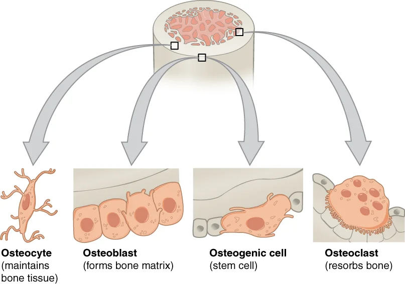
Bone is a dynamic connective tissue that provides structural support, protects vital organs, serves as a mineral reservoir, and facilitates movement. The primary cellular components of bone tissue include osteoblasts, osteocytes, osteoclasts, and osteogenic cells. Osteoblasts are responsible for forming new bone and are found in the growing portions of bone, including the periosteum and endosteum. These cells synthesize and secrete the collagen matrix and calcium salts. As the secreted matrix calcifies, osteoblasts become trapped within it and transform into osteocytes, the primary cells of mature bone and the most common type of bone cell. Each osteocyte is located in a space called a lacuna and maintains the mineral concentration of the matrix via enzyme secretion. [2]
Figure 1604 Bone cells
Long description
Four types of cells are found within bone tissue. Osteogenic cells are undifferentiated and develop into osteoblasts. When osteoblasts get trapped within the calcified matrix, their structure and function changes, and they become osteocytes. Osteoclasts develop from monocytes and macrophages and dif

Figure 1: Four types of cells are found within bone tissue.
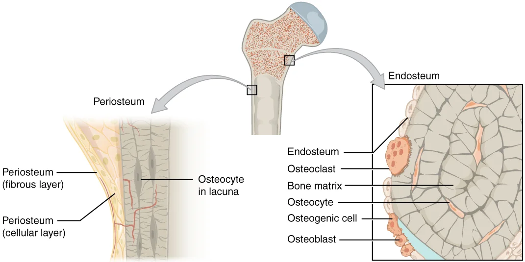
Figure 2607 Periosteum and Endosteum
Long description
The periosteum forms the outer surface of bone, and the endosteum lines the medullary cavity.

Figure 2: The periosteum forms the outer surface of bone, and the endosteum lines the medullary cavity.
Osteoclasts are multinucleated cells responsible for bone resorption or breakdown. They originate from monocytes and macrophages, not from osteogenic cells, and are found on bone surfaces. The ongoing balance between osteoblasts and osteoclasts is responsible for the constant reshaping of bone. Compact bone is dense and composed of osteons, while spongy bone is less dense and made up of trabeculae. [2]
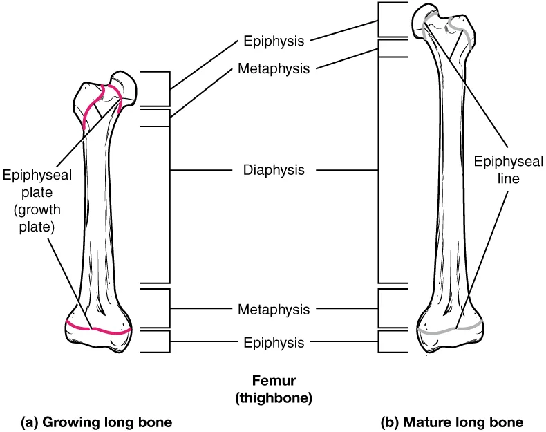
Ossification is the process by which bone forms, replacing cartilaginous or membranous tissue during development. There are two types of ossification: intramembranous, where bone develops directly from sheets of mesenchymal connective tissue, and endochondral, where bone develops by replacing hyaline cartilage. The epiphyseal plate is a layer of hyaline cartilage where ossification occurs in immature bones, enabling bones to grow in length. It is composed of four zones of cells and activity, with the reserve zone containing small chondrocytes that secure the epiphyseal plate to the osseous tissue of the epiphysis. When bone growth stops in early adulthood (approximately 18-21 years), the cartilage is replaced by osseous tissue and the epiphyseal plate becomes an epiphyseal line. [2] [1]
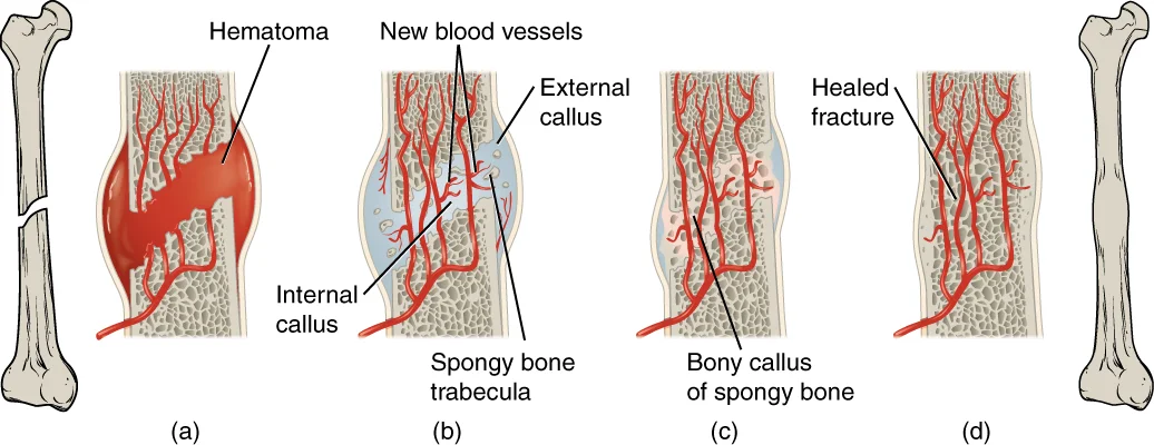
Figure 3613 Stages of Fracture Repair
Long description
The healing of a bone fracture follows a series of progressive steps: (a) A fracture hematoma forms. (b) Internal and external calli form. (c) Cartilage of the calli is replaced by trabecular bone. (d) Remodeling occurs.

Figure 3: The healing of a bone fracture follows a series of progressive steps: (a) A fracture hematoma forms.
Figure 4623 Epiphyseal Plate-Line
Long description
As a bone matures, the epiphyseal plate progresses to an epiphyseal line. (a) Epiphyseal plates are visible in a growing bone. (b) Epiphyseal lines are the remnants of epiphyseal plates in a mature bone.

Figure 4: As a bone matures, the epiphyseal plate progresses to an epiphyseal line.
Figure 5601 Bone Classification
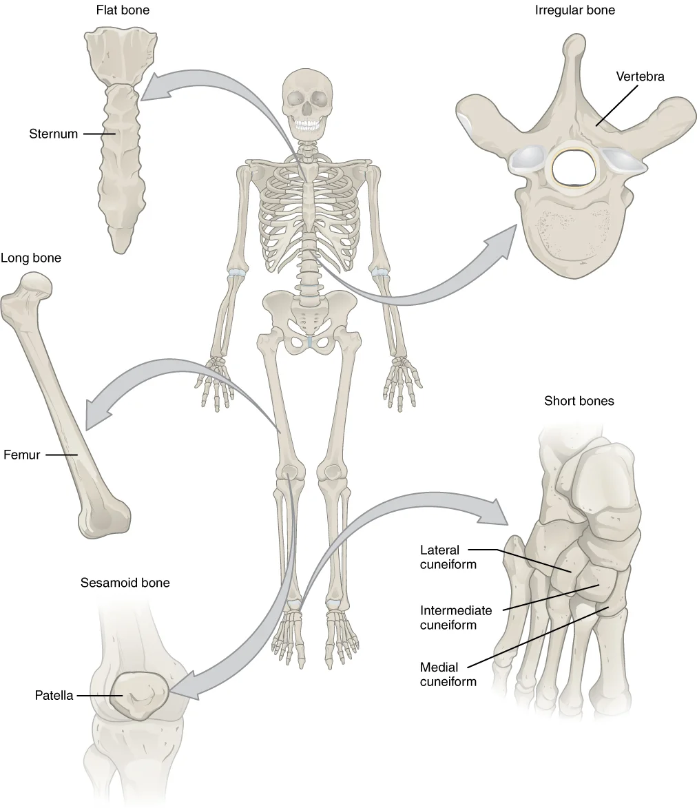
Long description
Bones are classified according to their shape.

Figure 5: Bones are classified according to their shape.
Growth in diameter, known as appositional growth, continues even after longitudinal growth ceases. This process is called modeling. Bone modeling primarily takes place during growth, while remodeling occurs when resorption of old or damaged bone takes place on the same surface where osteoblasts lay new bone to replace it. About 5 to 10 percent of the skeleton is remodeled annually. Various hormones influence bone growth and maintenance, including parathyroid hormone, calcitonin, growth hormone, thyroxine, and sex hormones. [1] [3]
Under light microscopy, bone tissue appears as a dense, calcified connective tissue with characteristic structures. The H&E staining reveals two main types of bone tissue: compact bone and spongy bone. Compact bone appears as a solid, dense layer with a uniform pink staining due to its high mineral content. Spongy bone, also known as cancellous bone, shows a lattice-like network of thin spicules called trabeculae, creating a honeycomb appearance with spaces in between. [2]
Figure 6621 Anatomy of a Flat Bone
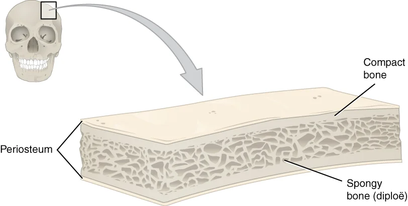
Long description
This cross-section of a flat bone shows the spongy bone (diploë) lined on either side by a layer of compact bone.

Figure 6: This cross-section of a flat bone shows the spongy bone (diploë) lined on either side by a layer of compact bone.
| Component | Description | Location |
|---|---|---|
| Osteocytes | Mature bone cells housed in lacunae | Embedded within both compact and spongy bone |
| Osteoblasts | Bone-forming cells that secrete osteoid | Surface of bone, particularly during growth and repair |
| Osteoclasts | Large cells that resorb bone | Surface of bone, especially in areas of remodeling |
| Trabeculae | Spicules of matrix forming a lattice | Spongy bone |
| Lacunae | Small cavities housing osteocytes | Both compact and spongy bone |
| Periosteum | Fibrous membrane covering outer bone surface | External surface of bone, except articular surfaces [2] |
| Endosteum | Delicate membranous lining | Medullary cavity [2] |
| Osteoid | Unmineralized bone matrix | Secreted by osteoblasts before mineralization [1] |
The compact bone appears as a dense, homogeneous pink layer. Within the spongy bone, you can see the trabeculae forming a lattice-like network. The spaces between trabeculae contain red marrow where hematopoiesis occurs.
Figure 7606 Spongy Bone
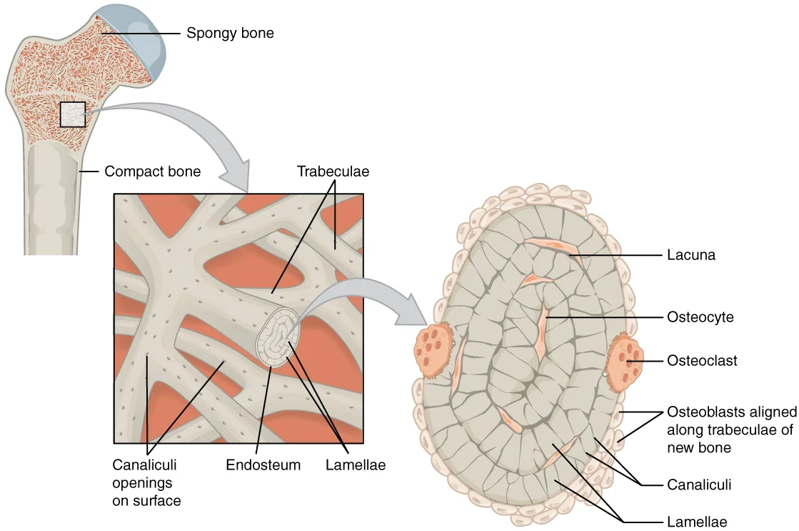
Long description
Spongy bone is composed of trabeculae that contain the osteocytes. Red marrow fills the spaces in some bones.

Figure 7: Spongy bone is composed of trabeculae that contain the osteocytes.
The trabeculae in spongy bone are not randomly arranged but form along lines of stress to provide strength to the bone. This arrangement allows spongy bone to be lighter than compact bone while still providing adequate support. The spaces in the trabeculated network balance the dense and heavy compact bone by making bones lighter so that muscles can move them more easily. [2]
Special stains are essential techniques in bone histology that highlight specific components of bone tissue. These stains allow us to differentiate between various structures and understand their composition and function. One of the most commonly used stains for bone is the von Kossa stain, which demonstrates calcium deposits. This stain is particularly useful for identifying areas of high bone mineralization, such as in osteons and the ca
Another important stain is the Masson's trichrome stain, which differentiates between collagen and mineralized bone. This stain helps visualize the osteoid (unmineralized bone matrix) and mature bone tissue. The Goldner's trichrome modification provides even better contrast between these components, with collagen appearing green and mineralized bone appearing red. These trichrome stains are particularly valuable for studying bone remodeling processes, as they clearly [2]
For examining cellular components, the hematoxylin and eosin (H&E) stain remains fundamental. Hematoxylin stains nuclei purple, while eosin stains the cytoplasm and extracellular matrix pink. In bone tissue, this stain allows identification of osteocytes trapped within lacunae, osteoblasts lining bone surfaces, and osteoclasts in resorption pits. The H&E stain is particularly useful for assessing overall bone cellularity and identifying normal versus pathological cellular arrangements. [2]
The tartrate-resistant acid phosphatase (TRAP) stain is particularly valuable for identifying osteoclasts, which are responsible for bone resorption. These cells show positive staining due to their high content of acid phosphatase enzymes. TRAP staining helps identify areas of active bone resorption, which is crucial in understanding conditions like osteoporosis and other metabolic bone diseases. In normal bone, osteoclasts are found at sites where bone remodeling is occurring, particularly on t [2]
Special stains can also reveal the effects of various hormones on bone tissue. For example, staining can demonstrate the increased osteoblastic activity stimulated by growth hormone, thyroxine, and sex hormones. These hormones promote bone matrix production and are responsible for the growth spurt during adolescence. Additionally, stains can show the effects of vitamin D on bone mineralization, as vitamin D is critical for calcium absorption and bone health.
In older adults, special stains can help identify age-related changes in bone, such as decreased mineralization and increased porosity. These changes contribute to the bone loss that occurs with aging and the increased risk of fractures. Regular exercise, particularly resistance training, has been shown to increase bone density, and special stains can demonstrate the increased bone deposition that occurs in response to mechanical stress.
The H&E stain reveals purple-stained osteocyte nuclei within the pink-stained mineralized matrix.
Figure 8Gray73
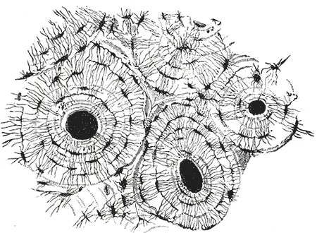
Long description
Transverse section of compact tissue bone. Magnified. (Sharpey.)

Figure 8: Transverse section of compact tissue bone (magnified).
The ultrastructure of bone reveals its complex organization at the electron microscopy level. The microscopic structural unit of compact bone is called an osteon, or Haversian system[1]. Each osteon consists of concentric rings of calcified matrix known as lamellae[1]. Running down the center of each osteon is the central canal, or Haversian canal, which contains blood vessels, nerves, and lymphatic vessels[1]. These vessels and nerves branch off at right angles thr [2]
Bone is composed of three main cell types: osteoblasts, osteocytes, and osteoclasts. Osteoblasts are responsible for bone formation and secrete bone tissue. Osteocytes are mature bone cells that maintain the bone matrix[1]. Osteoclasts are responsible for bone resorption[1]. These osteogenic cells differentiate into osteoblasts[1]. [2]
The process of bone development, or ossification (osteogenesis), begins around the sixth or seventh week of embryonic life. There are two osteogenic pathways: intramembranous ossification and endochondral ossification. During intramembranous ossification, compact and spongy bone develops directly from sheets of mesenchymal connective tissue. The flat bones of the face, most of the cranial bones, and the clavicles are formed via this process. [1]
In endochondral ossification, bone forms on a cartilaginous matrix. This process begins when mesenchymal cells differentiate into chondrocytes that form the cartilaginous skeletal precursor of the bones. As cartilage grows, capillaries penetrate it, transforming the perichondrium into the periosteum. Osteoblasts then form a periosteal collar of compact bone around the cartilage of the diaphysis. By the second or third month of fetal life, bone cell development and ossification create the primary ossification center. [2] [1]
Bones continue to grow in length until early adulthood, with the rate controlled by hormones. Growth occurs when osseous tissue is added to the diaphysis. When chondrocytes in the epiphyseal plate cease proliferation and bone replaces cartilage, longitudinal growth stops, leaving only the epiphyseal line. [1]
Bone histology reveals the dynamic nature of osseous tissue through its cellular components and matrix organization. The basic functional unit of compact bone is the osteon, also known as the Haversian system. Each osteon consists of concentric layers of bone matrix called lamellae, surrounding a central canal known as the Haversian canal. These central canals contain blood vessels and nerves that supply the bone tissue. Running perpendicular to the Haversian canals are Volkmann's canals, which [1]
Figure 9624 Diagram of Compact Bone-new
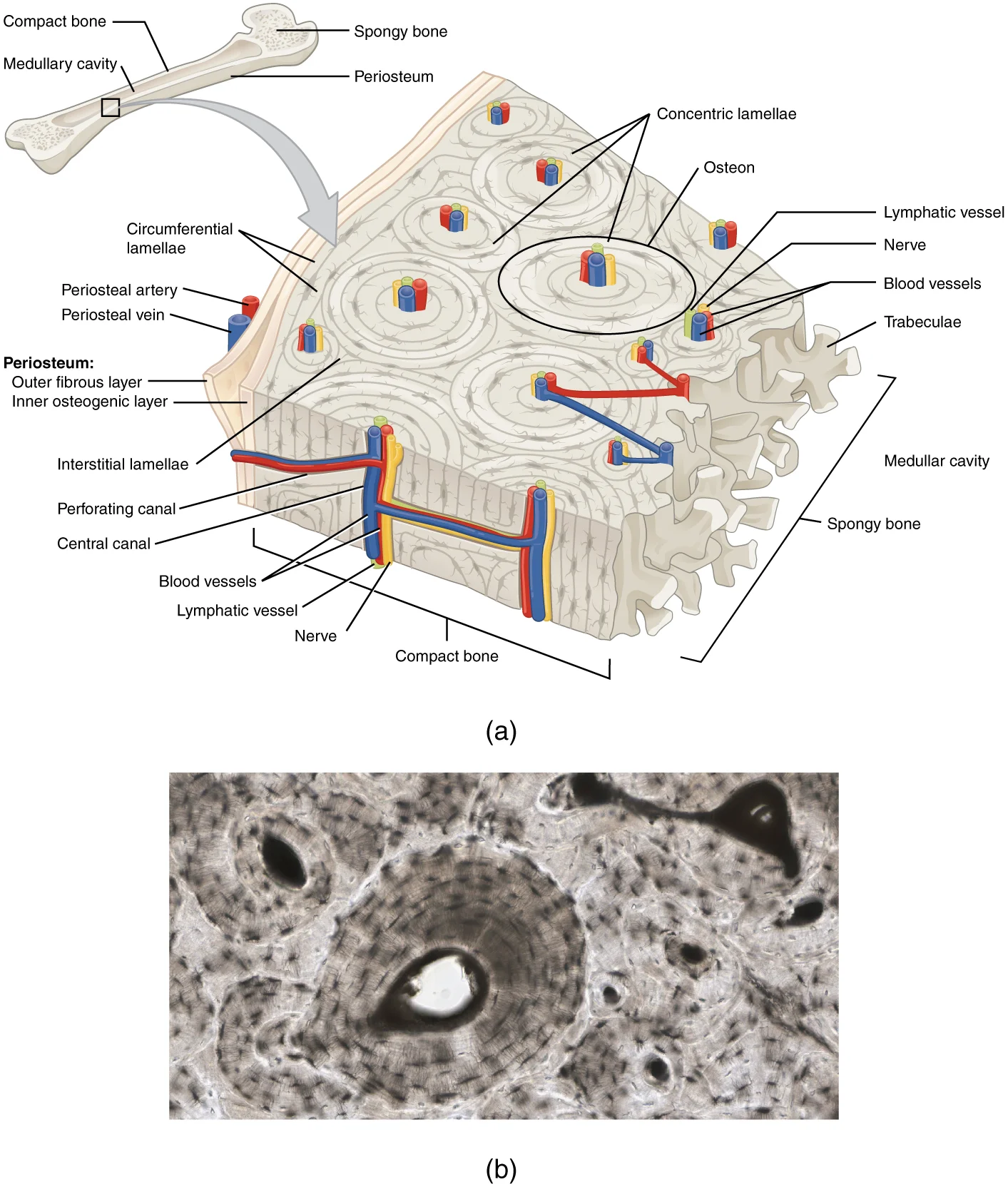
Long description
(a) This cross-sectional view of compact bone shows the basic structural unit, the osteon. (b) In this micrograph of the osteon, you can clearly see the concentric lamellae and central canals. LM × 40. (Micrograph provided by the Regents of University of Michigan Medical School © 2012)

Figure 9: (a) This cross-sectional view of compact bone shows the basic structural unit, the osteon.
Figure 10Gray77
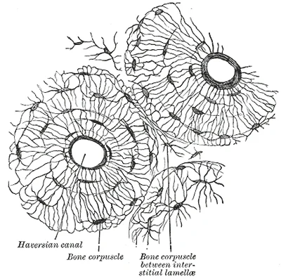
Long description
Transverse section of body of human fibula, decalcified. X 250.

Figure 10: Transverse section of body of human fibula, decalcified (x 250).
Figure 11Gray74
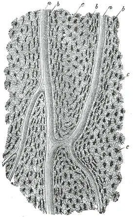
Long description
Section parallel to the surface from the body of the femur. X 100. a, Haversian canals; b, lacunæ seen from the side; c, others seen from the surface in lamellæ, which are cut horizontally.

Figure 11: Section parallel to the surface from the body of the femur (x 100).
Figure 12Gray75
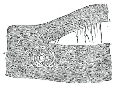
Long description
Perforating fibers, human parietal bone, decalcified. (H. Müller.) a, perforating fibers in situ; b, fibres drawn out of their sockets; c, sockets.

Figure 12: Perforating fibers, human parietal bone, decalcified (H.
Bone is a specialized connective tissue that serves multiple functions in the body, including support, protection, mineral storage, and blood cell production. The unique structure of bone directly enables these diverse functions.
The fundamental structural unit of bone is the osteon, also known as the Haversian system. Each osteon consists of concentric layers of bone matrix called lamellae, surrounding a central canal that contains blood vessels and nerves. The osteocytes, mature bone cells, are located within lacunae between these lamellae and extend processes through tiny channels called canaliculi. This network allows for communication and nutrient exchange between osteocytes and the blood supply. [2]
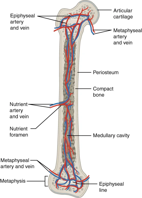
Figure 13609 Body Supply to the Bone
Long description
Blood vessels and nerves enter the bone through the nutrient foramen.

Figure 13: Blood vessels and nerves enter the bone through the nutrient foramen.
The matrix of bone is composed of both organic and inorganic components. The organic component consists primarily of collagen fibers that provide flexibility and tensile strength. The hydroxyapatite crystals give bones their hardness and compressive strength, while the collagen fibers prevent the bone from being brittle. This combination of materials creates a structure that is both strong and resilient, capable of withstanding mechanical stress. [2] [3]
Bone development begins with a cartilage model that serves as a template for bone formation. Chondroblasts produce a flexible matrix of hyaluronic acid, chondroitin sulfate, collagen fibers, and water. As these cells become surrounded by matrix, they are called chondrocytes. Cartilage is avascular, relying on diffusion for nutrient supply and waste removal, which contributes to its limited healing capacity[1]. During endochondral ossification, this cartilage model is gradually replaced by bone tissue through the action of osteoblasts, which secrete bone matrix, and osteoclasts, which resorb cartilage and bone.
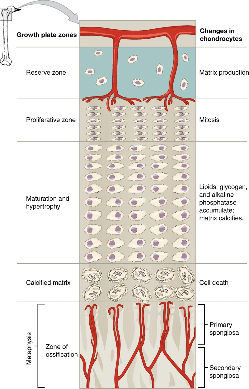
The growth plate, or epiphyseal plate, is responsible for longitudinal bone growth. It consists of several zones, including the proliferative zone where chondrocytes divide via mitosis, and the zone of maturation and hypertrophy where cells enlarge. The longitudinal growth of bone results from cellular division in the proliferative zone and maturation in the zone of maturation and hypertrophy[1]. This organized process allows bones to lengthen during development while maintaining their structural integrity.
Figure 14622 Longitudinal Bone Growth
Long description
The epiphyseal plate is responsible for longitudinal bone growth.

Figure 14: The epiphyseal plate is responsible for longitudinal bone growth.
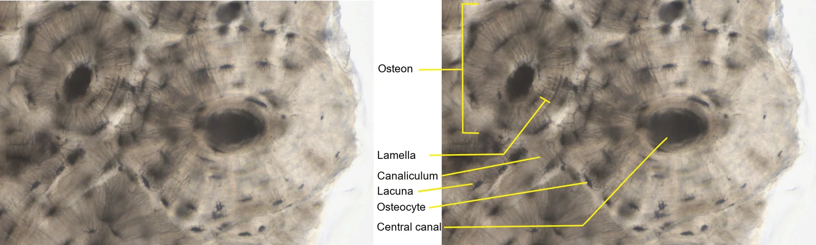
Each osteon's concentric structure provides maximum strength in response to mechanical loads, while the central canal ensures adequate blood supply to maintain bone health.
Figure 15Compact bone 093B
Long description
Photomicrograph taken from Michigan Histological and Vitual Microscopy Learning Resources website, https://histology.medicine.umich.edu/full-slide-list. Site originally compiled by Kent Christensen, Ph.D., J. Matthew Vel

Figure 15: Photomicrograph taken from Michigan Histological and Vitual Microscopy Learning Resources website, https://histology.medicine.umich.edu/full-slide-list.
Bone tissue can be identified on slides by its distinctive structural components and organization. The key features include the presence of osteocytes housed within lacunae, surrounded by calcified matrix arranged in concentric layers called lamellae. These lamellae form the basic structural units of bone tissue and are organized into functional units known as osteons or Haversian systems. [1]
When examining a bone slide, look for the characteristic pattern of concentric rings surrounding a central canal, which contains blood vessels and nerves. The osteocytes appear as small dark dots within the lacunae, and canaliculi (tiny channels) can be seen radiating from these lacunae, connecting them to each other and to the central canal. This network is essential for nutrient transport and waste removal in the avascular bone tissue. Once entrapped, these cells become osteocytes. [2] [1]
DefinitionOsteon
The fundamental functional unit of compact bone, consisting of concentric lamellae surrounding a central canal.
Several tissues may resemble bone on slides but can be distinguished by specific features. Cartilage, for example, lacks the organized lamellar structure and contains chondrocytes in lacunae rather than osteocytes. In developing bone, areas of cartilage may be present, but they can be identified by the absence of calcification and the presence of chondrocytes that haven't undergone the transformation to osteoblasts. During bone development, as more matrix is produced, the chondrocytes in the center of the cartilaginous model grow in size. As the matrix calcifies, nutrients can no longer reach the chondrocytes, resulting in their death and the disintegration of the surrounding cartilage. Blood vessels then invade the resulting spaces, carrying osteogenic cells with them, many of which become osteoblasts. [1]
| Feature | Bone | Cartilage |
|---|---|---|
| Cell type | Osteocytes | Chondrocytes |
| Matrix | Calcified, organized in lamellae | Uncalcified, amorphous |
| Vascularity | Vascular (except in mature bone tissue) | Avascular |
| Canaliculi | Present | Absent |
| Development | Forms from cartilage model | Direct connective tissue |
Bone identification on slides
- Examine slideLook for organized structure
- Question: Concentric lamellae present?
Yes
- Look for lacunae and canaliculiConfirm osteocytes
- Identify as bone
No
- Check for chondrocytesLook for cartilage
- Identify as cartilage
The ultrastructure of bone reveals its complex organization at the electron microscopy level. The microscopic structural unit of compact bone is called an osteon, or Haversian system[1]. Each osteon consists of concentric rings of calcified matrix known as lamellae[1]. Running down the center of each osteon is the central canal, or Haversian canal, which contains blood vessels, nerves, and lymphatic vessels[1]. These vessels and nerves branch off at right angles thr
The osteons appear as circular structures with concentric lamellae. Between the osteons, you can see the interstitial lamellae, which are remnants of previous osteons that have been partially resorbed during bone remodeling. The outermost layer of the bone is the periosteum, which contains blood vessels and osteoprogenitor cells. The inner surface facing the bone marrow is lined by endosteum, which also contains osteoprogenitor cells.
Osteoblasts are responsible for bone formation and secrete the organic components of bone matrix, primarily type I collagen. Once surrounded by matrix, osteoblasts differentiate into osteocytes, which maintain the bone tissue. [1]
The process of bone remodeling occurs throughout life and involves the coordinated action of osteoclasts and osteoblasts. This process maintains bone homeostasis and repairs microdamage. The remodeling cycle can be divided into four phases: activation, resorption, reversal, and formation. During activation, osteoclast precursors are recruited to the bone surface. In the resorption phase, osteoclasts resorb bone, creating resorption pits called Howship's lacunae. The reversal phase involves the r [1]
Clinical pearlOsteons
The basic functional unit of compact bone is the osteon (Haversian system), consisting of concentric lamellae around a central canal containing blood vessels. Each osteon typically measures 3-5 mm in length and 0.2-0.3 mm in diameter, with approximately 4-20 lamellae per osteon. The central canal houses vessels that supply nutrients to the osteocytes via canaliculi.
Exam trapWoven bone
Woven bone is the first bone formed during development and fracture healing, characterized by randomly arranged collagen fibers; it's weaker than lamellar bone and is eventually replaced. This immature bone type constitutes approximately 60% of fetal bone but is largely replaced by lamellar bone by age 5. Woven bone lacks organized collagen orientation and has a higher osteocyte density but reduced mechanical strength. [1]
Clinical pearlBone cells
Osteoblasts build bone, osteoclasts resorb bone; the balance between these cells determines bone mass. Osteoblasts originate from mesenchymal stem cells and secrete osteoid at a rate of approximately 1-2 μm/day. Osteocytes, derived from osteoblasts, comprise approximately 90-95% of all bone cells and act as mechanosensors. [1]
Clinical pearlRemodeling cycle
Bone remodeling occurs in units called basic multicellular units (BMUs), with a remodeling cycle of approximately 100-200 days in cortical bone. The process involves a resorption phase lasting about 2-3 weeks, followed by a reversal phase of 2-3 weeks, and a formation phase of 3-4 months. Approximately 5-10% of the adult skeleton is remodeled annually, with each BMU replacing about 25-100 mm³ of bone. [1]
Exam trapOsteoid
Osteoid is the unmineralized organic matrix produced by osteoblasts; normal mineralization requires adequate vitamin D, calcium, and phosphate levels. Osteoid typically constitutes approximately 30-35% of the bone volume before mineralization. Mineralization begins approximately 10-15 days after osteoid formation and is complete within 3-4 months. Impaired mineralization leads to osteomalacia, characterized by excessive unmineralized osteoid. [1]
Clinical pearlTrabecular bone
Trabecular (cancellous) bone has a higher surface area to volume ratio than cortical bone, making it more metabolically active and the primary site of bone remodeling. Trabecular bone comprises approximately 20% of the total bone mass but accounts for 80% of the total bone surface area. Trabeculae typically measure 50-500 μm in thickness and have a porosity of approximately 50-90%. [1]
Clinical pearlCanaliculi
Canaliculi are microscopic channels connecting osteocytes to each other and to the Haversian canal, allowing nutrient exchange and communication. Each osteocyte maintains connections with approximately 40-60 neighboring cells through canaliculi. These channels, typically 0.1-0.5 μm in diameter, contain extensions of osteocyte cytoplasm surrounded by perilacunar bone. The network of canaliculi facilitates the transport of nutrients, waste products, and signaling molecules throughout the bone matr [1]
Exam trapBone lining cells
Bone lining cells are flat, inactive osteoblasts that cover quiescent bone surfaces; they can be activated to become osteoblasts when needed. These cells form a thin layer approximately 1-2 cells thick and constitute approximately 4-6% of all bone cells. In adults, approximately 90% of bone surfaces are covered by lining cells, which regulate the passage of calcium and phosphate between bone and blood. These cells play a crucial role in initiating bone remodeling by detecting mechanical stress a [1]
Clinical pearlPeriosteum
The periosteum is a connective tissue membrane covering external bone surfaces. It contains blood vessels, nerves, and osteoprogenitor cells essential for bone growth and repair. It typically measures 0.1-0.5 mm in thickness and consists of an outer fibrous layer and an inner cambium layer. The periosteum is attached to bone by Sharpey's fibers and is approximately 2-3 times thicker in children than in adults. This membrane provides approximately 50% of the blood supply to cortical bone and is cr [1]
Clinical pearlEndosteum
The endosteum is a thin membrane lining internal bone cavities, containing osteoprogenitor cells that can differentiate into osteoblasts for bone remodeling. It typically measures 10-50 μm in thickness and lines the medullary cavity, Haversian canals, and Volkmann's canals. The endosteum contains approximately 1-2 layers of cells and is approximately 3-5 times more metabolically active than the periosteum. This membrane plays a crucial role in bone modeling and remodeling, particularly in respon [1]
Clinical pearlBone mineral
Bone mineral is primarily hydroxyapatite crystals, composed of calcium phosphate; this mineral gives bone its hardness and compressive strength. These crystals measure approximately 20-50 nm in length and 3-6 nm in width, with a surface area of approximately 100 m²/g. The mineral phase provides approximately 80% of the bone's compressive strength while the organic matrix [1]
Exam trapRuffled border
This structure increases the osteoclast's resorptive surface area by approximately 10-20 times. The ruffled border contains proton pumps that maintain a pH of approximately 4.5 in the resorption pit, allowing dissolution of bone mineral. The sealing zone, adjacent to the ruffled border, contains actin rings that create a [1]
Exam trapIncomplete ossification
A common mistake is assuming all bones are fully ossified at birth. In fact, the skull and clavicles are not fully ossified nor are the sutures of the skull closed at birth. This allows the skull and shoulders to deform during passage through the birth canal.
Exam trapCalcium sources misconception
Many students believe calcium is only found in dairy products. However, calcium is also present in green leafy vegetables, broccoli, and intact salmon and canned sardines with their soft bones. Nuts, beans, seeds, and shellfish provide calcium in smaller quantities. [3]
Exam trapVitamin D deficiency
Students often overlook vitamin D deficiency as a significant factor in bone health. Except for fatty fish like salmon and tuna, or fortified milk or cereal, vitamin D is not found naturally in many foods. The action of sunlight on the skin triggers the body to produce its own vitamin D, but many people, especially those of darker complexion and those living in northern latitudes, are deficient in vitamin D. [3]
Exam trapPTH function misunderstanding
A frequent error is oversimplifying parathyroid hormone (PTH) function. PTH stimulates osteoclast proliferation and activity, causing calcium to be released from bones into circulation. PTH also promotes reabsorption of calcium by kidney tubules, affecting calcium homeostasis. [3]
Exam trapSystem interdependence
Students often view bones in isolation rather than recognizing their interdependence with other systems. The skeletal system is affected by nutrients from the digestive system and hormones from the endocrine system. Even muscle use during exercise impacts bone health. [3]
Exam trapIntramembranous ossification timeline
Many students incorrectly assume intramembranous ossification completes before adolescence. In reality, it begins in utero during fetal development and continues into adolescence. The last bones to ossify via this process are the flat bones of the face, which reach their adult size at the end of the adolescent growth spurt. [1]
NoteClinical assessment
Understanding bone development is crucial in clinical assessment. Knowledge that skull sutures are not closed at birth helps clinicians recognize normal variations from pathological conditions. This understanding aids in diagnosing conditions like craniosynostosis, where premature fusion of sutures occurs.
NoteNutritional counseling
Bone histology knowledge informs nutritional counseling. Awareness that calcium comes from various sources beyond dairy allows for better dietary recommendations. Similarly, understanding vitamin D sources and production helps identify at-risk populations for deficiency.
NoteEndocrine disorders
Knowledge of PTH function is essential in managing endocrine disorders affecting bone. PTH stimulates osteoclast activity, releasing calcium from bones. This understanding helps in diagnosing and treating conditions like hyperparathyroidism, where excessive PTH leads to bone demineralization.
NoteExercise prescription
Understanding bone physiology informs exercise prescription. Knowledge that mechanical stress from muscle use impacts bone health helps in designing weight-bearing exercise programs to prevent osteoporosis. This is particularly important for patients with limited mobility or those on long-term steroid therapy.
Assessing bone health
- Assess dietary calcium intakeEvaluate sources beyond dairy
- Question: Adequate calcium intake?
No
- Recommend calcium-rich foodsGreen leafy vegetables, broccoli, salmon with bones
Yes
- Assess vitamin D statusConsider sun exposure, skin tone, latitude
- Question: Vitamin D sufficient?
No
- Consider supplementationEspecially for darker skin, northern latitudes
Yes
- Evaluate PTH functionCheck for signs of hyperparathyroidism
NoteImaging interpretation
Bone histology knowledge aids in interpreting imaging studies. Understanding that intramembranous ossification continues into adolescence helps radiologists differentiate normal developmental variations from pathological findings. This is particularly important in pediatric imaging.
NoteSurgical planning
Knowledge of bone development and structure informs surgical planning. Understanding that flat bones of the face complete ossification at the end of the adolescent growth spurt helps surgeons plan procedures for facial trauma or reconstruction. This timing consideration is crucial for optimal outcomes in pediatric and adolescent patients.
- Intramembranous ossification begins in utero and continues into adolescence, with facial bones completing development at the end of the adolescent growth spurt
- Bones are interdependent with other systems, affected by nutrients from digestion and hormones from the endocrine system
- Calcium sources include dairy, green leafy vegetables, broccoli, and fish with soft bones
- Vitamin D is primarily obtained through sunlight exposure, with deficiency common in darker-skinned individuals and those in northern latitudes
- PTH stimulates osteoclast activity, releasing calcium from bones and promoting calcium reabsorption by kidneys
Answer each one in your head first, then open the answer.
Which bones are the last to complete intramembranous ossification?
Show answerHide answer
The flat bones of the faceBesides dairy, what are three other sources of calcium?
Show answerHide answer
Green leafy vegetables, broccoli, and intact salmon and canned sardines with their soft bonesWhat is the primary source of vitamin D for most people?
Show answerHide answer
Sunlight exposure on the skinWhat effect does PTH have on osteoclasts?
Show answerHide answer
PTH stimulates osteoclast proliferation and activityHow does exercise affect bone health?
Show answerHide answer
Using muscles to engage in exercise has an impact on bones
- Betts JG, Young KA, Wise JA, et al. Bone Formation and Development. OpenStax, Anatomy and Physiology 2e (CC BY-NC-SA 4.0). Accessed .
- Betts JG, Young KA, Wise JA, et al. Bone Structure. OpenStax, Anatomy and Physiology 2e (CC BY-NC-SA 4.0). Accessed .
- Betts JG, Young KA, Wise JA, et al. Exercise, Nutrition, Hormones, and Bone Tissue. OpenStax, Anatomy and Physiology 2e (CC BY-NC-SA 4.0). Accessed .
Figure credits
- Figure 1: OpenStax College, via Wikimedia Commons, CC BY 3.0, CC BY 3.0.
- Figure 2: OpenStax College, via Wikimedia Commons, CC BY 3.0, CC BY 3.0.
- Figure 3: OpenStax College, via Wikimedia Commons, CC BY 3.0, CC BY 3.0.
- Figure 4: OpenStax College, via Wikimedia Commons, CC BY 3.0, CC BY 3.0.
- Figure 5: OpenStax College, via Wikimedia Commons, CC BY 3.0, CC BY 3.0.
- Figure 6: OpenStax College, via Wikimedia Commons, CC BY 3.0, CC BY 3.0.
- Figure 7: OpenStax College, via Wikimedia Commons, CC BY 3.0, CC BY 3.0.
- Figure 8: Henry Vandyke Carter, via Wikimedia Commons, Public domain, Public domain.
- Figure 9: OpenStax College, via Wikimedia Commons, CC BY 3.0, CC BY 3.0.
- Figure 10: Henry Vandyke Carter, via Wikimedia Commons, Public domain, Public domain.
- Figure 11: Henry Vandyke Carter, via Wikimedia Commons, Public domain, Public domain.
- Figure 12: Henry Vandyke Carter, via Wikimedia Commons, Public domain, Public domain.
- Figure 13: OpenStax College, via Wikimedia Commons, CC BY 3.0, CC BY 3.0.
- Figure 14: OpenStax College, via Wikimedia Commons, CC BY 4.0, CC BY 4.0.
- Figure 15: RWhitwam, via Wikimedia Commons, CC BY-SA 4.0, CC BY-SA 4.0.
Educational content for medical students and doctors. It is not a substitute for clinical judgement or local guidelines.