Muscle tissue
Muscle tissue consists of specialized cells that contract to produce movement.
Muscle tissue is a specialized form of tissue that has the ability to contract, producing movement in the body. There are three major types of muscle tissue: skeletal, cardiac, and smooth, each with distinct characteristics and functions.
Figure 1414 Skeletal Smooth Cardiac
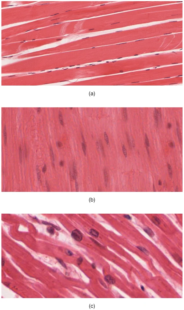
Long description
The body contains three types of muscle tissue: (a) skeletal muscle, (b) smooth muscle, and (c) cardiac muscle. (Micrographs provided by the Regents of University of Michigan Medical School © 2012)

Figure 1: The body contains three types of muscle tissue: (a) skeletal muscle, (b) smooth muscle, and (c) cardiac muscle.
Skeletal muscle is voluntary and responds to conscious stimuli. The cells are striated and multinucleated, appearing as long, unbranched cylinders. Each skeletal muscle is an organ consisting of various integrated tissues including skeletal muscle fibers, blood vessels, nerve fibers, and connective tissue. These muscles are wrapped in a sheath of dense, irregular connective tissue called the epimysium, which allows the muscle to contract and move powerfully while maintaining its structural integrity. [3]
Cardiac muscle is involuntary and found only in the heart. The cells, known as cardiomyocytes, are striated with a single centrally located nucleus. Compared to skeletal muscle, cardiac muscle cells are considerably shorter with much smaller diameters. Cardiac muscle fibers are branched and connected to one another at their ends by intercalated discs[1]. These intercalated discs contain two important structures: gap junctions and desmosomes[1]. Gap junctions form channels between adjacent cardiac muscle fibers, allowing depolarizing current to flow from one cell to the next, enabling quick transmission of action potentials and coordinated contraction[1]. Desmosomes anchor the ends of cardiac muscle fibers together so the cells do not pull apart during contraction[1]. [2][3]
Figure 2414c Cardiacmuscle
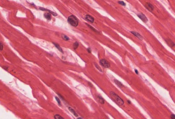
Long description
Cardiac muscle tissue is only found in the heart. LM × 1600. (Micrograph provided by the Regents of University of Michigan Medical School © 2012)

Figure 2: Cardiac muscle tissue is only found in the heart (lm × 1600).
The contractile elements in cardiac muscle are virtually identical to skeletal muscle, organized in sarcomeres along the length of the cell. Contraction in cardiac muscle is triggered by Ca++ ions that come from the sarcoplasmic reticulum and through voltage-gated calcium channels in the sarcolemma[1]. [2][3]
Smooth muscle is involuntary, with each cell being a spindle-shaped fiber containing a single nucleus. No striations are evident because the actin and myosin filaments do not align in the cytoplasm. [3]
Smooth muscle fibres are spindle-shaped, with a single central nucleus and no visible striations[1]. [2][3]
Blood vessels and nerves run within these layers. [1]
| Feature | Skeletal | Cardiac | Smooth |
|---|---|---|---|
| Fibre shape | Long, cylindrical, unbranched | Short, branched, cylindrical | Spindle-shaped, tapered ends |
| Nuclei | Multiple, peripheral | Single, central | Single, central |
| Striations | Prominent, regular | Prominent, regular | Absent |
| Intercalated discs | Absent | Present (dark lines) | Absent |
| Connective tissue layers | Epimysium, perimysium, endomysium | Endomysium only (plus perimysium around bundles) | Endomysium (fine) |
Fibre types on staining are best seen with histochemical methods, not routine H&E. Type I (slow oxidative) fibres stain dark with oxidative enzyme stains (e.g., NADH-TR) because they are rich in mitochondria and myoglobin; they appear red in fresh tissue. Type II (fast glycolytic) fibres stain pale with oxidative stains but dark with ATPase stains at alkaline pH; they appear white in fresh tissue. In a typical limb muscle, the two types are intermingled, giving a checkerboard pattern on ATPase staining. Type I fibres are smaller in diameter, while type II fibres are larger.
The fibres are polygonal and packed together, each surrounded by a thin endomysial line. Groups of fibres form fascicles bounded by the perimysium. The dark Type II fibres are scattered singly among pale Type I fibres, producing the characteristic mosaic pattern.
Special stains are essential for visualizing different components of muscle tissue that are not apparent with standard hematoxylin and eosin (H&E) staining. These stains highlight specific structures and cellular components, allowing for better identification and characterization of muscle types. [1]
DefinitionSpecial stains
Histological techniques that use specific dyes to highlight particular cellular structures or components in muscle tissue.
For skeletal muscle, special stains can demonstrate the striated pattern created by the organized arrangement of actin and myosin filaments within sarcomeres. The periodic acid-Schiff (PAS) stain may be used to visualize glycogen storage within muscle fibers, as cardiac muscle stores glycogen in its cytoplasm[1]. [2]
In cardiac muscle, special stains help identify intercalated discs, which are critical structures that mark junctions between adjoining cardiac cells[1]. These discs contain desmosomes and gap junctions that allow for synchronized contraction. Masson's trichrome stain can highlight the intercellular connective tissue that binds cardiac muscle cells together, which is necessary to withstand the forces exerted during contraction[1]. [2]
For smooth muscle, stains like Verhoeff's van Gieson can help distinguish it from other tissues by highlighting its spindle-shaped cells and lack of striations. Special stains can also demonstrate the presence of myoglobin in muscle tissue, a protein that stores oxygen and is found in both skeletal and cardiac muscle cells[1].
| Stain | Target structure | Application in muscle tissue |
|---|---|---|
| Masson's trichrome | Collagen | Highlights connective tissue between muscle fibers |
| PAS | Glycogen | Visualizes glycogen storage in cardiac muscle |
| Verhoeff's van Gieson | Elastic fibers | Helps identify smooth muscle in tissue sections |
| Iron hematoxylin | Myofibrils | Enhances visualization of striations in skeletal muscle |
The ultrastructure of skeletal muscle, as revealed by electron microscopy, reveals highly organized contractile units called sarcomeres. The sarcomere is the basic functional unit of skeletal muscle and extends from one Z-disc to the next Z-disc. Within each sarcomere, thick filaments composed of myosin project from the center toward, but not all the way to, the Z-discs. These myosin strands and their multiple heads are thicker and have more mass than the thin filaments, earning them the designation of thick filaments. [1]
The thin filaments are primarily composed of actin and are arranged in a regular pattern with the thick filaments. This precise arrangement of actin and myosin creates the characteristic striated appearance of skeletal muscle tissue. The striation pattern results from the regular alternation of these contractile proteins along with structural proteins that couple the contractile proteins to connective tissues. [3]
Surrounding each muscle fiber is the sarcolemma, which is the plasma membrane of the muscle cell. The sarcolemma contains specialized regions called motor end-plates where neurotransmitter receptors are located. Invaginations of the sarcolemma form T-tubules that carry action potentials deep into the muscle fiber interior. Adjacent to the T-tubules are the sarcoplasmic reticulum (SR), a specialized form of endoplasmic reticulum that stores calcium ions.
The sarcoplasm, or cytoplasm of the muscle fiber, contains numerous myofibrils, which are cylindrical organelles composed of many sarcomeres arranged end-to-end. These myofibrils are responsible for the contractile properties of muscle tissue. The highly organized arrangement of these structures at the ultrastructural level enables efficient muscle contraction and force generation.
Myosin filaments appear thicker and are centrally positioned, while actin filaments are thinner and extend toward the Z-discs.
Muscle tissue exhibits remarkable structure-function correlations that enable its specialized contractile properties. The fundamental unit of skeletal muscle is the muscle fiber, which can reach diameters of up to 100 μm. To ensure efficient excitation-contraction coupling in these large cells, specialized structures called T-tubules ("T" for transverse) periodically invaginate the sarcolemma. These T-tubules form triads with the sarcoplasmic reticulum (SR) membranes on either side, surrounding the myofibrils that contain the contractile proteins actin and myosin. This arrangement allows the action potential to rapidly reach the SR, triggering calcium ion release and subsequent muscle contraction. [1] [2]
The excitation process begins with depolarization of the membrane, which is immediately followed by repolarization to re-establish the negative membrane potential. At the neuromuscular junction, acetylcholine (ACh) released into the synaptic cleft is rapidly degraded by acetylcholinesterase (AChE), preventing unwanted extended muscle excitation and contraction. This precise timing ensures that muscle contractions are brief and controlled, allowing for rapid movements like finger snapping.
In cardiac muscle, the structure-function relationship is adapted for rhythmic, coordinated contractions. The total time from impulse initiation in the SA node until ventricular depolarization is approximately 225 ms[2].
Cardiac myocytes exhibit a distinct electrical pattern with rapid depolarization, followed by a plateau phase and repolarization, creating long refractory periods essential for effective blood pumping before a second contraction can occur. These cells normally do not initiate their own electrical potential but wait for an impulse to reach them. Damaged hearts or those stimulated by drugs may contract at higher rates but lose pumping effectiveness. [2]
Muscle tissue can be identified on slides by examining its distinctive cellular characteristics and organization. The three types of muscle tissue—skeletal, cardiac, and smooth—each have unique features that allow for identification.
Skeletal muscle fibers are long, cylindrical, multinucleated cells with peripheral nuclei. They exhibit a striated pattern of alternating light and dark bands when viewed under the microscope. The dark bands (A bands) contain the thick filaments of myosin, while the light bands (I bands) contain the thin filaments of actin. The Z discs bisect the I bands and define the sarcomere units, which are the functional contractile units of muscle.
Cardiac muscle cells are shorter and branched than skeletal muscle fibers, with a single central nucleus. They also display striations but have a less organized pattern than skeletal muscle. A defining characteristic of cardiac muscle is the presence of intercalated discs, which are specialized junctions that contain gap junctions and desmosomes. These discs allow for synchronized contraction and electrical communication between adjacent cells.
Smooth muscle cells are spindle-shaped with a single central nucleus and no striations. They are typically arranged in sheets or layers, often with nuclei that appear wavy or pleomorphic when viewed in cross-section. Unlike skeletal and cardiac muscle, smooth muscle cells lack the highly organized sarcomere structure.
When identifying muscle tissue, it's important to distinguish between these types. Skeletal muscle shows clear striations and multiple peripheral nuclei. Cardiac muscle shows striations but with intercalated discs and central nuclei. Smooth muscle lacks striations entirely and has a spindle-shaped appearance with a single central nucleus. [2]
The fibers are grouped into fascicles, separated by perimysium, and enclosed by epimysium. The nuclei are located at the periphery of each fiber, just beneath the sarcolemma.
The histological correlates of cardiac muscle include specialized conduction cells that coordinate the contraction of the heart. These cells are modified myocytes that prioritise electrical conduction over contractile force.
It arises from the AV node and proceeds through the interventricular septum. This anatomical course ensures that the electrical impulse is directed away from the atria and toward the apex of the heart before spreading upwards through the ventricular walls. [2]
DefinitionBundle of His
The atrioventricular bundle that transmits electrical impulses from the AV node through the interventricular septum to the bundle branches.
After passing through the septum, the atrioventricular bundle divides into two distinct pathways: the left and right bundle branches. These branches travel along the respective sides of the interventricular septum to deliver the impulse to the ventricular myocardium.
This asymmetry is a histological correlate of the functional demands of the heart; the left ventricle is much larger than the right, requiring a more robust conduction system to ensure efficient depolarization of its greater muscle mass. [2]
| Feature | Left Bundle Branch | Right Bundle Branch |
|---|---|---|
| Target Area | Left ventricle | Right ventricle |
| Relative Size | Considerably larger | Smaller |
In the right ventricle, a specialized muscular structure called the moderator band serves as a shortcut for electrical conduction. This arrangement allows the electrical impulse to bypass the main body of the ventricular wall and supply the right papillary muscles directly. [2]
The histological purpose of this connection is synchronisation. Because of the moderator band, each papillary muscle receives the electrical impulse at approximately the same time. This ensures that the papillary muscles begin to contract simultaneously, just before the rest of the myocardial contractile cells in the ventricles contract. [2]
Cardiac Impulse Pathway
- AV NodeOrigin of the impulse
- Bundle of HisProceeds through interventricular septum
- Question: Branching
Left
- Left Bundle BranchSupplies the larger left ventricle
Right
- Right Bundle BranchSupplies the right ventricle
- Moderator BandContains right bundle branch
- Right Papillary MusclesContract simultaneously
The septum is the central muscular wall separating the two ventricles. From here, the bundle divides into the left and right bundle branches. The left bundle branch is located on the left side of the septum and is visibly larger than the right branch, reflecting its role in supplying the larger left ventricle. The right bundle branch is seen on the right side of the septum, with a portion extending into the moderator band. The moderator band appears as a muscular bridge across the right ventricular cavity, terminating at the right papillary muscles. [2]
-
Cardiomyocytes typically have a single, central nucleus, though some may have two or more nuclei. This is in contrast to skeletal muscle fibers which are multinucleated. The central positioning of the nucleus in cardiac muscle allows for more efficient organization of myofibrils around it, optimizing the contractile function of these specialized cells.
-
Cardiac muscle has a slower onset of contraction compared to skeletal muscle because the sarcoplasmic reticulum stores few calcium ions, requiring most calcium to come from outside the cells. This dependence on extracellular calcium explains why cardiac muscle contraction is more susceptible to changes in serum calcium levels and why calcium channel blockers are effective in modulating cardiac contractility. [2]
-
Mitochondria are plentiful in cardiac muscle, comprising about 25-35% of the cell volume, providing energy for the continuous contractions of the heart. This high mitochondrial density reflects the heart's constant metabolic demands and its reliance on aerobic respiration, making cardiac tissue particularly vulnerable to hypoxia. [2]
These pathways consist of specialized conducting cells that are larger than ordinary myocardial cells and conduct impulses more rapidly, ensuring rapid and coordinated spread of the cardiac impulse. [2]
This specialized pathway ensures simultaneous activation of both atria, which is particularly important for maintaining coordinated atrial contraction and efficient atrial emptying into the ventricles. [2]
- Heart block refers to any interruption in the normal conduction pathway of the heart. These abnormalities can range from minor delays in conduction to complete cessation of impulse transmission, potentially leading to bradycardia and reduced cardiac output. [2]
SA nodal blocks often result in junctional escape rhythms, while AV nodal blocks may produce various escape rhythms depending on the level of block. [2]
These blocks typically produce wider QRS complexes on ECG as ventricular activation becomes less coordinated. [2]
These partial blocks may not always produce complete bundle branch block patterns on ECG but can still significantly alter ventricular activation sequences. [2]
AV nodal blocks are particularly common in elderly patients and those with underlying ischemic heart disease, while infra-Hisian blocks often indicate more significant conduction system disease. [2]
Exam trapConfusing depolarization with contraction
Students often mistakenly believe that electrical depolarization directly equals mechanical contraction. In reality, the wave of depolarization is the trigger for muscular contraction, but there is a brief delay between these events. [2]
Exam trapMisinterpreting ECG components
A common error is to assume that all visible waves on an ECG represent mechanical contraction. The P wave corresponds to atrial depolarization, not contraction, while the T wave represents ventricular repolarization, not contraction. [2]
Exam trapOverlooking regional differences in cardiac action potentials
Students frequently fail to recognize that contractile cells have different resting potentials in atria (approximately −80 mV) versus ventricles (approximately −90 mV), despite otherwise identical action potential components[2].
Exam trapMisunderstanding the plateau phase
Many students incorrectly attribute the plateau phase of cardiac action potentials solely to calcium influx. While Ca2+ entry is important, the plateau occurs because Ca2+ enters while few K+ channels are open, creating a balance of ion movements.
Exam trapConfusing conduction pathways with contraction
Students often mistake the moderator band's function for contraction rather than its role in allowing tension to develop on chordae tendineae prior to right ventricular contraction. [2]
Exam trapMisinterpreting Holter monitor duration
A common misconception is that Holter monitoring occurs over longer periods. In standard practice, continuous ambulatory electrocardiographs typically monitor heart electrical activity for 24 hours during the patient's normal routine. [2]
At the bedside, understanding muscle tissue physiology is fundamental for interpreting cardiac electrical activity. The 12-lead electrocardiograph uses 10 electrodes placed in standard locations on the patient's skin to assess heart function. Clinicians rely on recognizing specific ECG components such as the P wave (atrial depolarization), QRS complex (ventricular depolarization), and T wave (ventricular repolarization) to diagnose various cardiac conditions.
In the operating theatre, this knowledge guides cardiac surgeons when performing procedures involving the heart's conduction system. Understanding that the bundle branches descend to the apex where they connect with Purkinje fibers helps surgeons anticipate the heart's natural conduction pathway during interventions. The knowledge that depolarization begins in the right atrium and spreads across both atria before descending to the ventricles informs surgical approaches to certain arrhythmias.
On imaging, particularly with continuous ambulatory monitoring, clinicians use Holter monitors to detect intermittent cardiac electrical abnormalities that might not appear on standard 12-lead ECGs. These portable devices, worn by patients for typically 24 hours during normal activities, capture electrical activity that might be missed during brief in-office examinations.
In examinations, medical students must demonstrate understanding of cardiac muscle electrophysiology, including the differences between conductive and contractile cells. Students are expected to know that contractile cells have a stable resting phase at approximately −80 mV for atrial cells and −90 mV for ventricular cells. They must also recognize that the rapid depolarization period typically lasts 3–5 ms and is followed by a plateau phase where membrane potential declines relatively slowly due to Ca2+ influx with limited K+ efflux. [2]
Cardiac Muscle Conduction
- Depolarization beginsIn right atrium
- Spreads to atriaSuperior to inferior portions
- Question: Reaches AV node?
Yes
- Delays impulseAllows ventricular filling
- Impulse descendsThrough bundle branches
No
- Continues through atriaPumping blood into ventricles
- Reaches apexConnects with Purkinje fibers
- Ventricular contractionFrom apex upward
- The wave of depolarization is the trigger for muscular contraction, beginning in the right atrium
- Contractile cells have different resting potentials: atria at approximately −80 mV and ventricles at approximately −90 mV
- The rapid depolarization period typically lasts 3–5 ms, raising membrane potential to approximately +30 mV [2]
- The P wave corresponds to atrial depolarization, QRS complex to ventricular depolarization, and T wave to ventricular repolarization
- Holter monitors typically record heart electrical activity for 24 hours during normal patient activities
- The moderator band allows tension to develop on chordae tendineae prior to right ventricular contraction
Answer each one in your head first, then open the answer.
What triggers muscular contraction in the heart?
Show answerHide answer
The wave of depolarizationWhat is the resting potential of ventricular contractile cells?
Show answerHide answer
Approximately −90 mVWhich ECG component corresponds to ventricular repolarization?
Show answerHide answer
The T waveHow long does rapid depolarization typically last in cardiac muscle?
Show answerHide answer
3–5 msWhat is the typical duration of Holter monitoring?
Show answerHide answer
24 hours
- Betts JG, Young KA, Wise JA, et al. Skeletal Muscle. OpenStax, Anatomy and Physiology 2e (CC BY-NC-SA 4.0). Accessed .
- Betts JG, Young KA, Wise JA, et al. Cardiac Muscle and Electrical Activity. OpenStax, Anatomy and Physiology 2e (CC BY-NC-SA 4.0). Accessed .
- Betts JG, Young KA, Wise JA, et al. Muscle Tissue and Motion. OpenStax, Anatomy and Physiology 2e (CC BY-NC-SA 4.0). Accessed .
Figure credits
Educational content for medical students and doctors. It is not a substitute for clinical judgement or local guidelines.