Nervous tissue
Nervous tissue consists of neurons and glial cells that coordinate body functions.
Nervous tissue is composed of two types of cells, neurons and glial cells. Neurons are the primary type of cell that most anyone associates with the nervous system and are responsible for the computation and communication that the nervous system provides. They are electrically active and release chemical signals to target cells. Glial cells, or glia, are known to play a supporting role for nervous tissue. [1]
Glial cells, or neuroglia or simply glia, are the other type of cell found in nervous tissue. They are considered to be supporting cells, and many functions are directed at helping neurons complete their function for communication. Several types of glial cells are found in the nervous system, and they can be categorized by the anatomical division in which they are found. In the CNS, astrocytes, oligodendrocytes, microglia, and ependymal cells are found. Astrocytes are important for maintaining the chemical environment around the neuron and are crucial for regulating the blood-brain barrier. [1]
In the PNS, satellite cells are supporting cells for the neurons, and Schwann cells insulate peripheral axons. Oligodendrocyte cells produce myelin in the central nervous system (brain and spinal cord) while the Schwann cell produces myelin in the peripheral nervous system. [1][4]
A ganglion is a group of neuron cell bodies in the periphery. Ganglia can be categorized, for the most part, as either sensory ganglia or autonomic ganglia, referring to their primary functions. The most common type of sensory ganglion is a dorsal (posterior) root ganglion, whose neurons have sensory endings in the periphery that extend into the CNS through the dorsal nerve root. [1][2]
Figure 1Gray937
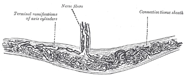
Long description
Nerve ending of Ruffini. (After A. Ruffini.)

Figure 1: Nerve ending of Ruffini (After A.
The other major category of ganglia are those of the autonomic nervous system, which is divided into the sympathetic and parasympathetic nervous systems. The sympathetic chain ganglia constitute a row of ganglia along the vertebral column that receive central input from the lateral horn of the thoracic and upper lumbar spinal cord. Three other autonomic ganglia that are related to the sympathetic chain are the prevertebral ganglia, which are located outside of the chain but have similar functions. They are referred to as prevertebral because they are anterior to the vertebral column. The neurons of these autonomic ganglia are multipolar in shape, with dendrites radiating out around the cell body where synapses from the spinal cord neurons are made. [2] [3]
The PNS is composed of the groups of neurons (ganglia) and bundles of axons (nerves) that are outside of the brain and spinal cord. Ganglia are of two types, sensory or autonomic. Sensory ganglia contain unipolar sensory neurons and are found on the dorsal root of all spinal nerves as well as associated with many of the cranial nerves. Autonomic ganglia are in the sympathetic chain, the associated paravertebral or prevertebral ganglia, or in terminal ganglia near or within the organs controlled by the autonomic nervous system. Not all axons from the central neurons terminate in the chain ganglia. Additional branches from the ventral nerve root continue through the chain and on to one of the collateral ganglia as the greater splanchnic nerve or lesser splanchnic nerve. For example, the greater splanchnic nerve at the level of T5 synapses with a collateral ganglion outside the chain before making the connection to the postganglionic nerves that innervate the stomach. [2] [3]
Figure 21318b Dorsal Root Ganglion
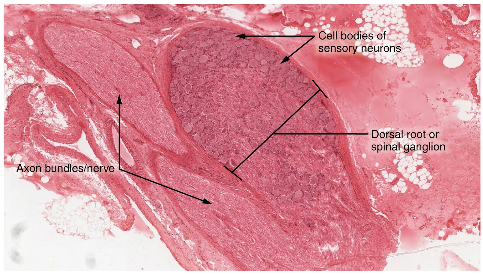
Long description
The cell bodies of sensory neurons, which are unipolar neurons by shape, are seen in this photomicrograph. Also, the fibrous region is composed of the axons of these neurons that are passing through the ganglion to be part of the dorsal nerve root (tissue source: canine). LM × 40. (Micrograph provid

Figure 2: The cell bodies of sensory neurons, which are unipolar neurons by shape, are seen in this photomicrograph.
Figure 3Gray936
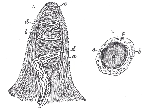
Long description
Papilla of the hand, treated with acetic acid. Magnified 350 times. A. Side view of a papilla of the hand. a. Cortical layer. b. Tactile corpuscle. c. Small nerve of the papilla, with neurolemma. d. Its two nervous fibers running with spiral coils around the tactile corpuscle. e. Apparent terminatio

Figure 3: Papilla of the hand, treated with acetic acid (magnified 350 times).
Figure 4Gray935
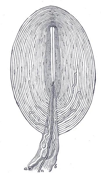
Long description
Pacinian corpuscle, with its system of capsules and central cavity. a. Arterial twig, ending in capillaries, which form loops in some of the intercapsular spaces, and one penetrates to the central capsule. b. The fibrous tissue of the stalk. n. Nerve tube advancing to the central capsule, there losi

Figure 4: Pacinian corpuscle, with its system of capsules and central cavity (a).
Figure 5Gray934
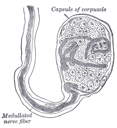
Long description
End-bulb of Krause. (Klein.). Labelled illustration from Wikimedia Commons, used under its open licence.

Figure 5: End-bulb of Krause (Klein.).
Figure 6416 Nervous Tissue-new
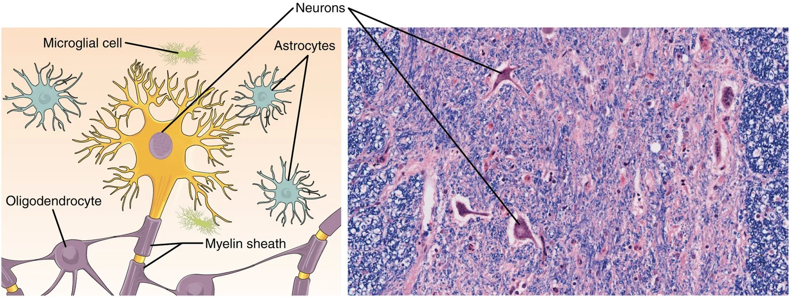
Long description
Nervous tissue is made up of neurons and neuroglia. The cells of nervous tissue are specialized to transmit and receive impulses. LM × 872. (Micrograph provided by the Regents of University of Michigan Medical School © 2012)

Figure 6: Nervous tissue is made up of neurons and neuroglia.
The astrocyte is named for its star shape, appearing star-shaped under the microscope. [1]
| Cell Type | Location | Appearance | Function |
|---|---|---|---|
| Neuron | Throughout nervous system | Large cell body with nucleus, dendrites, and axon | Transmit nerve impulses |
| Astrocyte | CNS | Star-shaped with multiple processes | Support neurons, maintain extracellular environment, contribute to blood-brain barrier |
| Satellite cell | PNS ganglia | Surround neuron cell bodies | Support neurons in periphery |
| Oligodendrocyte | CNS | Fewer processes than astrocytes | Form myelin sheaths around axons |
| Schwann cell | PNS | Wrapped around axons | Form myelin sheaths around axons |
Neurons are the primary functional cells of nervous tissue, characterized mainly by their ability to receive stimuli and respond by generating an electrical signal. They have three main components: the cell body (soma), dendrites, and axon. The cell body contains the nucleus and most organelles, while dendrites receive information from other neurons at specialized contact points called synapses. The axon is a single fiber that emerges from the cell body and propagates nerve impulses to target cells. [4]
Neurons can be classified based on their morphology and function. Multipolar neurons, the most common type, have several dendrites and a single prominent axon. Other classifications include bipolar neurons (with one dendrite and one axon) and unipolar neurons (with a single process that divides into peripheral and central branches). [4]
The dendrites appear as thin, branching extensions from the cell body, while the axon emerges as a single, longer process. The surrounding tissue contains smaller glial cells that provide support to the neurons. [1][2]
The myelin sheath has a distinctive appearance under light microscopy, resembling pastry wrapped around a hot dog. In the CNS, oligodendrocytes form myelin by wrapping around axon segments, with the cell body separate from the myelin sheath. [1]
Special stains are essential techniques in histology that allow for the visualization of specific components of nervous tissue that are not apparent with standard hematoxylin and eosin (H&E) staining. These stains highlight particular cellular structures, myelin sheaths, or neural connections that are crucial for understanding the organization and function of the nervous system.
Myelin is a lipid-rich sheath that surrounds the axon and by doing so creates a myelin sheath that facilitates the transmission of electrical signals[1]
The Golgi stain reveals the complete morphology of neurons, including their dendrites, cell bodies, and axons, by staining a random subset of neurons in their entirety. This staining method demonstrates the three-dimensional shape of neurons that makes the immense numbers of connections within the nervous system possible[1]. The Nissl stain, on the other hand, highlights the rough endoplasmic reticulum in neuronal cell bodies, allowing for the identification of neuronal cell bodies and their distribution throughout the nervous system.
Immunohistochemical staining is another powerful technique that uses antibodies to detect specific proteins in nervous tissue. For example, antibodies against glial fibrillary acidic protein (GFAP) can identify astrocytes, while antibodies against specific neuronal markers can identify different types of neurons. These stains are particularly useful for studying neuroglia, which play an essential role in supporting neurons and modulating their information propagation[1]. [4]
The myelin sheaths appear as thick, dark rings surrounding the lighter-stained axons. The large, pale-staining structures are neuronal cell bodies, and the smaller, dark-staining dots are glial cells. This stain allows for clear visualization of the myelinated fibers and their relationship to the cell bodies in this region of nervous tissue.
The ultrastructure of nervous tissue reveals the intricate organization of neurons and glial cells at the electron microscopy level. They possess distinct processes that can be classified based on their polarity and function. Using the standard model, each neuron has one axon and multiple dendrites, with information flowing from dendrites or cell bodies toward the axon. This directional flow is a key feature of neuronal organization. [1]
Neurons exhibit remarkable diversity and can be classified by various criteria, including the number of processes, location, discoverer, function, and neurotransmitter type. These specialized neurons demonstrate how structural classification relates to functional specialization within the nervous system. [1]
Glial cells provide essential support and insulation for neurons. In the central nervous system (CNS), oligodendrocytes, sometimes called "oligos," are responsible for insulating axons with myelin. The name oligodendrocyte reflects its structure—"oligo-" meaning few, "dendro-" meaning branches, and "-cyte" meaning cell. These cells extend several processes from their cell bodies, each surrounding an axon to provide myelin insulation. A single oligodendrocyte can myelinate multiple axons. Multipolar neurons have one axon and two or more dendrites. [1]
Myelin is a lipid-rich sheath that surrounds the axon. [1]
Nervous tissue is characterized by its unique cellular composition and specialized structures that enable rapid communication throughout the body. The primary cells of nervous tissue are neurons and glial cells. Neurons are the functional units responsible for transmitting electrical and chemical signals, while glial cells provide support, protection, and metabolic functions to neurons.
Neurons exhibit structural diversity based on their function and location. Multipolar neurons have multiple processes extending from their cell bodies, with one typically designated as the axon responsible for transmitting signals away from the cell body. This structural flexibility allows neurons to adapt their signaling capabilities based on physiological demands, which is particularly important in the dynamic environment of the nervous system.
Glial cells include microglia, which are smaller than most other glial cells and may originate from white blood cells called macrophages that become part of the central nervous system (CNS) during early development. Microglia are the cells in the CNS that can ingest and digest diseased or damaged cells or the pathogens that cause disease in normal, healthy tissue, and are therefore also referred to as CNS-resident macrophages. This phagocytic function is crucial for maintaining neural homeostasis and responding to injury or infection within the protected environment of the CNS. [1]
The autonomic nervous system consists of sympathetic and parasympathetic divisions that often project to the same organs to regulate their function. For example, the heart receives two inputs—one increasing heart rate and the other decreasing it. This dual innervation allows for precise control of organ function, with the sympathetic system promoting "fight or flight" responses and the parasympathetic system promoting "rest and digest" functions. [2]
Synapses of the autonomic system are classified as either cholinergic, meaning that acetylcholine (ACh) is released, or adrenergic, meaning that norepinephrine is released. [3]
Nervous tissue consists of neurons and glial cells, which can be distinguished by their characteristic morphology and staining properties. Neurons are the functional units of the nervous system, consisting of a cell body (soma), dendrites, and an axon. The cell body contains the nucleus and Nissl substance, which appears as basophilic granules under light microscopy. Dendrites are typically shorter, branched processes that receive signals, while axons are longer, unbranched processes that transmit signals away from the cell body.
Glial cells provide support and insulation for neurons. There are six types of glial cells: four in the CNS (astrocytes, oligodendrocytes, microglia, and ependymal cells) and two in the PNS (Schwann cells and satellite cells). Oligodendrocytes in the CNS and Schwann cells in the PNS produce myelin, which appears as a lipid-rich sheath around axons. Myelin facilitates the transmission of electrical signals along the axon and stains differently from neuronal cytoplasm. [1]
When examining a slide of nervous tissue, look for the following key features:
- Neurons with large, euchromatic nuclei and prominent nucleoli
- Basophilic Nissl substance in the cell body and dendrites
- Myelinated axons appearing as segmented structures with nodes of Ranvier
- Glial cells interspersed between neurons, appearing smaller with darker nuclei
Figure 71319B Nerve Mag
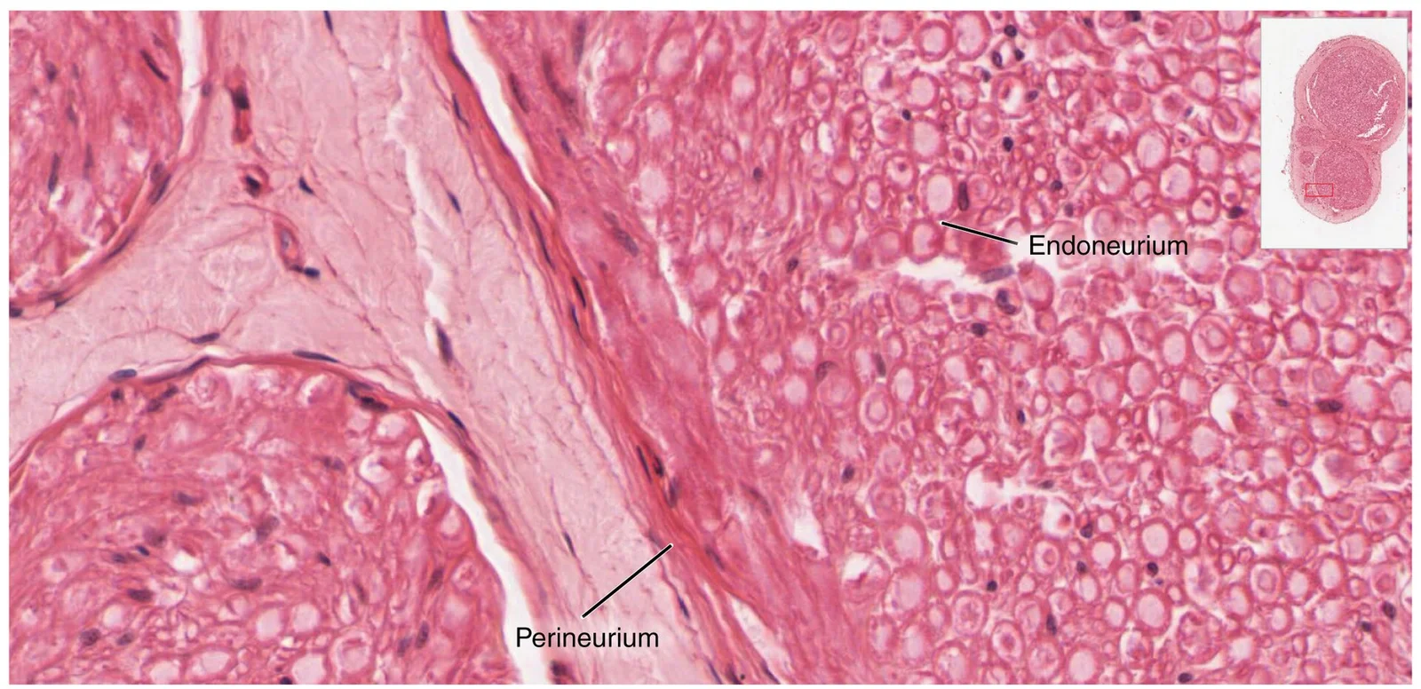
Long description
Zoom in on this slide of a nerve trunk to examine the endoneurium, perineurium, and epineurium in greater detail (tissue source: simian). LM × 1600. (Micrograph provided by the Regents of University of Michigan Medical School © 2012)

Figure 7: Zoom in on this slide of a nerve trunk to examine the endoneurium, perineurium, and epineurium in greater detail (tissue source: simian) (lm × 1600).
Common look-alikes and how to distinguish them:
- Connective tissue: Contains collagen fibers and fibroblasts, which appear more organized and lack the characteristic neuronal morphology. Connective tissue cells typically have smaller, denser nuclei.
- Epithelial tissue: Arranged in sheets with uniform polarity, unlike the irregular arrangement of neurons. Epithelial cells are tightly packed with minimal extracellular space.
- Muscle tissue: Contains elongated cells with striations (skeletal muscle) or spindle-shaped cells (smooth muscle), which are distinct from the irregular shape of neurons.
Figure 8Gray938
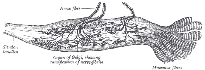
Long description
Organ of Golgi (neurotendinous spindle) from the human tendo calcaneus. (After Ciaccio.)

Figure 8: Organ of Golgi (neurotendinous spindle) from the human tendo calcaneus (After Ciaccio.).
Surrounding these neurons are smaller glial cells, including oligodendrocytes responsible for myelination. The myelinated axons appear as light-staining regions with dark nodes of Ranvier, which represent gaps in the myelin sheath where ion channels are concentrated.
Histopathology of degeneration
Nervous tissue degeneration is classified by which cellular compartment fails first. In Wallerian degeneration, the axon distal to a transection swells and fragments; the myelin sheath breaks into ovoids that are phagocytosed by macrophages. Proximal to the lesion, the axon retracts and forms a terminal bulb. In the peripheral nervous syste
The senile plaque is an extracellular deposit of β-amyloid surrounded by dystrophic neurites and reactive microglia. [1]
Histopathology of tumours
Primary nervous tissue tumours are graded by the WHO (grades 1–4). Glioblastoma (grade 4) shows pseudopalisading necrosis — tumour cells arranged around zones of necrosis — and microvascular proliferation with glomeruloid vessels. [1]
Clinical pearlKey distinction
Wallerian degeneration spares the cell body initially; neuronal degeneration (e.g., Alzheimer) begins in the perikaryon. Tumours are graded by necrosis and vascular proliferation, not by cell size.
- Neurons have polarity with information flowing unidirectionally from dendrites to cell body to axon, establishing a clear pathway for neural communication. This directional flow is fundamental to understanding neural signaling and is a key concept in neurophysiology. [1]
- Unipolar neurons are technically "pseudo-unipolar" in humans; true unipolar cells are found only in invertebrate animals. Unipolar cells are exclusively sensory neurons, and their cell bodies are always found in ganglia. [1]
- Bipolar neurons are relatively rare, primarily found in specialized sensory structures like the olfactory epithelium (where smell stimuli are detected) and as part of the retinal structure. These neurons have exactly two processes extending from opposite ends of the cell body—one axon and one dendrite. [1]
- Multipolar neurons constitute the majority of neurons in the human nervous system, characterized by having one axon and two or more dendrites (typically many more). With the exception of unipolar sensory ganglion cells and specific bipolar cells, virtually all other neurons are multipolar. [1]
- Schwann cells are responsible for myelinating peripheral axons, with each Schwann cell wrapping around only one axon segment, unlike oligodendrocytes which myelinate multiple axon segments. The nucleus and cytoplasm of Schwann cells are located on the edge of the myelin sheath, creating a distinct histological appearance. [1]
- Myelin sheaths can extend for 1-2 millimeters in length, which represents 100-1000 times the diameter of the axon itself (which ranges from 1-20 micrometers). If drawn to scale, myelinated neurons would be enormous—potentially covering an entire room wall due to this disproportionate length-to-diameter ratio. [1]
- There are precisely 31 pairs of spinal nerves: 8 cervical (C1-C8), 12 thoracic (T1-T12), 5 lumbar (L1-L5), 5 sacral (S1-S5), and 1 pair of coccygeal nerves. These nerves are numbered from superior to inferior positions and emerge through intervertebral foramina at their respective levels. [2]
- Spinal nerves emerge through intervertebral foramina, with C1 having a unique emergence point between the first cervical vertebra and the occipital bone, while C8 emerges between C7 and T1. [2]
- The cervical plexus (formed by spinal nerves C1-C5) gives rise to nerves supplying the posterior neck and head, as well as the important phrenic nerve which innervates the diaphragm at the base of the thoracic cavity. This plexus is crucial for both sensory and motor functions of the neck and diaphragm. [2]
- The brachial plexus is formed by spinal nerves C4 through T1, which reorganize to give rise to the nerves of the arms. [2]
- Adrenaline and epinephrine refer to the same molecule, with epinephrine being the preferred term in the United States and adrenaline in Great Britain[1]. This terminology difference persists because "adrenalin" was once a registered proprietary drug name in the US. [3]
- Norepinephrine and noradrenaline are also alternative names for the same molecule, following similar naming conventions as their related compound[1]. These neurotransmitters play crucial roles in the sympathetic nervous system and stress response. [3]
Exam trapMyelin misconception
Myelin is produced by glial cells, not by the neuron itself, and contains gaps called nodes of Ranvier that are essential for saltatory conduction.
Exam trapNerve plexus confusion
Peripheral nerves are not direct continuations of spinal nerves but represent reorganization of axons into networks called plexuses, which occur at four specific locations along the vertebral column.
Exam trapPreganglionic vs postganglionic
Preganglionic fibers are myelinated and relatively short, while postganglionic fibers are unmyelinated and longer due to greater distance to target effectors. [3]
Exam trapNeuron terminology
The term "postganglionic neuron" is technically incorrect as only the fiber is postganglionic; the cell body remains in the ganglion.
Exam trapAdrenal medulla exception
Preganglionic fibers projecting to the adrenal medulla are still classified as preganglionic despite not terminating in a ganglion, as they stimulate chromaffin cells to release signaling molecules into the bloodstream.
Exam trapAutonomic synapse classification
Autonomic synapses are classified as cholinergic (acetylcholine release) or adrenergic (norepinephrine release), referring to both the signaling molecule and receptor type. [3]
Exam trapCranial nerve functions
The oculomotor nerve controls four extraocular muscles, lifts the upper eyelid when eyes point up, and causes pupillary constriction, while the trochlear and abducens nerves each control different extraocular muscles.
NoteClinical assessment
Knowledge of nerve pathways is essential for neurological examination, such as testing facial nerve function by assessing facial expressions controlled by the facial nerve.
NotePain management
Understanding nerve plexuses is crucial for regional anesthesia techniques, such as blocking the sciatic nerve (from the sacral plexus) to anesthetize the lower extremity. [2]
Sciatica Assessment
- Patient presents with radiating leg painAsk about pain distribution, aggravating factors, and associated neurological symptoms
- Question: Is there evidence of nerve root compression?
Yes
- Perform neurological examinationTest motor strength, sensation, and reflexes in affected limb
- Order appropriate imagingMRI or CT to identify herniated disc or spinal stenosis
No
- Consider alternative diagnosesEvaluate for musculoskeletal causes or referred pain
- Diagnose and treat accordinglyMay include conservative management, epidural steroid injections, or surgical intervention
NoteAutonomic nervous system applications
Knowledge of sympathetic and parasympathetic pathways informs treatment of conditions like Horner's syndrome (disruption of sympathetic pathways) or neurogenic bladder (disruption of sacral parasympathetic outflow).
NotePharmacological interventions
Understanding cholinergic and adrenergic synapses guides drug therapy, such as using acetylcholinesterase inhibitors to enhance cholinergic transmission in myasthenia gravis or beta-blockers to block adrenergic receptors in hypertension.
NoteSurgical planning
Knowledge of nerve anatomy is critical in surgical procedures, such as preserving the facial nerve during parotidectomy or avoiding injury to the oculomotor nerve during orbital surgery.
NoteDiagnostic imaging
Interpretation of neuroimaging requires understanding of myelin's role in nerve conduction, as demyelinating conditions like multiple sclerosis appear as white matter lesions on MRI.
- Myelin, produced by glial cells, contains nodes of Ranvier essential for saltatory conduction
- Peripheral nerves reorganize into plexuses at four locations along the vertebral column
- Preganglionic fibers are myelinated and short; postganglionic fibers are unmyelinated and longer
- The adrenal medulla is a special case where preganglionic fibers stimulate chromaffin cells to release hormones
- Autonomic synapses are classified as cholinergic (acetylcholine) or adrenergic (norepinephrine)
- Cranial nerves have specific functions: oculomotor controls eye movements and pupillary constriction
- Sciatica results from compression or irritation of the sciatic nerve from the sacral plexus
Answer each one in your head first, then open the answer.
What is the function of the nodes of Ranvier in myelinated axons?
Show answerHide answer
They are important to the way that electrical signals travel down the axonWhich nerve plexus gives rise to the sciatic nerve?
Show answerHide answer
The sacral plexusWhat is the difference between preganglionic and postganglionic fibers in the sympathetic system?
Show answerHide answer
Preganglionic fibers are myelinated and short; postganglionic fibers are unmyelinated and longerWhich cranial nerve is responsible for pupillary constriction?
Show answerHide answer
The oculomotor nerve (cranial nerve III)What neurotransmitter is released at cholinergic synapses?
Show answerHide answer
Acetylcholine
- Betts JG, Young KA, Wise JA, et al. Nervous Tissue. OpenStax, Anatomy and Physiology 2e (CC BY-NC-SA 4.0). Accessed .
- Betts JG, Young KA, Wise JA, et al. The Peripheral Nervous System. OpenStax, Anatomy and Physiology 2e (CC BY-NC-SA 4.0). Accessed .
- Betts JG, Young KA, Wise JA, et al. Divisions of the Autonomic Nervous System. OpenStax, Anatomy and Physiology 2e (CC BY-NC-SA 4.0). Accessed .
- Betts JG, Young KA, Wise JA, et al. Nervous Tissue Mediates Perception and Response. OpenStax, Anatomy and Physiology 2e (CC BY-NC-SA 4.0). Accessed .
Figure credits
- Figure 1: Henry Vandyke Carter, via Wikimedia Commons, Public domain, Public domain.
- Figure 2: OpenStax, via Wikimedia Commons, CC BY 4.0, CC BY 4.0. resized.
- Figure 3: Henry Vandyke Carter, via Wikimedia Commons, Public domain, Public domain.
- Figure 4: Henry Vandyke Carter, via Wikimedia Commons, Public domain, Public domain.
- Figure 5: Henry Vandyke Carter, via Wikimedia Commons, Public domain, Public domain.
- Figure 6: OpenStax College, via Wikimedia Commons, CC BY 3.0, CC BY 3.0. resized.
- Figure 7: OpenStax, via Wikimedia Commons, CC BY 4.0, CC BY 4.0. resized.
- Figure 8: Henry Vandyke Carter, via Wikimedia Commons, Public domain, Public domain.
Educational content for medical students and doctors. It is not a substitute for clinical judgement or local guidelines.