Blood and bone marrow
Blood and bone marrow: cellular composition and functional histology
Blood and bone marrow form the hematopoietic system responsible for producing blood cells and maintaining the skeletal framework. The bone marrow is a specialized connective tissue found within the medullary cavities of bones and consists of two types: red marrow, which is hematopoietically active, and yellow marrow, which is primarily adipose tissue. In adults, red marrow is primarily located in the axial skeleton and proximal ends of long bones, while yellow marrow predominates in the diaphyse [1]
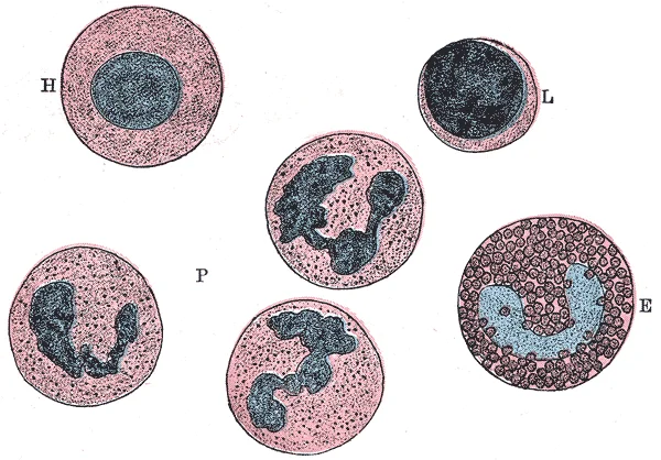
Figure 1Gray454
Long description
Varieties of leucocytes found in human blood. Highly magnified.

Figure 1: Varieties of leucocytes found in human blood (highly magnified).
The bone marrow architecture consists of a network of reticular cells forming stroma that supports hematopoietic stem cells (HSCs). These HSCs are retained in the bone marrow through interactions with the CXCR4 chemokine receptor and its ligand stromal cell-derived factor 1α (SDF-1α), which is produced by osteoblasts, endothelial cells, and nestin-positive mesenchymal stem cells. The balance between bone resorption by osteoclasts and bone formation by osteoblasts is crucial for maintaining bone [1][2]
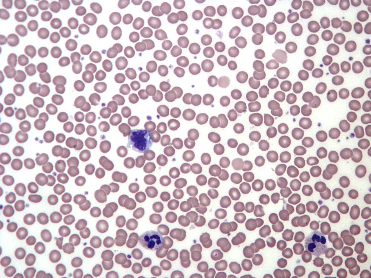
Blood smear examination reveals the various circulating blood cells, including erythrocytes, leukocytes, and platelets. Normal findings include abnormal findings may include leukocytosis (elevated white blood cell count), anemia (reduced red blood cell count), or thrombocytopenia (reduced platelet count). [1]
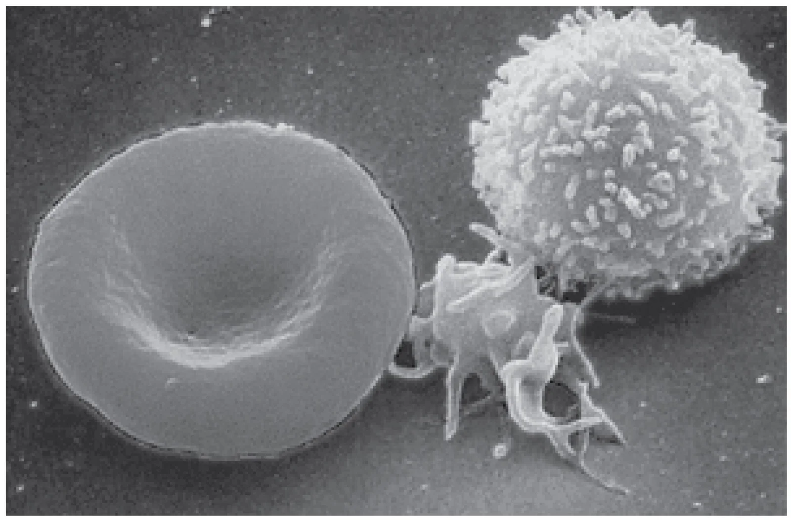
Figure 21900 Blood cells
Long description
A single drop of blood contains millions of red blood cells, platelets, and white blood cells. One of each type is shown here, isolated from a scanning electron micrograph.

Figure 2: A single drop of blood contains millions of red blood cells, platelets, and white blood cells.
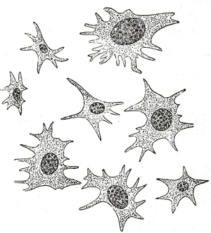
Figure 3Gray456
Long description
Blood platelets. Highly magnified. (After Kopsch.)

Figure 3: Blood platelets (highly magnified).
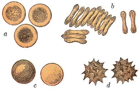
Figure 4Gray453
Long description
Human red blood corpuscles. Highly magnified. a. Seen from the surface. b. Seen in profile and forming rouleaux. c. Rendered spherical by water. d. Rendered crenate by salt solution.

Figure 4: Human red blood corpuscles (highly magnified).
The hematopoietic lineages originate from hematopoietic stem cells and differentiate into myeloid and lymphoid lineages. The myeloid lineage gives rise to erythrocytes, megakaryocytes (which produce platelets), granulocytes, and monocytes, while the lymphoid lineage produces lymphocytes. [1]
This vascular network not only provides nutrients and oxygen but also plays a role in regulating blood supply and bone growth. [1]
Figure 5Normal Adult Blood Smear
Long description
Blood smear from a normal healthy adult. Stained with Giemsa. Note the uniform staining and size of cells.

Figure 5: Blood smear from a normal healthy adult (stained with giemsa).
The histological examination of blood and bone marrow relies on standard staining techniques, primarily Hematoxylin and Eosin (H&E), which provide contrast between nuclear and cytoplasmic components. In bone tissue, the H&E appearance reveals distinct cellular layers and structural units. The primary cell of mature bone is the osteocyte, which is the most common type of bone cell. These cells are located within spaces called lacunae and are surrounded by calcified bone tissue. Osteocytes maintain the mineral concentration of the matrix via enzyme secretion and communicate with one another through long cytoplasmic processes that extend through canaliculi. Adjacent to these structures, osteoblasts are found in the growing portions of bone, including the periosteum and endosteum. Osteoblasts synthesize and secrete the collagen matrix and calcium salts; as this matrix calcifies, the osteoblast becomes trapped and transforms into an osteocyte. A third category, the osteogenic cell, is undifferentiated with high mitotic activity and serves as the only bone cell capable of division. These immature cells reside in the deep layers of the periosteum and the marrow, differentiating into osteoblasts to replenish the population. [1]
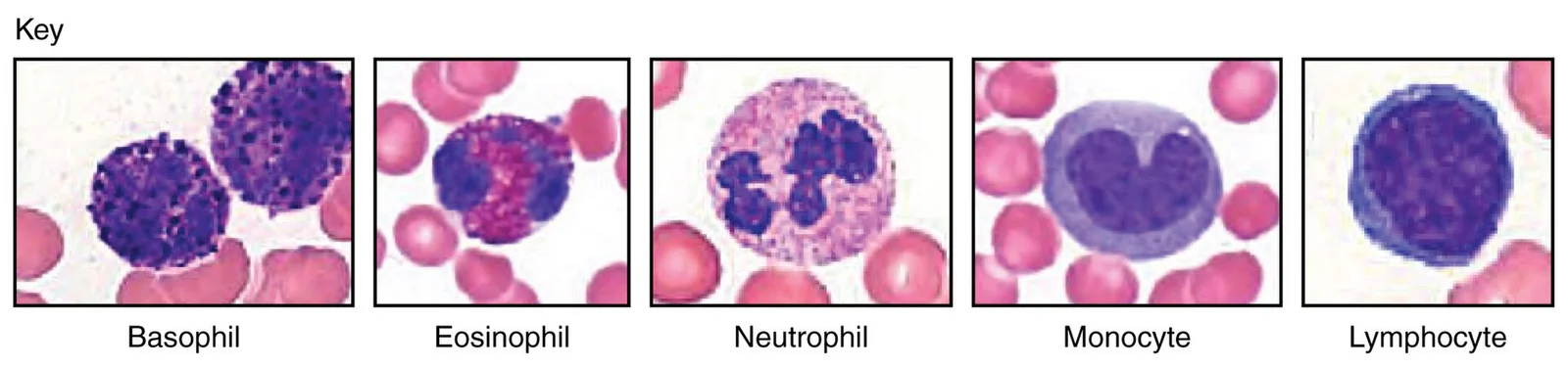
Figure 61916 Leukocyte Key
Long description
(Micrographs provided by the Regents of University of Michigan Medical School © 2012)

Figure 6: (Micrographs provided by the Regents of University of Michigan Medical School © 2012).
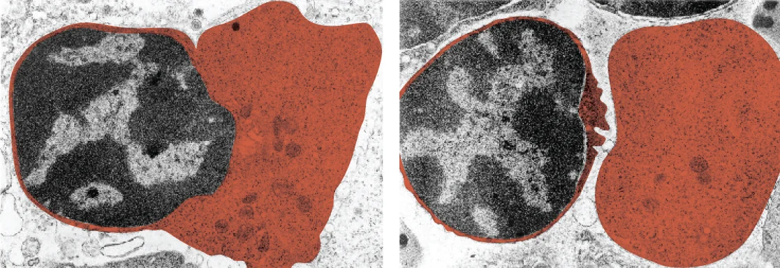
Figure 70320 RBC Extruding Nucleus Micrograph
Long description
Mature red blood cells lack a nucleus. As they mature, erythroblasts extrude their nucleus, making room for more hemoglobin. The two panels here show an erythroblast before and after ejecting its nucleus, respectively. (credit: modification of micrograph provided by the Regents of University of Mich

Figure 7: Mature red blood cells lack a nucleus.
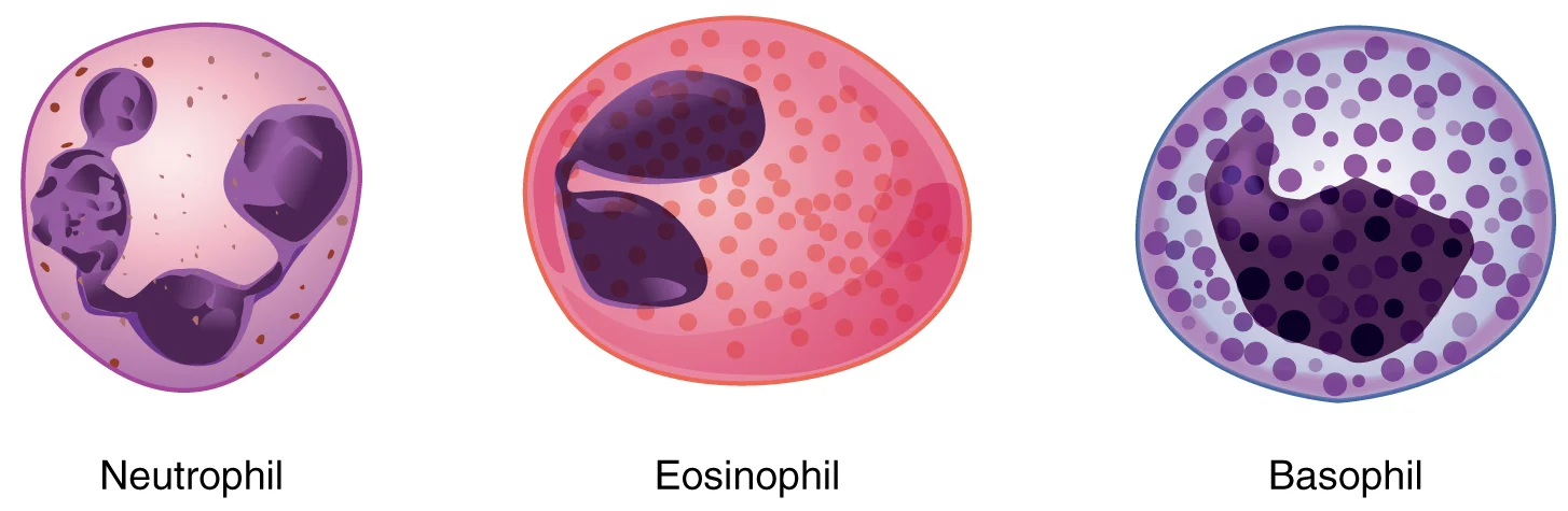
The white cell types are distinguished by their nuclear morphology and cytoplasmic granules. Neutrophils exhibit multi-lobed nuclei, while lymphocytes have large, round nuclei with scant cytoplasm. Monocytes are larger with kidney-shaped nuclei, and eosinophils and basophils are identified by their specific granule staining patterns.
Figure 81907 Granular Leukocytes
Long description
A neutrophil has small granules that stain light lilac and a nucleus with two to five lobes. An eosinophil’s granules are slightly larger and stain reddish-orange, and its nucleus has two to three lobes. A basophil has large granules that stain dark blue to purple and a two-lobed nucleus.

Figure 8: A neutrophil has small granules that stain light lilac and a nucleus with two to five lobes.
| Cell Type | H&E Appearance | Location / Relation |
|---|---|---|
| Osteocyte | Most common bone cell; located in lacuna; maintains mineral concentration | Surrounded by bone tissue; communicates via canaliculi |
| Osteoblast | Forms new bone; found in growing portions; secretes collagen and calcium salts | Located in periosteum and endosteum; becomes trapped as matrix calcifies |
| Osteogenic Cell | Undifferentiated; high mitotic activity; only dividing bone cell | Found in deep layers of periosteum and marrow; differentiates into osteoblasts |
| Red Blood Cell | Biconcave disc; anucleate in mature state | Background layer in blood smear |
| Platelet | Small anucleate fragment | Scattered among larger cells in blood smear |
| Neutrophil | Multi-lobed nucleus | Interspersed in blood smear |
| Lymphocyte | Large round nucleus; scant cytoplasm | Interspersed in blood smear |
| Monocyte | Kidney-shaped nucleus; larger size | Interspersed in blood smear |
The vascular architecture within bone also influences the microscopic landscape. Bone blood vessels are lined with specialized endothelial cells that possess specific morphologic and functional properties. These vessels show high expression of CD31 and Emcn. In contrast, sinusoidal vessels in the diaphysis form a dense, highly branched capillary network in the bone marrow cavity. The hematopoietic stem cell resides in a specialized microenvironment, or niche, which regulates its function through intrinsic cues and surrounding signals. This microenvironment is critical for lifelong blood cell production and response to injury or infection. [3]
Special stains are histochemical techniques that highlight specific cellular components or tissue elements that are not visible with standard hematoxylin and eosin (H&E) staining. In hematology and bone marrow pathology, these stains are essential for identifying and characterizing different cell types, detecting abnormal inclusions, and identifying infectious organisms.
DefinitionSpecial stain
A histochemical technique that highlights specific cellular components or tissue elements not visible with standard H&E staining.
One of the most commonly used special stains in bone marrow examination is the Perls' Prussian blue stain, which is used to identify iron storage. This stain detects ferric iron (Fe³⁺) in tissues, appearing as a blue color when iron is present. In normal bone marrow, iron is stored as ferritin and hemosiderin in macrophages, which can be visualized as blue granules[1].
| Stain | Purpose | Positive Result |
|---|---|---|
| Perls' Prussian blue | Detects iron storage | Blue granules (ferric iron) |
| Periodic acid-Schiff (PAS) | Highlights carbohydrates, glycogen | Magenta/pink color |
| Myeloperoxidase (MPO) | Identifies granulocyte precursors | Black/brown precipitate [1] |
| Sudan black B | Detects lipids | Black granules [1] |
| Reticulin silver stain | Evaluates bone marrow architecture | Black reticular fibers |
The reticulin silver stain is particularly important for assessing bone marrow architecture. This stain highlights reticular fibers, which are type III collagen fibers produced by fibroblasts. In normal bone marrow, reticulin fibers form a delicate supporting network. Increased reticulin fiber deposition, known as reticulin fibrosis, can be seen in conditions like myelofibrosis and can be graded as mild, moderate, or severe based on the pattern and density of fiber deposition[1].
The reticulin fibers appear as fine black fibers forming a delicate network throughout the bone marrow. These fibers provide structural support to the hematopoietic cells and adipocytes. The hematopoietic cells, including erythroid precursors, granulocytic precursors, and megakaryocytes, are embedded within this network. The adipocytes appear as empty white spaces. The reticulin fibers are particularly concentrated around blood vessels and the bony trabeculae, providing structural integrity to the bone marrow[1].
Another important special stain is the Periodic acid-Schiff (PAS) stain, which is used to highlight carbohydrates and glycogen. In bone marrow examination, PAS staining can help identify abnormal inclusions in cells, such as those seen in some leukemias or storage disorders. The stain produces a magenta or pink color in positive areas, making it easier to detect these inclusions against the background of the cell cytoplasm[1].
The myeloperoxidase (MPO) stain is particularly valuable for identifying granulocyte precursors. This cytoplasmic enzyme is present in granulocytes and their precursors but not in lymphocytes or erythroid cells. The MPO stain produces a black or brown precipitate in the cytoplasm of positive cells, helping to distinguish granulocytic lineage cells from other cell types in the bone marrow[1].
Sudan black B is a lipid-soluble dye that binds to lipids, appearing as black granules in positive cells. This stain is useful for identifying lipid inclusions in cells, such as those that might be seen in certain metabolic disorders or in the identification of some types of leukemia cells[1].
The ultrastructure of blood and bone marrow reveals intricate cellular and vascular components that are essential for hematopoiesis and bone function. Under electron microscopy, we can observe the detailed organization of these tissues at the cellular level.
DefinitionHematopoietic stem cells
Hematopoietic stem cells (HSCs) are multipotent cells that give rise to all blood cell lineages and reside in specialized bone marrow niches. [2]
Bone marrow contains hematopoietic stem and progenitor cells (HSPCs) that adhere to osteopontin via β1 integrins such as VLA-4. Osteopontin is expressed by osteoblasts and acts as a negative regulator of HSC pool size within the bone marrow niche. The bone marrow hematopoietic niche is hypoxic, and HSCs in hypoxic niches have greater hematopoietic-repopulating ability than those in normoxic environments. This hypoxic environment stabilizes hypoxia inducible factor 1α (HIF-1α), which upregulates erythropoietin production, cell proliferation and survival genes, and vascular endothelial growth factor. [2]
Notch signaling in postnatal bone is associated with increased angiogenesis and osteogenesis[1]. During bone development and regeneration, type H vessels play a crucial role in replacing cartilage matrix with bone[1]. [3]
The hypoxic environment of the niche is represented by the dense cellular arrangement around the vessels.
The bone marrow architecture consists of a network of reticular cells forming stroma that supports hematopoietic stem cells (HSCs). These HSCs are retained in the bone marrow through interactions with the CXCR4 chemokine receptor and its ligand stromal cell-derived factor 1α (SDF-1α), which is produced by osteoblasts, endothelial cells, and nestin-positive mesenchymal stem cells. The balance between bone resorption by osteoclasts and bone formation by osteoblasts is crucial for maintaining bone
The vascular system within bone is organized into distinct types that serve specific functions.
The coupling of angiogenesis and osteogenesis occurs at the molecular level through various signaling pathways.
Disruption of niche interactions can result in HSC mobilization - the release of HSCs from the bone marrow to peripheral circulation. This process can be achieved through chemotherapy, hematopoietic growth factors, chemokines, small-molecule chemokine receptor inhibitors, or antibodies against HSC niche interactions. Multiple mechanisms have been proposed to explain HSPC trafficking and mobilization, though it remains unclear whether these represent truly independent pathways or different aspects of a unifying mechanism. [2]
Blood and bone marrow can be identified on microscopic slides by recognizing their characteristic cellular components and extracellular matrix. Bone tissue contains a relatively small number of cells entrenched in a matrix of collagen fibers that provide a surface for inorganic salt crystals to adhere. These salt crystals form when calcium phosphate and calcium carbonate combine to create hydroxyapatite, which incorporates other inorganic salts like magnesium hydroxide, fluoride, and sulfate as it crystallizes on the collagen fibers. The hydroxyapatite crystals give bones their hardness and strength, while the collagen fibers give them flexibility so that they are not brittle. [1]
Figure 9424 Blood A Fluid Connective Tissue-new
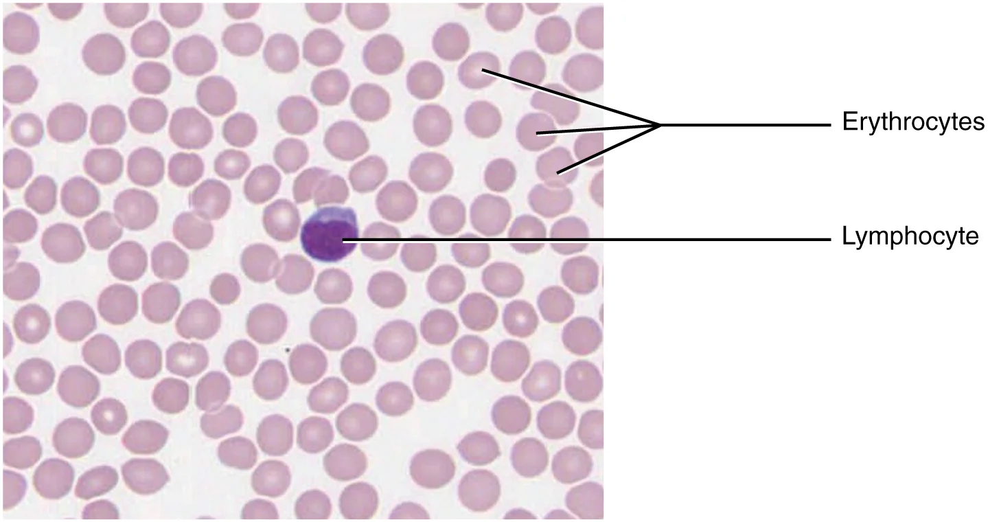
Long description
Blood is a fluid connective tissue containing erythrocytes and various types of leukocytes that circulate in a liquid extracellular matrix. LM × 1600. (Micrograph provided by the Regents of University of Michigan Medical School © 2012)

Figure 9: Blood is a fluid connective tissue containing erythrocytes and various types of leukocytes that circulate in a liquid extracellular matrix (lm × 1600).
Figure 10Gray455

Long description
Human colorles blood corpuscle, showing its successive changes of outline within ten minutes when kept moist on a warm stage. (Schofield.)

Figure 10: Human colorles blood corpuscle, showing its successive changes of outline within ten minutes when kept moist on a warm stage (Schofield.).
Figure 11Gray72
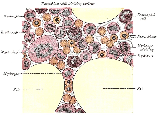
Long description
Human bone marrow. Highly magnified. Labelled illustration from Wikimedia Commons, used under its open licence.

Figure 11: Human bone marrow (highly magnified).
On a bone marrow slide, you should identify hematopoietic stem cells (HSCs) and various progenitor cells. The bone marrow niche contains specialized endothelial cells (type H ECs) that play a role in modulating the activities of osteoblasts, thereby coupling osteogenesis with angiogenesis during skeletal development and bone regeneration. These type H vessels can be identified by their specific markers and location within the bone marrow. [3]
When examining bone tissue, look for the characteristic markings where vessels and nerves penetrate the bone. The size and shape of these markings reflect the size of the vessels and nerves that penetrate the bone at these points. In bone marrow slides, you may also observe preosteoclast-derived PDGF-BB, which promotes the migration and differentiation of mesenchymal stem cells and bone marrow-derived endothelial progenitor cells via binding to its receptor PDGF receptor β. [1] [3]
DefinitionType H vessels
Specialized endothelial cells that couple angiogenesis with osteogenesis during skeletal development and bone regeneration.
The bone marrow contains hematopoietic cells at various stages of development, including erythroid precursors, granulocytes, and megakaryocytes. Type H vessels can be identified as specialized capillaries with high expression of specific markers, often found near active bone formation sites. The vessels appear as tubular structures lined with endothelial cells, surrounded by hematopoietic cells and osteoblasts.
| Cell Type | Characteristics |
|---|---|
| Hematopoietic stem cells (HSCs) | Primitive cells capable of self-renewal and differentiation into all blood lineages |
| Osteoblasts | Bone-forming cells derived from mesenchymal stem cells |
| Osteoclasts | Large multinucleated cells responsible for bone resorption |
| Type H endothelial cells | Specialized vessels that couple angiogenesis with osteogenesis |
| Preosteoclasts | Precursors to osteoclasts that can secrete PDGF-BB |
The microscopic structural unit of compact bone is called an osteon, or Haversian system. Each osteon is composed of concentric rings of calcified matrix called lamellae. Running down the center of each osteon is the central canal, or Haversian canal, which contains blood vessels, nerves, and lymphatic vessels. [1]
Bone marrow serves as the primary hematopoietic stem cell (HSC) niche in mammals and is composed of stromal cells and an extracellular matrix of collagens, fibronectin, and proteoglycans. HSCs are thought to be tethered to osteoblasts, other stromal cells, and the extracellular matrix in this stem cell niche through various adhesion molecule interactions. The hypoxic nature of the hematopoietic bone marrow niche may regulate the CXCR4/SDF-1α signaling axis, which is important for HSPC retention and mobilization. [2]
This decline is associated with decreases in angiocrine, pro-osteogenic factors and osteoprogenitors and correlates with age-related bone loss.
Bone Marrow Histology
- Bone Marrow StructureComposed of stromal cells and extracellular matrix
- HSC NicheHSCs tethered to osteoblasts and stromal cells
- Question: Type H Vessels Present?
Yes
- Support bone formation
- Express RANKL and MMP9
No
- Associated with bone loss
- Seen in osteoporosis
Each osteon contains a central canal (Haversian canal) running through its center, which houses blood vessels and nerves.
- Osteoblasts express VCAM-1 and regulate hematopoietic stem cell (HSC) function and retention in the bone marrow niche. [2]
- Targeting the interaction between VLA-4 and VCAM-1 with antibodies or small molecule inhibitors results in HSPC mobilization. [2]
- The Eph-ephrin A3 signaling axis increases adhesion to fibronectin and VCAM-1; its disruption mobilizes HSPCs. [2]
- Osteoclast activity increases with RANK ligand treatment, correlating with moderate hematopoietic progenitor cell (HPC) mobilization. [2]
- Physiological stress models (bleeding or lipopolysaccharide treatment) increase bone marrow niche osteoclasts and HPC mobilization. [2]
- Inhibition of osteoclasts with calcitonin or genetic knockout of PTPε reduces HPC mobilization response to G-CSF. [2]
- Non-resorbing osteoclast lineage cells secrete PDGF-BB to recruit endothelial and osteoblast precursor cells during bone modeling. [3]
- Type H vessels have a dense arrangement of osteoprogenitors expressing Osterix, which can differentiate into osteoblasts and osteocytes. [3]
- Runx2+ early osteoprogenitors and collagen type 1α+ osteoblast cells are densely arranged around CD31+ vessels in the metaphysis and endosteum. [3]
- Osteoprogenitors and osteoblasts are not present near diaphyseal sinusoidal type L vessels. [3]
- Vessel-associated osteoclasts can stimulate endothelial cells to help digest the cartilage template during endochondral ossification. [3]
- HIF-1α is essential for bone formation and regeneration, with endothelial HIF-1α being a significant promoter of type H vessel formation. [3]
- Type H endothelial cells express high levels of HIF-1α in young mice, which decreases with age and is associated with age-dependent bone loss. [3]
- VEGFA is considered a master regulator of angiogenesis and promotes endothelial cell migration and proliferation. [3]
- VEGFA secretion can be induced by HIF-1α, Noggin, PDGF-BB, and Runx2. [3]
- In endochondral bone formation, VEGF secretion from hypertrophic chondrocytes promotes vascular invasion of cartilage. [3]
- Type H vessels digest the cartilage matrix, which may be a mechanism of cartilage degeneration in osteoarthritis. [3]
- Halofuginone reduces aberrant type H vessels and attenuates articular cartilage degeneration and subchondral bone sclerosis. [3]
- Low-intensity pulsed ultrasound enhances spinal fusion by increasing type H vessel angiogenesis and osteoblast numbers. [3]
Exam trapMisunderstanding the bone marrow niche
The bone marrow niche is not a static structure but a dynamic microenvironment with multiple cell types interacting to regulate hematopoiesis. Osteoblasts, osteoclasts, endothelial cells, and mesenchymal stem cells all play crucial roles in maintaining hematopoietic stem cell (HSC) function and localization.
Exam trapOveremphasizing osteoclast role in HSC mobilization
While osteoclasts can influence HSC mobilization, they are not the primary mechanism in clinical settings. [2]
Exam trapAssuming bisphosphonates impair HSC mobilization
Bisphosphonates, which inhibit osteoclast activity, do not impair HSC mobilization by G-CSF. In fact, some studies show bisphosphonate treatment may actually increase mobilization.
Exam trapIgnoring the sympathetic nervous system connection
G-CSF mobilization is not solely a cytokine effect but involves altered sympathetic nervous system signaling to osteoblasts. This signaling pathway is crucial for understanding the mechanism of HSC mobilization.
Exam trapOverlooking the vitamin D receptor pathway
The vitamin D receptor is an important regulator of HSC localization and is necessary for G-CSF-induced suppression of osteoblast function. [2]
Exam trapAssuming CXCR4/SDF-1α is the exclusive pathway
While the CXCR4/SDF-1α pathway is central to HSC retention and mobilization, it is not the exclusive target for HSPC mobilization. Other molecular mechanisms likely contribute to the process.
Exam trapNeglecting circadian rhythms in clinical practice
Circadian rhythms regulate niche SDF-1α production and HSPC CXCR4 expression in an oscillating manner. Clinical mobilization should be performed at the trough of SDF-1α and CXCR4 expression (early night for humans), while transplantation may be optimal at peak expression (early morning). [2]
NoteClinical HSC mobilization
Understanding the bone marrow niche is crucial for clinical hematopoietic stem cell (HSC) mobilization and transplantation. The standard mobilizing regimen uses granulocyte colony-stimulating factor (G-CSF), which works through multiple mechanisms including suppression of osteoblasts and alteration of the CXCR4/SDF-1α pathway. [2]
NoteTiming of procedures
Knowledge of circadian rhythms affecting the bone marrow niche has practical implications for transplantation timing. Clinical mobilization should be performed at the trough of SDF-1α and CXCR4 expression (early night for humans), while transplantation may be optimal at peak expression (early morning). [3]
HSC Mobilization Mechanisms
- G-CSF administrationStandard mobilizing regimen
- Question: Alters sympathetic nervous system?
Yes
- β2-adrenergic signalingUpregulates vitamin D receptor on osteoblasts
- Osteoblast suppressionReduces SDF-1α production
No
- Direct cytokine effectsOther mechanisms may be involved
- Reduced SDF-1αDecreases retention of HSCs in niche
- HSC mobilizationCells enter peripheral blood for collection
NoteTargeting the niche for therapy
Understanding the cellular mechanisms of the bone marrow niche provides targets for enhancing HSC collection for transplantation. Future therapies may aim to specifically modulate osteoblast function, SDF-1α production, or other niche components to improve mobilization efficiency. [2]
NoteBone marrow imaging and assessment
Knowledge of type H vessels (defined by high expression of CD31 and Endomucin) and their association with osteogenesis is important for interpreting bone marrow imaging and assessing bone health in patients with hematologic disorders. [3]
NoteUnderstanding treatment side effects
Clinical use of G-CSF for mobilization can cause bone pain due to effects on the bone marrow niche, including osteoblast suppression and altered osteoclast activity. Understanding these mechanisms helps explain and potentially manage side effects.
NoteExamination applications
Medical students must understand the bone marrow niche for examinations covering hematology, oncology, and transplantation. Key concepts include the role of SDF-1α/CXCR4 in HSC retention, the effects of G-CSF on osteoblasts, and the timing of procedures based on circadian rhythms.
- The bone marrow niche is a dynamic microenvironment with multiple cell types regulating hematopoiesis
- Osteoblast suppression, not osteoclast activity, is the primary mechanism of G-CSF-induced HSC mobilization
- The CXCR4/SDF-1α pathway is central but not exclusive to HSC retention and mobilization
- Circadian rhythms affect SDF-1α production and CXCR4 expression, influencing optimal timing for procedures
- Type H vessels (CD31hiEmcnhi) are closely associated with osteogenesis and bone formation
- Vitamin D receptor signaling is necessary for G-CSF-induced suppression of osteoblast function
- Bisphosphonates do not impair HSC mobilization and may enhance it
- Understanding niche mechanisms provides targets for improving HSC collection for transplantation
Answer each one in your head first, then open the answer.
What is the primary mechanism of G-CSF-induced HSC mobilization?
Show answerHide answer
Suppression of osteoblasts and reduction in SDF-1α productionWhen does osteoclast increase typically occur after G-CSF treatment in humans?
Show answerHide answer
6 to 8 days after treatmentWhat is the optimal timing for clinical HSC mobilization based on circadian rhythms?
Show answerHide answer
Early night (trough of SDF-1α and CXCR4 expression)How are type H vessels defined?
Show answerHide answer
By high expression of CD31 and Endomucin (CD31hiEmcnhi)
- Betts JG, Young KA, Wise JA, et al. Bone Structure. OpenStax, Anatomy and Physiology 2e (CC BY-NC-SA 4.0). Accessed .
- Hoggatt J, Pelus LM. Mobilization of hematopoietic stem cells from the bone marrow niche to the blood compartment. Stem cell research & therapy 2011;2(2):13. doi:10.1186/scrt54. Accessed .
- Peng Y, Wu S, Li Y, Crane JL. Type H blood vessels in bone modeling and remodeling. Theranostics 2020;10(1):426-436. doi:10.7150/thno.34126. Accessed .
Figure credits
- Figure 1: Henry Vandyke Carter, via Wikimedia Commons, Public domain, Public domain.
- Figure 2: OpenStax College, via Wikimedia Commons, CC BY 3.0, CC BY 3.0. resized.
- Figure 3: Henry Vandyke Carter, via Wikimedia Commons, Public domain, Public domain.
- Figure 4: Henry Vandyke Carter, via Wikimedia Commons, Public domain, Public domain.
- Figure 5: Keith Chambers, via Wikimedia Commons, CC BY-SA 3.0, CC BY-SA 3.0.
- Figure 6: OpenStax College, via Wikimedia Commons, CC BY 3.0, CC BY 3.0. resized.
- Figure 7: OpenStax, via Wikimedia Commons, CC BY 4.0, CC BY 4.0.
- Figure 8: OpenStax College, via Wikimedia Commons, CC BY 3.0, CC BY 3.0.
- Figure 9: OpenStax College, via Wikimedia Commons, CC BY 3.0, CC BY 3.0.
- Figure 10: Henry Vandyke Carter, via Wikimedia Commons, Public domain, Public domain.
- Figure 11: Henry Vandyke Carter, via Wikimedia Commons, Public domain, Public domain.
Educational content for medical students and doctors. It is not a substitute for clinical judgement or local guidelines.