Cardiovascular histology
Study of heart structure at microscopic level
The heart wall consists of three distinct layers: the endocardium, myocardium, and epicardium. The endocardium is the innermost layer, composed of endothelial cells and connective tissue, providing a smooth lining for the cardiac chambers and valves. The myocardium is the thickest layer of the heart wall, composed of cardiac muscle cells responsible for the heart's contractile function. The epicardium is the outermost layer, also known as the visceral layer of the pericardium, containing adipose tissue including epicardial adipose tissue (EAT) which can increase in certain conditions. [3]
Cardiac muscle cells, or cardiomyocytes, are specialized striated muscle cells that form the myocardium. These cells are interconnected by intercalated discs, which contain gap junctions allowing for rapid electrical conduction throughout the heart muscle. Purkinje fibres are specialized cardiac muscle fibres located in the subendocardial layer that conduct electrical impulses rapidly through the ventricles, ensuring coordinated contraction. These fibres are larger than ordinary cardiac muscle cells, with fewer myofibrils and more glycogen, making them pale-staining on routine histology.
The heart contains lymphatic vessels that help maintain fluid balance by draining excess interstitial fluid from the cardiac tissue. These vessels begin as blind-ended capillaries in the myocardium and converge into larger vessels that eventually empty into the systemic circulation. Lymphatic drainage is particularly important in the subendocardial region, where oedema can impair conduction.
Cardiac muscle has unique metabolic characteristics due to the heart's constant need for energy. Unlike skeletal muscle, cardiac muscle has no ATP reserve and must generate energy continuously. Energy is stored in cardiac muscle cells in three forms: the first is phosphocreatine (PCr), which can rapidly donate its high-energy phosphates to produce ATP from ADP, with relatively modest energy used only during very rapid bursts of exercise. The adult heart requires high energy for contractile function, and disruptions in myocardial fuel metabolism and bioenergetics contribute to cardiovascular disease. Ketone bodies generated in the liver enter the bloodstream and are used by other organs, such as the brain, kidneys, skeletal muscle, and heart. [3]
The endocardium lines the chambers and covers the valves. It has three sublayers: the endothelium (simple squamous epithelium), a subendothelial layer of loose connective tissue, and a subendocardial layer containing blood vessels, nerves, and Purkinje fibres. The subendocardial layer is continuous with the interstitial connective tissue of the myocardium. In the atria, the endocardium is thicker and contains more elastic fibres than in the ventricles, reflecting the lower pressure and greater distensibility of the atria. The endocardium is avascular in its innermost part, receiving oxygen and nutrients by diffusion from the chamber blood. [1]
The myocardium is the thickest layer of the heart wall, composed of cardiac muscle cells. The muscle fibres are arranged in spiralling bundles that wrap around the chambers, allowing the heart to squeeze blood effectively. In the ventricles, the myocardium is arranged in three layers: an outer oblique, a middle circular, and an inner longitudinal layer. The atrial myocardium is thinner and has two layers. Cardiac muscle cells are branched, striated, and contain one or two centrally placed nuclei. They are connected end-to-end by intercalated discs, which contain desmosomes for mechanical adhesion and gap junctions for electrical coupling. The myocardium is richly supplied with capillaries, with each muscle cell lying within a few micrometres of a capillary, reflecting the high oxygen demand of the heart.
The epicardium is the visceral layer of the serous pericardium. It consists of a mesothelial surface overlying a layer of connective tissue containing blood vessels, nerves, and adipose tissue. Epicardial adipose tissue (EAT) is normally present, but its increase leads to cardiac steatosis and to an increase in mass in both ventricles, resulting in ventricular hypertrophy, contractile dysfunction, apoptosis, fibrosis, and impaired left ventricular diastolic function. The epicardium also contains the coronary vessels and their branches, which supply the myocardium. In older adults, the epicardial fat layer may thicken, and in obesity or metabolic disease, ectopic lipid deposition of toxic fatty acid species (i.e., diacylglycerol, ceramide) can occur in extra-adipose tissue such as the heart, leading to an increase of EAT. [3]
Cardiac muscle cells are involuntary, striated, and have a single central nucleus. They contain abundant mitochondria, accounting for about 25–35% of cell volume, reflecting the heart's dependence on aerobic metabolism. The sarcoplasmic reticulum is less developed than in skeletal muscle, and the T-tubules are larger and located at the Z-discs. Purkinje fibres are modified cardiac muscle cells that form the atrioventricular bundle and its branches. They are larger than ordinary cardiomyocytes, with pale cytoplasm due to abundant glycogen and few myofibrils. They conduct impulses at a velocity of about 1.5–4 m/s, much faster than ordinary cardiac muscle (0.3–0.5 m/s), ensuring rapid and coordinated ventricular contractionikuha.
Figure 1414c Cardiacmuscle
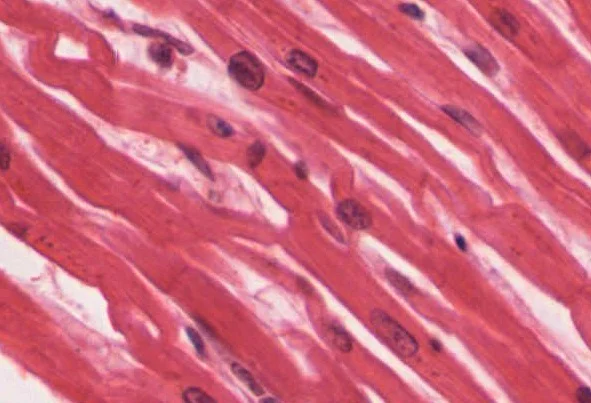
Long description
Cardiac muscle tissue is only found in the heart. LM × 1600. (Micrograph provided by the Regents of University of Michigan Medical School © 2012)

Figure 1: Cardiac muscle tissue is only found in the heart (lm × 1600).
The cardiac lymphatic system begins as blind-ended capillaries in the myocardium and subendocardium. These drain into collecting vessels that run alongside the coronary arteries in the epicardiumhol. The lymph then passes through epicardial lymph nodes and eventually drains into the right lymphatic duct or thoracic duct. The lymphatic system removes excess interstitial fluid, proteins, and immune cells from the myocardium. In conditions of impaired lymphatic drainage, such as after cardiac surgery or in heart failure, myocardial oedema can develop, impairing contractile function and electrical conduction. The lymphatic vessels are particularly dense in the subepicardial region, where they accompany the coronary vasculature.
The cardiovascular system is composed of specialised tissues designed to withstand pressure, transport blood, and coordinate electrical impulses. Under light microscopy with haematoxylin and eosin (H&E) staining, these structures are distinguished by their cellular arrangement and the presence of specific extracellular matrix components.
The heart wall consists of three distinct layers. The outermost layer is the epicardium (visceral pericardium), followed by the thick muscular myocardium, and the innermost endocardium, which lines the chambers and valves.
| Layer | H&E Appearance | Primary Cell Types |
|---|---|---|
| Endocardium | Simple squamous epithelium (endothelium) and subendothelial connective tissue | Endothelial cells, fibroblasts |
| Myocardium | Striated muscle fibres with central nuclei and intercalated discs | Cardiac myocytes, macrophages |
| Epicardium | Mesothelium and loose connective tissue with adipocytes | Mesothelial cells, adipocytes, fibroblasts |
The conduction system consists of specialised cardiac muscle cells that are larger, paler staining, and contain fewer myofibrils than contractile myocytes. These include the sinoatrial node, atrioventricular node, and Purkinje fibres. Purkinje fibres are located in the subendocardial layer, appearing as large, pale cells with abundant glycogen.
Vessel walls generally share a three-layered architecture: the tunica intima (innermost), tunica media (middle), and tunica adventitia (outermost).
| Vessel Type | Tunica Intima | Tunica Media | Tunica Adventitia |
|---|---|---|---|
| Elastic Artery | Endothelium and internal elastic lamina | Thick, concentric fenestrated elastic laminae | Thin, contains vasa vasorum |
| Muscular Artery | Endothelium and prominent internal elastic lamina | Thick layer of smooth muscle cells | Connective tissue with external elastic lamina |
| Arteriole | Endothelium and thin subendothelial layer | 1 to 2 layers of smooth muscle | Very thin or absent |
| Vein | Endothelium and thin subendothelial layer | Thin smooth muscle and collagen | Thickest layer; longitudinal collagen fibres |
| Venule | Endothelium | Sparse smooth muscle or pericytes | Loose connective tissue |
In the heart wall, the endocardium is the innermost lining, adjacent to the thick, eosinophilic myocardium, which is wrapped by the epicardium. Within the vessel walls, the tunica intima is the innermost layer, followed by the muscular tunica media and the outer tunica adventitia.
Capillaries are the smallest vessels, consisting only of a single layer of endothelial cells and a basal lamina, lacking a tunica media and adventitia. They are classified into three types:
- Continuous: Complete endothelial lining (e.g., muscle, lungs).
- Fenestrated: Small pores in the endothelium (e.g., kidney, endocrine glands).
- Sinusoidal: Large gaps between cells for passage of proteins and cells (e.g., liver, spleen).
Heart valves are folds of endocardium. They consist of a core of dense connective tissue (fibrosa) and a layer of elastic fibres (spongiosa), covered by endothelium on both sides.
Vessel Hierarchy
- ArteriesThick media, high pressure
- ArteriolesResistance vessels
- CapillariesExchange vessels; no media
- VenulesCollection vessels
- VeinsThin media, capacitance vessels
In cardiovascular histology, routine haematoxylin and eosin (H&E) staining reveals general architecture but cannot distinguish the specific extracellular matrix components, elastic fibres, or amyloid deposits that define cardiac and vascular pathology. Special stains are therefore essential to characterise the vessel wall layers, the cardiac interstitium, and the nature of any pathological deposits. The most commonly used stains target collagen, elastin, mucopolysaccharides, fibrin, and amyloid, each answering a different diagnostic question.
The Verhoeff–van Gieson (VVG) stain is the standard method for demonstrating elastic fibres. It combines Verhoeff’s haematoxylin (which stains elastin black or dark blue) with van Gieson’s picrofuchsin (which stains collagen red and muscle yellow). In a normal muscular artery, the internal elastic lamina appears as a thick, wavy black line between the intima and media, and the external elastic lamina as a similar but thinner line between the media and adventitia. In the aorta, the media shows multiple parallel black elastic lamellae alternating with red collagen and smooth muscle. This stain is invaluable for assessing arteriosclerosis, where elastic fragmentation and duplication occur, and for identifying the loss of elastic lamellae in aneurysmal disease.
Masson’s trichrome stains collagen blue or green, muscle red, and nuclei black. It is used to quantify myocardial fibrosis: in a section of left ventricle, normal myocytes appear red, while interstitial collagen appears blue. In hypertensive heart disease or after myocardial infarction, the blue collagen replaces the red myocytes in a patchy or diffuse pattern. The stain also highlights the adventitial collagen of vessels and the fibrous cap of atherosclerotic plaques. A modification, the picrosirius red stain, enhances collagen birefringence under polarised light, allowing the distinction of thick type I collagen (yellow-orange) from thin type III collagen (green), which is useful in remodelling studies.
Alcian blue at pH 2.5 stains acidic mucopolysaccharides and glycosaminoglycans a blue colour. In cardiovascular tissue, it demonstrates the ground substance of the arterial media, which is increased in cystic medial degeneration (as seen in Marfan syndrome and aortic dissection). Here, the blue-staining pools of mucoid material separate the elastic lamellae, weakening the wall. It also stains the myxoid matrix of cardiac myxomas, a primary atrial tumour, where the abundant blue ground substance surrounds scattered stellate cells.
Martius Scarlet Blue (MSB) and phosphotungstic acid haematoxylin (PTAH) are used to demonstrate fibrin. MSB stains fibrin bright red, red blood cells yellow, and collagen blue. It is used to identify fibrinoid necrosis in vessels, as in malignant hypertension or vasculitis, and to confirm the presence of organising thrombus. PTAH stains fibrin deep blue and is also useful for striated muscle cross-striations, helping to assess myocyte integrity in infarcted tissue.
Congo red is the classic stain for amyloid. Under ordinary light, amyloid deposits appear salmon-pink; under polarised light, they show apple-green birefringence. In cardiac amyloidosis, the deposits surround individual myocytes and may involve the vessel walls. The stain is essential because amyloid infiltration of the heart causes restrictive cardiomyopathy, and the diagnosis is often missed on routine H&E where amyloid appears as amorphous eosinophilic material.
The choice of stain depends on the clinical question. For a suspected dissection, VVG and Alcian blue are combined to show elastic fragmentation and mucoid degeneration. For heart failure with suspected fibrosis, Masson’s trichrome quantifies collagen. For a cardiac mass, Alcian blue and Congo red help distinguish myxoma from amyloid. In every case, the special stain must be interpreted alongside the H&E morphology)Skip to content
The ultrastructure of cardiovascular tissues reveals intricate details visible only through electron microscopy.
In blood vessels, vascular smooth muscle cells form the medial layer. These cells can exist in two phenotypes: a contractile phenotype and a more synthetic phenotype. The switch to a synthetic phenotype occurs due to hemodynamic changes, particularly in chronic kidney disease (CKD). Under electron microscopy, these cells appear elongated with abundant myofilaments in the contractile state, while in the synthetic state, they exhibit increased rough endoplasmic reticulum and Golgi apparatus, reflecting their synthetic capabilities.
The extracellular matrix (ECM) of blood vessels consists primarily of collagen fibers, elastic fibers, and proteoglycans. Collagen molecules in cardiovascular tissues, such as in bioprosthetic heart valves (BHVs), can sustain damage as early as after 20 million cycles of mechanical stress, equivalent to approximately 6 months of in vivo valve operation. Electron microscopy reveals the organized collagen meshwork that provides structural integrity. When damaged, this meshwork shows delamination, structural rearrangements, and destruction, which are irreversible since BHVs lack the capacity for ECM regeneration and remodeling. [2]
Cardiomyocytes, the contractile cells of the heart, contain numerous mitochondria arranged in rows between myofibrils. Under electron microscopy, these mitochondria appear as double-membraned organelles with cristae. Insulin resistance affects cardiomyocyte metabolism by promoting glucose and fatty acid uptake while inhibiting fatty acid utilization as an energy source. This metabolic shift is visible at the ultrastructural level through changes in mitochondrial density and distribution.
In the context of insulin resistance, molecular pathways involving mTOR and S6K1 become activated, leading to serine phosphorylation of IRS-1 and subsequent decline in PI3K activity. These intracellular signaling changes can be observed through immunoelectron microscopy techniques that identify phosphorylated proteins within cardiomyocytes.
Note
Ultrastructural changes in cardiovascular tissues reflect both physiological adaptations and pathological processes, providing insights into disease mechanisms at the cellular level.
The cardiovascular system is a closed hydraulic circuit whose every histological layer is built to match its mechanical and metabolic demands. The heart wall, from endocardium to epicardium, illustrates this principle: the endocardium is a thin, smooth endothelial lining that minimises friction and thrombus formation, while the myocardium is a thick, spirally arranged mass of cardiac muscle fibres whose intercalated discs allow rapid, synchronised contraction. The epicardium, a serous membrane, carries the coronary vessels and fat that nourish and cushion the beating heart. Each layer's thickness and cellular composition directly reflect the pressure and workload it endures.
The endothelium itself is a single layer of squamous cells lining the entire vascular tree. Its structure–function correlation is profound: in arteries, where pressure is high, the endothelium sits on a thick internal elastic lamina and a media of smooth muscle and elastic fibres; in veins, where pressure is low, the media is thinner and the lumen wider, with valves preventing backflow. Capillaries, only one endothelial cell thick, have no media or adventitia, allowing rapid exchange of oxygen, nutrients and waste across a distance of less than 1 µm. This is the textbook example of form following function: the thinner the wall, the faster the diffusion.
The heart valves—atrioventricular and semilunar—are avascular structures composed of dense connective tissue covered by endothelium. Their function is unidirectional flow, and their histology reflects this: the fibrous core (the spongiosa and fibrosa layers) resists the shear stress and bending deformations experienced during each cardiac cycle. Native valves can self-repair because their extracellular matrix (ECM) contains living fibroblasts that remodel collagen fibres. In contrast, bioprosthe [2]
In the ventricular wall, identify from inside out: the endocardium (thin, dark line), the myocardium (bulk, striated fibres with central nuclei), and the epicardium (connective tissue with coronary vessels). In the valve leaflet, note the three layers: the fibrosa (dense collagen, facing the outflow side), the spongiosa (loose connective tissue, central), and the ventricularis (elastic fibres, facing the inflow side).
The conduction system—sinoatrial node, atrioventricular node, bundle of His and Purkinje fibres—is histologically distinct: modified cardiac muscle cells with fewer myofibrils and more glycogen. Their function is rapid electrical propagation, and their structure—large, pale cells with sparse contractile elements—ensures that they conduct impulses faster than ordinary myocardium, coordinating atrial and ventricular contraction. This is a direct structure–function correlation: less contractile machinery means more speed.
Finally, the vasa vasorum—small vessels in the adventitia of large arteries and veins—supply the outer half of thick vessel walls, because diffusion from the lumen alone cannot nourish a wall thicker than about 0.5 mm. The presence of vasa vasorum in the aorta but not in the femoral artery reflects the greater wall thickness of the proximal aorta. Thus, every histological feature, from endothelial thickness to valve ECM composition, is a mechanical and metabolic solution to the haemodynamic problem it faces.
Figure 22102 Comparison of Artery and Vein
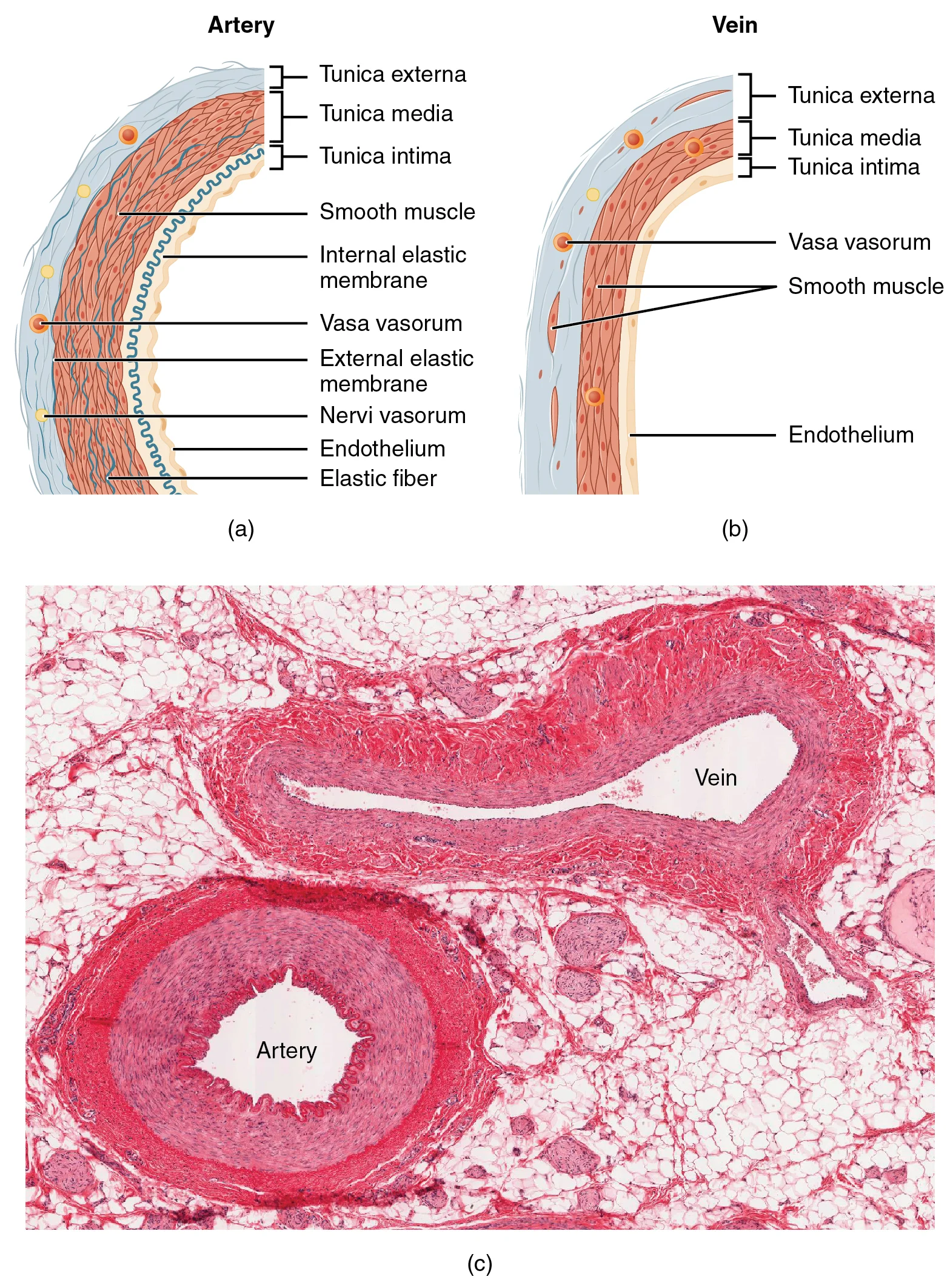
Long description
(a) Arteries and (b) veins share the same general features, but the walls of arteries are much thicker because of the higher pressure of the blood that flows through them. (c) A micrograph shows the relative differences in thickness. LM × 160. (Micrograph provided by the Regents of the University of

Figure 2: (a) Arteries and (b) veins share the same general features, but the walls of arteries are much thicker because of the higher pressure of the blood that flows through them.
Cardiovascular histology is characterised by the organisation of the heart wall and the structure of the blood vessels. Identification relies on recognising the concentric layers of the vessel walls and the specialised architecture of the myocardium.
The heart wall consists of three primary layers: the endocardium, the myocardium, and the epicardium. On a slide, the myocardium is the most prominent layer, consisting of cardiac muscle cells (cardiomyocytes). These cells are branched and contain centrally located nuclei. A key identifying feature is the intercalated disc, a specialised junction between cells that appears as a dark transverse line.
The endocardium is the innermost layer, consisting of a thin layer of endothelium and subendothelial connective tissue, lining the heart chambers. Moving outward, the myocardium is the thick middle layer of muscle. The epicardium is the outermost visceral layer of the serous pericardium, consisting of a simple squamous epithelium (mesothelium) and connective tissue.
Blood vessels are identified by their three-layered wall structure: the tunica intima, tunica media, and tunica adventitia.
- Tunica Intima: The innermost layer, consisting of a single layer of endothelial cells resting on a basement membrane.
- Tunica Media: The middle layer, primarily composed of smooth muscle cells and elastic fibres. In elastic arteries, this layer is thick and contains prominent elastic lamellae.
- Tunica Adventitia: The outermost layer, composed of collagen and elastic fibres that anchor the vessel to surrounding tissues.
It is common to confuse cardiac muscle with skeletal muscle or smooth muscle.
| Feature | Cardiac Muscle | Skeletal Muscle | Smooth Muscle |
|---|---|---|---|
| Striations | Present | Present | Absent |
| Nuclei | Central, single | Peripheral, multiple | Central, single |
| Branching | Present | Absent | Absent |
| Control | Involuntary | Voluntary | Involuntary |
Another common point of confusion is the distinction between arteries and veins. Arteries typically have a thicker tunica media and a more circular lumen, whereas veins have a thinner tunica media, a larger, often collapsed lumen, and a more prominent tunica adventitia.
Clinical pearlIdentifying the Myocardium
Look for the combination of branching cells, central nuclei, and the dark lines of intercalated discs to distinguish cardiac muscle from skeletal muscle.
Vessel Identification
- Examine the wall layersIdentify intima, media, and adventitia
- Question: Is the tunica media thick and muscular?
Yes
- ArteryCheck for elastic lamellae to distinguish elastic from muscular arteries
No
- VeinLook for a collapsed lumen and thinner muscular layer
Atherosclerosis is characterized by endothelial dysfunction, lipid deposition in the subendothelial layer, and an intense lipid-driven inflammatory reaction. These processes lead to the activation of resident cells such as vascular interstitial cells (VICs) or smooth muscle cells, resulting in a fibroproliferative response with ultimate tissue mineralization. The lipid deposits primarily consist of oxidized low-density lipoprotein (LDL), which is a characteristic marker of atherosclerosis. [2]
In an insulin resistant state, the PI3K pathway is affected while the MAP kinase pathway remains intact, causing a mitogenic effect of insulin in endothelial cells that leads to atherosclerosis. The integrity of the functional endothelium is fundamental to vascular health, with nitric oxide (NO) being the most potent endogenous vasodilator in the body. A reduction in NO bioavailability is a hallmark of endothelial dysfunction, which contributes to cardiovascular diseases including atherosclerosis. [3]
Blood glucose levels greater than 90 mg/dl can lead to atherosclerosis in the carotid artery. Furthermore, a 1 unit increase in total glycosylated hemoglobin (HbA1C) can increase the risk of cardiovascular disease by up to 18%. [3]
Hypertension shares risk factors with atherosclerosis including the metabolic syndrome, diabetes mellitus, smoking, and hyperlipidemia. Approximately 30% of subjects with a diagnosis of hypertension have insulin resistance. [3]
The coexistence of hypertension in diabetic patients greatly enhances the likelihood of developing cardiovascular disease. Abnormalities in vasodilatation, blood flow, and the renin-angiotensin-aldosterone system (RAAS) can be linked to both hypertension and insulin resistance. An additional cause of hypertension in insulin-resistant patients is over-activity of the sympathetic nervous system, which promotes myocyte hypertrophy, interstitial fibrosis, and reduced contractile function, accompanied by increased myocyte apoptosis. [3]
The activation of RAAS and hyperinsulinemia may synergistically stimulate the MAPK pathway, which exerts damaging effects on the vascular wall by inducing endothelial dysfunction and promoting atherosclerosis. Additionally, the activation of RAAS (Ang II and aldosterone) and overnutrition contributes to endothelial dysfunction through an increase in reactive oxygen species (ROS) production mediated by nicotinamide adenine dinucleotide phosphate (NADPH)-oxidase, a mechanism that also contributes to hypertension and other cardiovascular diseases. [3]
In the atherosclerosis section, you can see the characteristic lipid deposition in the subendothelial layer, with foam cells present - these are atherosclerosis-specific cell types. In the hypertension section, the vessel wall demonstrates thickening with increased smooth muscle cells and collagen deposition. The lumen appears narrowed, and there may be evidence of myocyte hypertrophy in the surrounding tissue.
Clinical pearlAlbuminuria as CVD risk factor
Increased albuminuria or proteinuria is a potent risk factor for CVD in both diabetic and nondiabetic patients with CKD, and the incidence of cardiovascular events decreases with the institution of antiproteinuric measures, in particular renin-angiotensin system (RAS) blockade. [1]
Exam trapRAS blockade and albuminuria
The pathomechanistic link between albuminuria and CVD may not be direct: systemic but particularly intrarenal hemodynamic effects of RAS blockers affect progression of CKD and thus indirectly of CVD. Therefore, data supporting RAS blockers in albuminuric patients are reasonably strong for preventing progression of CKD and less so for CVD protection. [1]
Clinical pearlMagnesium and vascular calcification
Magnesium, frequently reduced in serum in CKD, inhibits vascular calcification by interfering with hydroxyapatite crystal formation and can halt vascular calcification progress in advanced CKD. [1]
Exam trapDysmagnesemia as target
Electrolyte imbalances like dysmagnesemia are common in CKD and contribute to poor patient outcome; therefore, electrolyte imbalances are potential targets for managing coronary artery calcification. [1]
Clinical pearlAngiotensin receptor/neprilysin inhibitors
In symptomatic patients on combination therapy with ACE inhibitors (or angiotensin1-receptor blockers), β-blockers, and MRAs, if the ACE inhibitor was well tolerated, administration of an angiotensin receptor/neprilysin inhibitor is recommended. Sacubitril, given with valsartan, inhibits neprilysin, prolonging the half-life of vasoactive peptides such as BNP. [1]
Clinical pearlLCZ 696 efficacy
For LCZ 696 (sacubitril and valsartan), a reduction in overall mortality, cardiovascular mortality, and hospitalization compared with enalapril was demonstrated, and this effect was also seen in patients with CKD stages 3 to 5. [1]
Clinical pearlTAVI in elevated operative risk
Transcatheter aortic valve implantation is recommended as a safe and effective treatment option in patients <75 years old at elevated operative risk (Society of Thoracic Surgeons Score >4%). [1]
Clinical pearlTAVI vs surgery in low risk
In patients at low risk, all-cause mortality after 24 months decreases by 12% and stroke incidence by 19% compared with surgical aortic valve replacement, independent of preoperative risk. [1]
Exam trapCKD and TAVI outcomes
Impaired kidney function affects mortality and risk for dialysis after transcatheter aortic valve implantation. Long-term risk for death and need for introduction of kidney replacement therapy were increased by 51% and 56%, respectively. [1]
Clinical pearlAcute kidney injury after valve procedures
Acute kidney injury after transcatheter aortic valve implantation (7%) was less prevalent than surgical aortic valve replacement (12%). [1]
Exam trapCKD and valve intervention risk
The degree of CKD is associated with increased adverse outcomes risk after interventions and surgery as well as bearing an enhanced intermediate and long-term risk, in particular in patients >75 years of age. In the latter group, aortic valve transfemoral aortic valve implantation should be considered the superior method. [1]
Clinical pearlStructural valve disease and lipids
High risk of SVD was associated with an increased ratio of LDL/high-density lipoprotein, reflecting the balance between proatherogenic and antiatherogenic lipoproteins. [2]
Exam trapSVD and metabolic dysregulation
Dysregulated hemodynamic parameters of BHVs correlated with insulin resistance, increased lipoprotein-associated phospholipase A2 activity, and higher levels of subtilisin-kexin type 9 proprotein convertase. [2]
Clinical pearlOxidative destruction of bioprostheses
BHV-infiltrating macrophages and neutrophils promote SVD through generation of reactive oxygen species released during phagocytosis, inducing oxidative destruction of the prosthesis. Explanted BHVs demonstrated high concentrations of tyrosine oxidation products. [2]
Exam trapOxidation and collagen destruction
Incubation of glutaraldehyde-treated bovine pericardium under oxidizing conditions led to collagen destruction, loss of glutaraldehyde cross-links, and increased susceptibility to collagenase degradation. [2]
Clinical pearlInsulin resistance pathways to CVD
Insulin resistance contributes to generate CVD via two independent pathways: (1) atheroma plaque formation and (2) ventricular hypertrophy and diastolic abnormality. Both effects lead to heart failure. [3]
Clinical pearlCKD and CVD mechanisms
In addition to traditional risk factors, two major mechanisms contribute to the development of CVD in CKD: the kidney can release hormones, enzymes, and cytokines in response to kidney injury or insufficiency, leading to characteristic changes in the vasculature; and CKD-associated mediators as well as hemodynamic alterations contribute to cardiac damage. [1]
Exam trapValve disease decision-making in CKD
Because valve disease is a common comorbidity in patients with CKD, after echocardiographic evaluation, the decision to treat valve disease with surgery or intervention should be on the basis of the temporary guidelines of the American Heart Association, American College of Cardiology, and European Society of Cardiology. [1]
Exam trapConfusing valve structures
Students often mistake the histological components of heart valves, particularly between the three layers of the aortic valve: fibrosa, spongiosa, and ventricularis. The fibrosa contains dense collagen, the spongiosa has proteoglycans, and the ventricularis is elastin-rich.
Exam trapMisunderstanding calcification mechanisms
A common error is attributing all valve calcification solely to atherosclerosis. While LDL deposition and oxidation occur in bioprosthetic heart valves (BHV), the process is distinct from atherosclerosis and may involve LDL trapping by glycosaminoglycans and oxidation by infiltrating neutrophils and macrophages. [2]
Exam trapIgnoring mechanical stress effects
Students frequently overlook how mechanical stress influences valve pathology. In the HeartMate XVE left ventricular assist device, inflow valves experience higher stress values and are more frequently calcified compared to outflow valves. Calcification initiates in leaflet margins where mechanical stress is maximum. [2]
Exam trapOverlooking stent expansion impact
Another misconception is that incomplete stent expansion is inconsequential. However, incomplete expansion by ≥2 mm (≥9.1%) induces relocation of high stress regions and significantly affects transcatheter heart valve (THV) durability, despite 10-15% expansion being considered acceptable in clinical practice. [2]
Exam trapAssuming statins prevent SVD
Students often incorrectly assume statins are effective in preventing structural valve degeneration (SVD). While some retrospective studies suggested benefit, the largest observational study (1193 subjects) found no benefit of lipid-lowering therapy in slowing BHV degeneration at 1, 5, and 10 years after surgery. [2]
Exam trapNeglecting immune response
Many students fail to recognize the role of immune responses in SVD development. In some subjects, extremely rapid SVD development is associated with allergic responses to BHV components, and controlling this immune response can significantly alleviate SVD. [2]
NoteClinical assessment of valve disease
Knowledge of cardiovascular histology is essential for understanding the progression of valve diseases. For example, recognizing that calcification in BHVs initiates in leaflet margins where mechanical stress is maximum helps clinicians assess which valves are at higher risk of dysfunction.
NoteSurgical planning
Histological understanding informs surgical decisions, particularly regarding valve replacement options. The knowledge that less immunogenic homografts demonstrate longer functioning in children and young subjects compared to more immunogenic xenografts guides prosthetic selection. [2]
NoteInterventional procedures
Cardiovascular histology knowledge is crucial for interventional cardiologists. Understanding that incomplete stent expansion by ≥2 mm (≥9.1%) significantly affects THV durability influences procedural techniques to ensure proper deployment.
NotePathology interpretation
Histological expertise allows pathologists to distinguish between different types of valve degeneration. Recognizing that LDL oxidation in BHVs may be mediated by infiltrating neutrophils and macrophages helps differentiate this process from native aortic valve dysfunction. [2]
NoteResearch and development
Histological knowledge drives innovation in valve design. The understanding that high mechanical stress promotes calcification informs the development of new prosthetic materials and designs to distribute stress more evenly.
Bioprosthetic Heart Valve Degeneration Assessment
- Patient with BHV implantationNote time since implantation
- Question: Significant valve dysfunction?
Yes
- Assess for rapid SVDConsider allergic response to BHV components
- Evaluate stent expansionCheck for incomplete expansion ≥2 mm
- Consider tissue engineering optionsFor younger patients
No
- Regular monitoringAnnual echocardiography
- Lipid managementDespite limited evidence for statin benefit
NoteExamination preparation
Understanding cardiovascular histology is fundamental for medical examinations. Questions often focus on distinguishing normal valve histology from pathological changes, recognizing the layers of cardiac valves, and understanding the mechanisms of bioprosthetic valve degeneration.
- The three layers of aortic valves have distinct histological features: fibrosa (dense collagen), spongiosa (proteoglycans), and ventricularis (elastin-rich)
- Calcification in BHVs initiates in leaflet margins where mechanical stress is maximum
- Incomplete stent expansion by ≥2 mm (≥9.1%) significantly affects THV durability
- LDL oxidation in BHVs may be mediated by infiltrating neutrophils and macrophages
- The largest observational study (1193 subjects) found no benefit of statins in slowing BHV degeneration
- Allergic responses to BHV components can cause extremely rapid SVD development
- Less immunogenic homografts demonstrate longer functioning in children and young subjects
Answer each one in your head first, then open the answer.
Which layer of the aortic valve contains dense collagen?
Show answerHide answer
FibrosaWhere does calcification initially occur in BHVs?
Show answerHide answer
Leaflet marginsWhat is the threshold of incomplete stent expansion that significantly affects THV durability?
Show answerHide answer
≥2 mm (≥9.1%)Which cells mediate LDL oxidation in BHVs?
Show answerHide answer
Neutrophils and macrophages
- Jankowski J, Floege J, Fliser D, et al. Cardiovascular Disease in Chronic Kidney Disease: Pathophysiological Insights and Therapeutic Options. Circulation 2021;143(11):1157-1172. doi:10.1161/circulationaha.120.050686. Accessed .
- Kostyunin AE, Yuzhalin AE, Rezvova MA, et al. Degeneration of Bioprosthetic Heart Valves: Update 2020. Journal of the American Heart Association 2020;9(19):e018506. doi:10.1161/jaha.120.018506. Accessed .
- Ormazabal V, Nair S, Elfeky O, et al. Association between insulin resistance and the development of cardiovascular disease. Cardiovascular diabetology 2018;17(1):122. doi:10.1186/s12933-018-0762-4. Accessed .
Figure credits
Educational content for medical students and doctors. It is not a substitute for clinical judgement or local guidelines.