Respiratory histology
Study of respiratory system microscopic structures and functions
The respiratory system is composed of several specialized structures that work together to facilitate gas exchange between the body and the environment. The trachea extends from the larynx toward the lungs and is formed by 16 to 20 stacked, C-shaped pieces of hyaline cartilage. The trachea is lined with pseudostratified ciliated columnar epithelium, which is continuous with the larynx. The trachea branches into the right and left primary bronchi at the carina, which are also lined by pseudostratified ciliated columnar epithelium containing goblet cells. [1] [3]
The bronchi continue to branch into a bronchial tree, with bronchioles branching from the tertiary bronchi. Bronchioles, which are about 1 mm in diameter, further branch until they become the tiny terminal bronchioles, of which there are more than 1000 in each lung. The lining of the conducting zone is composed mostly of pseudostratified ciliated columnar epithelium with goblet cells. As the bronchioles become smaller and nearer the alveoli, the epithelium thins and becomes simple squamous epithelium. As the nasopharynx becomes the oropharynx, the epithelium changes from pseudostratified ciliated columnar epithelium to stratified squamous epithelium. [1][3]
The alveolar wall consists of three major cell types: type I alveolar cells, type II alveolar cells, and alveolar macrophages. Type II alveolar cells are interspersed among the type I cells and secrete pulmonary surfactant, a substance composed of phospholipids and proteins that reduces the surface tension of the alveoli. Alveolar macrophages are phagocytic cells that remove debris and pathogens from the alveoli. [1]
The endothelium of the surrounding capillaries, together with the alveolar epithelium, forms the respiratory membrane, which is a blood-air barrier through which gas exchange occurs by simple diffusion. The nasal cavity extends from the nostrils to the nasopharynx and is about 12-14 cm in length, with three turbinates providing a mucosal surface area of about 160 cm2. About 80-90% of the human nasal cavity is covered with respiratory mucosa, which warms inhaled air and participates in first line defense by filtering air and removing particles such as allergens and microorganisms. [1][3]
Under the light microscope, the respiratory system is best appreciated by examining its conducting and respiratory zones separately, as their histology differs fundamentally. In a standard haematoxylin and eosin (H&E) preparation, the mucosa, submucosa and cartilage or smooth muscle layers are the key landmarks. The epithelium lining the lumen is the first structure to identify; its type and height change predictably along the airway tree.
The trachea and main bronchi show a pseudostratified columnar ciliated epithelium with goblet cells, sitting on a prominent basement membrane. Beneath this lies a lamina propria rich in elastic fibres, then a submucosa containing seromucous glands. The C-shaped hyaline cartilage rings are clearly seen in the adventitia, with smooth muscle (trachealis) bridging their open ends. In smaller bronchi, the cartilage becomes irregular plates, and the smooth muscle forms a complete circumferential layer [1]
As airways narrow into bronchioles, the epithelium becomes simple columnar then simple cuboidal, goblet cells disappear, and cartilage is absent. The muscularis mucosa is relatively thick, and in terminal bronchioles, Clara cells (club cells) appear as dome-shaped, non-ciliated cells that secrete components of surfactant and detoxify inhaled substances. The respiratory bronchiole is the first airway with alveoli budding from its wall, marking the transition to the respiratory zone. [1]
| Layer | Trachea / Main bronchus | Bronchus | Bronchiole |
|---|---|---|---|
| Epithelium | Pseudostratified columnar ciliated with goblet cells | Pseudostratified columnar ciliated, fewer goblet cells | Simple columnar to cuboidal; Clara cells in terminal bronchioles |
| Lamina propria | Elastic fibres, lymphocytes | Elastic fibres | Thin, elastic |
| Submucosa | Seromucous glands | Seromucous glands, fewer | Absent |
| Cartilage | C-shaped hyaline rings | Irregular plates | Absent |
| Smooth muscle | Trachealis (posterior) | Complete circumferential layer | Prominent, spiralling |
The alveolar wall is the site of gas exchange. In H&E sections, it appears as a thin, delicate septum lined by two cell types. Its nuclei are inconspicuous, appearing as small, flattened profiles along the septum. The alveolar type II cell (septal cell) is cuboidal, often found at the corners of alveoli, with a rounded nucleus and foamy cytoplasm due to surfactant-containing lamellar bodies; these cells constitute about 5% of the surface area but are more numerous by cell count. Alveolar macrophages are also seen within the alveolar spaces, appearing as large cells with ingested pigment or debris.
The alveolar walls appear as thin pink lines; along them, note the flattened nuclei of type I cells, the larger rounded nuclei of type II cells protruding into the airspace, and occasional dark macrophages within the lumen.
By light microscopy, this membrane is too thin to resolve individually, but its total thickness is under 1 micrometre in most areas, allowing efficient gas exchange. The alveolar septum also contains connective tissue with elastic fibres and occasional fibroblasts, which provide structural support and recoil.
Figure 12310 Structures of the Respiratory Zone
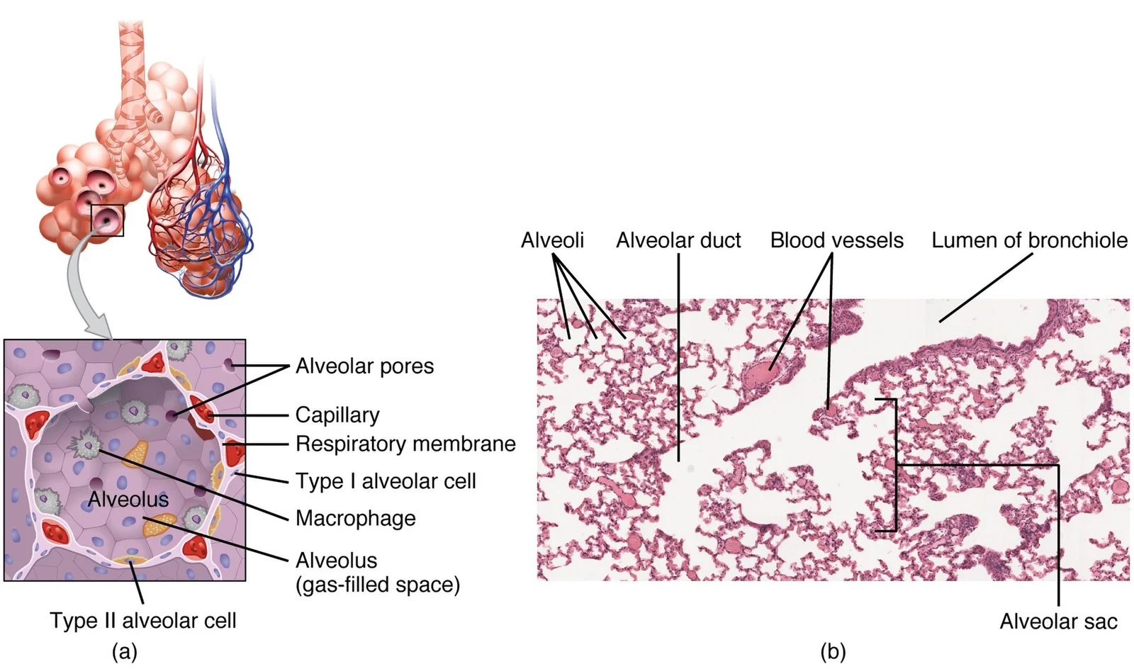
Long description
(a) The alveolus is responsible for gas exchange. (b) A micrograph shows the alveolar structures within lung tissue. LM × 178. (Micrograph provided by the Regents of University of Michigan Medical School © 2012)

Figure 1: (a) The alveolus is responsible for gas exchange.
Alveolar type II cells are the progenitors of type I cells; after injury, they proliferate and differentiate to restore the alveolar lining. They also synthesise and secrete pulmonary surfactant, a phospholipid-protein complex that reduces surface tension and prevents alveolar collapse at end-expiration. Surfactant production begins in fetal life, but adequate levels for effective lung compliance are not reached until about the eighth month of pregnancy; a full complement of functional alveoli appears only around 8 years of age. [2] [3]
The study of respiratory histology relies heavily on specific staining techniques to differentiate the cellular components of the airways and gas-exchange surfaces. While standard hematoxylin and eosin (H&E) staining provides a general overview of tissue architecture, special stains are essential for identifying specific cell types, extracellular matrix components, and pathological changes that are not easily visible with routine methods. For medical students, understanding which stain highlights which structure is fundamental to interpreting histological slides in examinations and clinical practice.
The primary lining of the conducting zone, including the nasal cavity, trachea, and bronchi, is pseudostratified ciliated columnar epithelium. This epithelium is interspersed with goblet cells, which are specialized columnar cells responsible for producing mucus. In standard H&E sections, goblet cells appear as clear, vacuolated cells because the mucus they contain is washed out during processing. To visualize these cells and their secretory products more clearly, mucicarmine or Periodic Acid-Schiff (PAS) stains are often used. These stains highlight the glycoprotein content of the mucus, allowing for the quantification of goblet cell density, which is a key metric in assessing airway inflammation and remodeling in conditions like asthma or chronic obstructive pulmonary disease. The cilia themselves, which beat in a coordinated motion to sweep mucus and trapped debris towards the throat, are difficult to see with light microscopy but can be inferred from the brush border appearance at the apical surface of the columnar cells. [1] [3]
The respiratory zone is defined by the presence of alveoli, which are lined by type I alveolar cells. These cells form a simple squamous epithelium that is extremely thin, attached to a delicate basement membrane. The functional significance of this thinness is that it forms part of the respiratory membrane, which is approximately 0.5 μm thick, allowing for efficient gas exchange via simple diffusion. Special stains such as silver stains (e.g., Gomori’s silver impregnation) are traditionally used to highlight the delicate reticular fibers and the basement membrane of the alveoli. This technique allows for the visualization of the alveolar septa and the capillary network, which is crucial for identifying emphysema, where the destruction of alveolar walls leads to enlarged air spaces. The silver stain outlines the honeycomb-like structure of the lung parenchyma, making it easier to detect the loss of septal integrity compared to H&E, where the thinness of type I cells can sometimes make the alveolar structure appear fragmented or unclear. [1]
The nasal cavity also contains olfactory epithelium, which is distinct from the respiratory epithelium found in the conchae and meatuses. Olfactory epithelium is a pseudostratified columnar epithelium containing olfactory receptor neurons, supporting cells, and Bowman’s glands[1]. Special stains for olfactory tissue often focus on the identification of the basal cells and the nerve fibers. While routine H&E shows the general stratification, immunohistochemical stains for olfactory markers can be used in research and advanced diagnostic settings to distinguish olfactory regions from respiratory regions, particularly in the context of intranasal drug delivery studies where the integrity of the mucosa is critical[1].
The basement membrane is distinct beneath the epithelial layer. Bowman's glands are located in the lamina propria of the olfactory mucosa. [3]
In clinical pathology, the pattern of staining can indicate specific diseases. For instance, an increase in the number of goblet cells, visible as a thickened mucin layer with PAS staining, is a hallmark of chronic airway inflammation. Conversely, the loss of alveolar septa, best visualized with silver stains, is diagnostic of emphysema. Understanding these patterns allows for the correlation of histological findings with clinical symptoms such as dyspnea or chronic cough. The thinness of the respiratory membrane, measured at 0.5 μm, is a critical parameter that is preserved in healthy tissue but can be altered in interstitial lung diseases, where thickening of the membrane impairs gas exchange.
The ultrastructure of the respiratory system reveals intricate details visible only through electron microscopy.
The respiratory epithelium consists of several specialized cell types. Ciliated columnar cells are the predominant cell type, featuring numerous motile cilia that project from their apical surface. These cilia beat in a coordinated manner, creating a current that moves mucus and trapped particles toward the pharynx. The cilia are anchored in basal bodies and contain the classic 9+2 microtubule arrangement characteristic of eukaryotic cilia.
Goblet cells are interspersed among the ciliated cells and contain numerous secretory granules filled with mucinogen. These cells produce and secrete the mucus that forms a protective layer over the epithelial surface. [1]
Basal cells serve as stem cells within the epithelium, providing a regenerative capacity. These cells are located at the basement membrane and can differentiate into other epithelial cell types as needed.
It provides structural support and contains anchoring fibrils that connect to the underlying connective tissue.
Beneath the epithelium, the lamina propria contains numerous blood vessels, nerves, and glands. The submucosal glands in the lower respiratory tract express the cystic fibrosis transmembrane conductance regulator (CFTR) protein at the apical site of secretory cells, which regulates chloride secretion. In contrast, Bowman's glands in the olfactory region do not express CFTR, and their secretion mechanisms remain unknown. [3]
The nasal cavity receives innervation from both the olfactory and trigeminal nerves. The trigeminal nerve, the fifth cranial nerve, has three branches: ophthalmic, maxillary, and mandibular. The ophthalmic and maxillary branches innervate the nasal cavity, with the nasopalatine nerve serving the posterior portion and the ethmoid nerve innervating the anterior nasal mucosa and external nasal surface. [3]
The respiratory system is intricately designed to facilitate gas exchange while protecting the body from pathogens and particulate matter. The external nose consists of several key regions: the root located between the eyebrows, the bridge connecting the root to the dorsum nasi (the length of the nose), and the apex which forms the tip. The nostrils (nares) are laterally formed by the alae, cartilaginous structures that help shape the nasal openings. The philtrum connects the apex of the nose to the upper lip. [1]
The nasopharynx serves exclusively as an airway and is flanked by the conchae of the nasal cavity. It contains the pharyngeal tonsils (adenoids), which are aggregates of lymphoid reticular tissue covered with ciliated epithelium that traps and destroys pathogens. These tonsils are prominent in children but tend to regress with age. The uvula, a small bulbous structure at the apex of the soft palate, helps prevent food and liquid from entering the nasal cavity during swallowing. [1] [3]
The larynx, a cartilaginous structure connecting the pharynx to the trachea, regulates air volume entering and leaving the lungs. Its major cartilages include the thyroid cartilage (anterior, forming the "Adam's apple"), the epiglottis (superior), and the cricoid cartilage (inferior). The thyroid cartilage is the largest laryngeal cartilage, while the cricoid forms a ring with a wider posterior region. [1]
The bronchi function primarily as air passageways while their mucous membranes trap debris and pathogens. This dual function ensures efficient air movement while maintaining respiratory defense mechanisms.
The nasopharynx appears posterior to the nasal cavity, demonstrating its position as a purely air-conducting passage. The trachea continues inferiorly to the bronchi, which branch into the lungs.
When examining respiratory histology slides, key features include the presence of ciliated pseudostratified columnar epithelium with goblet cells, which characterizes the respiratory tract. This clearance mechanism is crucial for maintaining respiratory health, as it removes trapped particles and pathogens. [3]
The olfactory mucosa presents distinct features with olfactory sensory neurons (OSN) that are non-myelinated and enwrapped by specialized ensheathing cells. These neurons form glomeruli (fila olfactoria) which project in bundles to the olfactory bulb. The olfactory epithelium appears thicker than respiratory epithelium and contains Bowman's glands, which are not present in other parts of the respiratory tract. [3]
DefinitionRespiratory Epithelium
Pseudostratified columnar epithelium with cilia and goblet cells that lines most of the respiratory tract.
Look-alikes include olfactory epithelium, which can be distinguished by the presence of olfactory sensory neurons and Bowman's glands, whereas respiratory epithelium contains ciliated cells and goblet cells but lacks these specialized structures. The cilia in respiratory epithelium appear as dense, hair-like projections on the apical surface, while goblet cells appear as mucin-filled cells interspersed among the ciliated cells. In pathological conditions, such as chronic inflammation, the number of goblet cells may increase, leading to a thicker mucus layer that may appear more basophilic on histological slides.
The basement membrane separates the epithelium from the underlying connective tissue. In the subepithelial region, blood vessels and nerves can be observed, with the olfactory nerve showing unique features as it does not form a single bundle like other nerves.
| Feature | Description |
|---|---|
| Epithelial Type | Pseudostratified columnar with cilia and goblet cells |
| Mucus Viscosity | 1.6 ± 1.5 Pa·s |
| Mucus Clearance Time | 10-20 minutes |
| Olfactory Glomeruli | Each projects to 10-20 mitral/tufted cells |
| Olfactory Neuron Lifespan | About one month [3] |
| Mucus pH Range | 5.5 up to 7.8 [3] |
This balance is maintained by the composition of mucus, including its pH and ionic strength, which contribute to its viscosity of 1.6 ± 1.5 Pa·s.
The respiratory system's histological features correspond to specific pathological changes that can be identified under microscopy. The trachea and bronchi are lined with pseudostratified ciliated columnar epithelium containing goblet cells, which secrete mucus to trap inhaled particles. In chronic bronchitis, this epithelium undergoes hyperplasia and increased goblet cell number, leading to excessive mucus production. The cartilage rings in the trachea provide structural support; their calcific [1]
The bronchioles, lacking cartilage, are lined with simple ciliated columnar or cuboidal epithelium. In asthma, these bronchioles show thickening of the basement membrane, hypertrophy of bronchial smooth muscle, and infiltration of inflammatory cells such as eosinophils. The alveoli, the primary sites of gas exchange, are lined primarily with type I alveolar cells, which constitute up to 97 percent of the alveolar surface area and are highly permeable to gases, and type II alveolar cells, which secrete pulmonary surfactant, a substance composed of phospholipids and proteins that reduces the surface tension of the alveoli. In pulmonary edema, the alveolar spaces fill with [1]
The pulmonary vasculature includes arteries, veins, and capillaries. The pulmonary arteries carry deoxygenated blood and have thinner walls than systemic arteries. The pleura, a serous membrane covering the lungs, consists of a visceral and parietal layer with a potential space between them. [1]
The cartilage appears as hyaline tissue with perichondrium, providing structural support. The submucosal glands contain mucous cells that secrete mucus. The smooth muscle layer is arranged spirally, allowing for bronchoconstriction and dilation.
- The transition from conducting to respiratory zones is marked strictly by the appearance of alveolar outpocketings along the walls of the respiratory bronchioles.
- Cartilage plates and submucosal seromucous glands are entirely absent in bronchioles, whereas smooth muscle forms a prominent, continuous layer relative to the airway diameter.
- Club cells populate terminal bronchioles as dome-shaped, non-ciliated structures responsible for secreting surfactant components and detoxifying airborne xenobiotics.
- Type I alveolar cells form the broad, squamous surface optimized for gas exchange, whereas Type II alveolar cells are cuboidal septal cells characterized by surfactant-producing lamellar bodies.
- Pulmonary arteries run alongside the bronchial tree with branches typically positioned superior and anterior to the corresponding airway, while pulmonary veins course separately within the interlobular septa.
- The lung parenchyma floats in water and crepitates upon palpation due to trapped alveolar air, and collapses significantly upon opening the thoracic cavity due to intrinsic elastic recoil.
One common mistake is confusing the regions of the pharynx. The pharynx is divided into three major regions: the nasopharynx, the oropharynx, and the laryngopharynx. Students often misidentify the boundaries between these regions or confuse their functions. [1]
Another frequent error is misunderstanding the composition of mucus. Mucus consists predominantly of proteins of the mucin family and water. Many students incorrectly believe mucus is primarily composed of lipids or carbohydrates. The different types of mucins include secreted, oligomeric gel-forming mucins like MUC2, MUC5AC, MUC5B, MUC19, and secreted monomeric non-gel-forming mucin like MUC7.
Students often struggle with the concept of mucus layers. Mucus can be divided into two different layers: the periciliary layer adjacent to the epithelial cells and the upper gel-like layer. The pericilliary layer is of low viscosity and reaches nearly as high as the tips of motile cilia. The upper, more viscous layer in intestinal mucus is between 0.5 and up to 5 µm thick, but it is thinner in nasal mucus. [3]
A common misconception is about drug diffusion through mucus. Small particles like protein degradation products and viruses move quite freely in mucus. The mesh space of mucin ranges from 20 to 200 nm and is clearly wide enough for small molecules and particles, while bigger particles are slowed down by the mesh. Students often incorrectly assume all drugs diffuse through mucus at the same rate. [3]
Exam trapMucin dominance in disease
In healthy airways, MUC5AC is the most prominent mucin present. However, in chronic diseases like cystic fibrosis or chronic obstructive pulmonary disease, the ratio changes and MUC5B becomes the dominant mucin. [3]
Students frequently misunderstand the properties of mucus. Mucus displays non-Newtonian properties, meaning it possesses both viscous (fluid) and elastic (solid) properties, termed viscoelastic. If shear stress is applied to mucus, its viscoelasticity ensures decreasing viscosity with increasing shear stress, but recovery of mucus is only partial after removing stress.
Another common error is confusing the components of mucosa-associated lymphoid tissue (MALT). MALT is composed of diffusively distributed lymphatic tissue along the lamina propria, containing T-cells, IgA-, IgM- and IgG-secreting plasma cells, few B-cells and dendritic cells, as well as macrophages and specialized Microfold cells. Students often overlook the specialized Microfold cells that are crucial for antigen sampling.
Respiratory histology knowledge is essential in understanding drug delivery systems, particularly nasal drug delivery. The nasal cavity is highly suitable for drug delivery as the nasal mucosa presents an efficient absorption and a very good permeability of both small molecule drugs and biopharmaceuticals. A promising strategy is the delivery of drugs from the nose to the brain (N2B) to bypass the blood-brain barrier and blood cerebrospinal-fluid barrier. [3]
Nasal to Brain Drug Delivery
- Drug administrationNasal delivery via devices like ViaNase™
- Question: Drug particle size?
9-11 µm
- Optimal for nasal deposition
Smaller
- Can diffuse through mucus mesh
- Transport pathwaysOlfactory and trigeminal nerve routes
- AdvantagesLower dosage needed, reduced systemic toxicity, high patient compliance
At the bedside, understanding respiratory histology is crucial for vaccination strategies. Vaccination through the nasal route is of special interest in pediatrics since nasally administered vaccination is able to induce a systemic immune response. M cells transcytose vaccine antigens very fast and transfer them to underlying lymphatic tissue, priming the immune system and inducing protection against pathogens. An easy method is dispersing an ordinary commercially available vaccine for intramuscular administration into the nasal cavity. [3]
In the operating theatre, knowledge of respiratory histology guides surgical approaches. Understanding the structure of the pharynx—a tube formed by skeletal muscle and lined by mucous membrane that is continuous with that of the nasal cavities—is essential for procedures involving this region. The three major regions—nasopharynx, oropharynx, and laryngopharynx—must be clearly differentiated during surgical planning.
On imaging, understanding mucus properties helps interpret findings. The two-layered structure of mucus—the pericilliary layer and the upper gel-like layer—can be visualized in certain imaging modalities. Changes in mucus composition, such as the shift from MUC5AC to MUC5B dominance in diseases like cystic fibrosis or chronic obstructive pulmonary disease, can be detected through specialized imaging techniques.
In examinations, students must demonstrate knowledge of respiratory histology components. The mucosa-associated lymphoid tissue (MALT) is an important part of every mucosa to protect the organism from infections. Nose-specific MALT is called nasopharynx-associated lymphoid tissue (NALT). Understanding that MALT can act independently of the systemic immune system and that immune responses at one mucosa may result in IgA production on another mucosal site in a different organ is frequently tested. [3]
The pharynx is divided into three major regions: nasopharynx, oropharynx, and laryngopharynx. Mucus consists predominantly of mucin proteins and water, with MUC5AC being most prominent in healthy airways. Mucus has a two-layer structure: pericilliary layer and upper gel-like layer. Nasal drug delivery can bypass the blood-brain barrier via the N2B route. Mucosa-associated lymphoid tissue (MALT) provides immune protection at mucosal surfaces. Nasal vaccination can induce systemic immune responses, particularly valuable in pediatrics. Mucus mesh space (20-200 nm) allows diffusion of small particles while hindering larger ones.
Answer each one in your head first, then open the answer.
What are the three major regions of the pharynx?
Show answerHide answer
nasopharynx, oropharynx, and laryngopharynxWhich mucin is most prominent in healthy airways?
Show answerHide answer
MUC5ACWhat is the advantage of N2B drug delivery over crossing the blood-brain barrier?
Show answerHide answer
Lower dosage needed, reduced systemic toxicity, high patient complianceWhat are the two layers of mucus?
Show answerHide answer
pericilliary layer and upper gel-like layerWhat is nose-specific MALT called?
Show answerHide answer
nasopharynx-associated lymphoid tissue (NALT)
- Betts JG, Young KA, Wise JA, et al. Organs and Structures of the Respiratory System. OpenStax, Anatomy and Physiology 2e (CC BY-NC-SA 4.0). Accessed .
- Betts JG, Young KA, Wise JA, et al. Embryonic Development of the Respiratory System. OpenStax, Anatomy and Physiology 2e (CC BY-NC-SA 4.0). Accessed .
- Gänger S, Schindowski K. Tailoring Formulations for Intranasal Nose-to-Brain Delivery: A Review on Architecture, Physico-Chemical Characteristics and Mucociliary Clearance of the Nasal Olfactory Mucosa. Pharmaceutics 2018;10(3):E116. doi:10.3390/pharmaceutics10030116. Accessed .
Figure credits
Educational content for medical students and doctors. It is not a substitute for clinical judgement or local guidelines.