Gastrointestinal histology
Study of microscopic structure of digestive system tissues
Most aspects of GI physiology are under neural control, which is exerted via extrinsic nerves (which include both primary afferent and autonomic fibers that ultimately connect the gut tissues with the central nervous system [CNS]) and a vast network of intrinsic enteric neurons (1–5 × 10^8) and glial cells that are organized into t [3]
The oesophageal epithelium consists of stratified squamous epithelium designed to protect against mechanical damage from ingested food. The oesophagus contains mucous glands that secrete mucus to lubricate the passage of food and protect the esophageal lining from abrasion.
The stomach contains specialized glands that secrete acid, enzymes, and mucus. These glands are distributed throughout the stomach lining and are essential for initial food breakdown and sterilization.
The duodenum is the first part of the small intestine and contains villi and crypts that increase the surface area for absorption.
The jejunum, like the duodenum, features villi and crypts that maximize nutrient absorption. The villi are finger-like projections covered by simple columnar epithelium containing absorptive cells and goblet cells. [4]
The ileum is the final section of the small intestine and contains villi, crypts, and prominent Peyer patches. The Peyer patches in the ileum are particularly well-developed and serve as important sites for immune response to ingested pathogens.
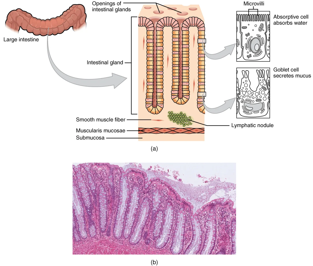
The colon mucosa lacks villi but contains numerous crypts lined primarily with absorptive cells and abundant goblet cells that secrete mucus. The high density of goblet cells in the colon helps protect the intestinal wall and facilitates the smooth passage of fecal material.
DefinitionEnteric nervous system
The enteric nervous system (ENS) consists of millions of neurons and glial cells organized into interconnected ganglia embedded within the gut wall, capable of autonomously regulating gastrointestinal tissue dynamics and gut homeostasis. [2]
The ENS has the ability to autonomously command gastrointestinal tissue dynamics and gut homeostasis, devoid of input from the brain or spinal cord, earning it the sobriquet 'second brain'. As the greatest division of the autonomic nervous system and rivalling the spinal cord in terms of complexity, ENS components form integrated circuits which independently or in concert with extrinsic parasympathetic and sympathetic innervation regulate a myriad of gut processes including bowel motility, transmucosal movement of fluids, immune responses, and local blood flow.
In vertebrates, the majority of enteric neurons and glia develop from neural crest cells originating from the vagal level of the neural tube which invade the foregut and rostro-caudally migrate culminating in the quasi-uniform colonization of the gastrointestinal tract. The constellation of intrinsic primary afferent neurons, interneurons, and motor neurons, with distinct neurochemical coding and functional roles are surrounded by 1–7 times as many enteric glial cells (EGCs) and generally configured within two major ganglionated and interconnected plexus layers: the myenteric and submucosal plexus. Enteric neuron cell bodies are located in the myenteric and submucosal ganglia while their processes extend throughout the external muscle layers, submucosa and mucosa. [2]
NoteBarrett oesophagus
Barrett oesophagus is a condition where the normal stratified squamous epithelium of the oesophagus is replaced by columnar epithelium, typically occurring in response to chronic gastroesophageal reflux disease.
NoteCoeliac disease
Coeliac disease is an autoimmune disorder triggered by ingestion of gluten that leads to villous atrophy and crypt hyperplasia in the small intestine, primarily affecting the duodenum and jejunum.
NotePolyps
Polyps are abnormal tissue growths that project from the mucous membrane of the gastrointestinal tract, with colonic polyps being common and ranging from benign hyperplastic polyps to adenomatous polyps with malignant potential.
The image is a composite of transverse sections through each organ, oriented with the lumen at the top and the serosa at the bottom.
The gut wall is built from four concentric layers. From the lumen outward these are: the mucosa, submucosa, muscularis propria (muscularis externa), and adventitia or serosa. [1]
| Layer | H&E appearance | Key cell types |
|---|---|---|
| Mucosa – epithelium | Varies by region: stratified squamous (oesophagus), columnar with gastric pits (stomach), villi and crypts (small intestine), flat surface with crypts (large intestine) | Absorptive cells, goblet cells, enteroendocrine cells, Paneth cells (small intestine), parietal and chief cells (stomach) [1] |
| Mucosa – lamina propria | Pale, loose connective tissue with capillaries and immune cells | Fibroblasts, lymphocytes, plasma cells, eosinophils |
| Mucosa – muscularis mucosae | Thin pink band of smooth muscle | Smooth muscle cells |
| Submucosa | Denser pink connective tissue, larger vessels | Fibroblasts, vessels, submucosal plexus neurons |
| Muscularis propria | Two thick pink layers, inner circular, outer longitudinal | Smooth muscle cells, myenteric plexus neurons |
| Serosa/adventitia | Thin connective tissue, mesothelium if serosa | Mesothelial cells (serosa), fat cells |
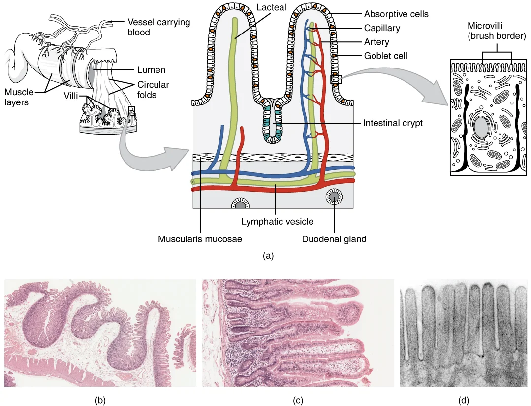
The small intestine is distinguished by villi (finger-like projections of mucosa) and crypts of Lieberkühn (glandular invaginations at the villus bases). [1]
Parietal cells secrete acid and intrinsic factor. [1]
Paneth cells sit at the base of small intestinal crypts and secrete antimicrobial peptides. [1]
Routine haematoxylin and eosin (H&E) staining reveals the general architecture of the gastrointestinal tract but cannot distinguish many of the specialised cell types, connective tissue components, or microorganisms that are clinically relevant. Special stains are therefore used to highlight specific structures, allowing the histologist to identify features that would otherwise be invisible or ambiguous. The choice of stain depends on the question being asked: whether it is to demonstrate mucin, argentaffin cells, fibrous tissue, organisms, or the brush border of enterocytes.
The periodic acid–Schiff (PAS) reaction is one of the most versatile stains in gastrointestinal histology. It oxidises vicinal glycol groups in carbohydrates to aldehydes, which then react with Schiff reagent to produce a magenta colour. In the stomach, PAS vividly stains the neutral mucins of the surface foveolar cells and the mucous neck cells of the gastric glands. In the small intestine, it highlights the glycocalyx on the microvilli of enterocytes and the mucin of goblet cells. In the colon, goblet cell mucin is strongly PAS-positive, and the stain is useful for assessing the thickness of the mucus layer. When combined with diastase digestion (PAS-D), the stain distinguishes glycogen (digested) from other mucins (undigested), which is helpful in identifying glycogen-rich cells or deposits.
Alcian blue, at pH 2.5, stains acidic mucins (sialomucins and sulphomucins) a blue colour, whereas neutral mucins remain unstained. This is particularly valuable in the gastrointestinal tract because different regions produce different mucin types. For example, the gastric surface epithelium secretes neutral mucins (PAS-positive, Alcian blue-negative), while the goblet cells of the small and large intestine secrete acidic mucins (Alcian blue-positive). A combined Alcian blue–PAS stain can therefore differentiate neutral from acidic mucins in a single section: neutral mucins appear magenta, acidic mucins blue, and a mixture of both appears purple. This is useful in conditions such as Barrett's oesophagus, where intestinal-type goblet cells (acidic mucin) can be distinguished from gastric-type foveolar epithelium (neutral mucin).
The Grimelius silver stain and the Fontana–Masson stain are used to demonstrate enteroendocrine (argentaffin and argyrophil) cells. These cells are scattered throughout the gastrointestinal epithelium and contain secretory granules that reduce silver salts. The Fontana–Masson method stains argentaffin cells (e.g. enterochromaffin cells containing serotonin) black, because they can reduce silver directly without an external reducing agent. The Grimelius method requires an external reducer and demonstrates argyrophil cells, which include a wider range of endocrine cells. In the gastric antrum, G cells (gastrin-producing) are argyrophil but not argentaffin, so they require the Grimelius technique. In the small intestine, enterochromaffin cells are argentaffin and will be black with Fontana–Massoncars. These stains are rarely used in routine diagnosis today, having been largely replaced by immunohistochemistry, but they remain useful for teaching and for confirming the endocrine nature of a tumour.
Masson's trichrome stain is valuable for demonstrating connective tissue and fibrosis in the gastrointestinal wall. It stains collagen blue or green, muscle red, and nuclei dark. In the bowel wall, it clearly delineates the muscularis mucosae, the submucosa, and the muscularis externa, and it can highlight increased collagen deposition in conditions such as Crohn's disease (transmural fibrosis) or in chronic radiation injury. The stain also helps to distinguish smooth muscle from fibrous tissue in biopsy specimens where the architecture is distorted.
The Warthin–Starry silver stain and the Giemsa stain are used to demonstrate microorganisms. Helicobacter pylori, the spiral bacterium colonising the gastric mucus layer, is best seen with a modified Giemsa stain or with the Warthin–Starry silver stain, both of which render the organism dark against a pale background. The organisms lie within the surface mucus or closely apposed to the apical membrane of surface epithelial cellscars. The Giemsa stain is simple and reliable, and it also demonstrates mast cells, which contain metachromatic granules that appear purple. Mast cells are normally present in the lamina propria of the gastrointestinal tract and increase in number in some inflammatory conditions.
The ultrastructure of the gastrointestinal tract reveals the intricate cellular and subcellular components that enable its diverse functions. Electron microscopy provides detailed visualization of these structures, allowing us to understand the specialized adaptations of different regions.
The absorptive cells (enterocytes) of the small intestine exhibit remarkable ultrastructural specializations. The apical surface is covered with microvilli, approximately 1-2 μm in length and 0.1 μm in diameter, which increase the absorptive surface area by approximately 20-fold. Each microvillus contains a core of actin filaments anchored in the terminal web, a dense network of cytoskeletal proteins that provides structural support. The glycocalyx, a fuzzy coat of glycoproteins and carbohydrates, covers the microvilli and facilitates nutrient absorption.
Figure 12418 Histology Small IntestinesN
Long description
(a) The absorptive surface of the small intestine is vastly enlarged by the presence of circular folds, villi, and microvilli. (b) Micrograph of the circular folds. (c) Micrograph of the villi. (d) Electron micrograph of the microvilli. From left to right, LM x 56, LM x 508, EM x 196,000. (credit b-

Figure 1: (a) The absorptive surface of the small intestine is vastly enlarged by the presence of circular folds, villi, and microvilli.
The lateral membranes of adjacent enterocytes are connected by specialized junctional complexes. Tight junctions near the apex seal the paracellular space, controlling the passage of substances between cells. Below these, adherens junctions and desmosomes provide mechanical strength. The basolateral membrane contains numerous infoldings and houses ion pumps and transporters essential for nutrient absorption.
Gastric parietal cells display unique ultrastructural features that enable acid secretion. They contain an extensive network of intracellular canaliculi lined with microvilli, which dramatically increase the surface area for proton pump (H+/K+-ATPase) activity. Numerous mitochondria are present to provide the ATP required for acid secretion. When stimulated, the canaliculi expand, and tubulovesicles containing proton pumps fuse with the canalicular membrane.
Chief cells contain abundant rough endoplasmic reticulum (rER) and large zymogen granules filled with pepsinogen. The rER synthesizes the pepsinogen precursor, which is processed and stored in the granules until release. Paneth cells at the base of intestinal crypts contain large secretory granules with antimicrobial peptides such as defensins.
The gastrointestinal microbiota includes diverse microorganisms with characteristic ultrastructures.
Their cell walls contain chitin, distinguishing them from bacterial cells. [4]
The basement membrane underlying the epithelium is composed of a network of collagen, laminin, and other glycoproteins. It provides structural support and serves as a selective barrier. In the lamina propria, the extracellular matrix contains various fibers (collagen, reticular, elastic) and ground substance that support the epithelium and house immune cells, blood vessels, and nerve fibers.
The cytoplasm contains numerous organelles such as mitochondria, endoplasmic reticulum, and Golgi apparatus, reflecting the high metabolic activity of these cells.
The histology of the gastrointestinal tract is designed to facilitate a progression from digestion and absorption to waste storage and excretion. This is achieved through a coordinated transition of the mucosal lining and the underlying wall layers, with each segment specialized for its specific function.
The mucosa consists of epithelium, lamina propria, and muscularis mucosae. [1]
The anal canal represents a critical histological transition zone where the specialised lining of the digestive tract meets the protective lining of the external environment. This transition is essential for maintaining both digestive functions and protection against external pathogens and mechanical stress.
This transition occurs gradually over approximately 1-2 cm to provide mechanical protection against the abrasion associated with the passage of firm faecal matter. The epithelial cells in this region are rich in glycogen, which may contribute to maintaining a healthy microbial environment.
As the canal descends, the lining transitions again at the anal verge. Here, the non-keratinised stratified squamous epithelium of the anal canal meets the keratinised stratified squamous epithelium of the perianal skin. This transition zone is approximately 5-6 mm in length and marks the boundary between the internal and external anal sphincters.
The anal canal contains specialized structures that contribute to its function. Above this line, the epithelium is derived from embryonic endoderm and has the same blood supply and nerve innervation as the rest of the gut. Below the pectinate line, the epithelium is derived from ectoderm and has a different blood supply and is sensitive to pain.
NoteProtective Adaptation
The shift from a single layer of absorptive cells to multiple layers of squamous cells reflects the change in function from nutrient and water absorption to purely protective barrier maintenance.
Anal Canal Epithelial Transition
- RectumSimple columnar epithelium
- Anal Canal (Upper)Transition to non-keratinised stratified squamous epithelium
- Anal Canal (Lower)Non-keratinised stratified squamous epithelium
- Question: Anal Verge reached?
Yes
- Perianal SkinKeratinised stratified squamous epithelium
No
- Continue descent
Chief cells secrete pepsinogen. [1]
DefinitionKey identification feature
The presence of villi and microvilli is characteristic of the small intestine, distinguishing it from the stomach which has gastric pits and glands.
When examining the colon, you'll notice the absence of villi, abundant goblet cells, and the presence of tenia coli in the muscularis externa. The appendix has similar features to the colon but contains abundant lymphoid follicles.
| Segment | Key Features | Identifying Characteristics [4] |
|---|---|---|
| Stomach | Gastric pits, gastric glands, parietal cells, chief cells | Presence of parietal cells (acid-secreting) and chief cells (pepsinogen-secreting) |
| Small intestine | Villi, microvilli, crypts of Lieberkühn, goblet cells | Abundant villi and microvilli for absorption |
| Large intestine | No villi, abundant goblet cells, crypts, tenia coli | Absence of villi, presence of tenia coli in muscular layer |
| Appendix | Similar to colon but with lymphoid follicles | Abundant lymphoid follicles in the lamina propria |
Look-alikes can sometimes cause confusion. For example, the esophagus has a stratified squamous epithelium with no glands in the lamina propria, distinguishing it from the stomach which has simple columnar epithelium with gastric glands. The duodenum contains Brunner's glands in the submucosa, which are not present in other parts of the small intestine. [1]
Figure 22421 Histology of the Large IntestineN
Long description
(a) The histologies of the large intestine and small intestine (not shown) are adapted for the digestive functions of each organ. (b) This micrograph shows the colon’s simple columnar epithelium and goblet cells. LM x 464. (credit b: Micrograph provided by the Regents of University of Michigan Medic

Figure 2: (a) The histologies of the large intestine and small intestine (not shown) are adapted for the digestive functions of each organ.
Exam trapCommon mistake
Confusing the stomach with the duodenum - remember that only the duodenum contains Brunner's glands in the submucosa.
When examining slides of normal gastrointestinal tissue, you should also identify the different cell types present. In the mucosa, you'll find absorptive cells, goblet cells, enteroendocrine cells, and Paneth cells (in the small intestine). The lamina propria contains capillaries, lymphatics, and immune cells. The muscularis mucosae consists of an inner circular and outer longitudinal layer of smooth muscle.
The mucosa is the innermost layer, consisting of epithelium, lamina propria, and muscularis mucosae. The submucosa lies beneath the mucosa and contains blood vessels, nerves, and in the duodenum, Brunner's glands. The muscularis externa has an inner circular and outer longitudinal layer of smooth muscle, with the myenteric plexus (Auerbach's plexus) between them. The outermost layer is the serosa, consisting of connective tissue covered by mesothelium. [1]
The gastrointestinal tract harbors a complex ecosystem of microorganisms that significantly contribute to systemic metabolism and impact health and disease. Over 1000 different microbial species can reside in the human gastrointestinal tract, with a total of 1057 intestinal species identified, including 92 species of Eukarya, 8 of Archaea, and 957 of Bacteria. These microorganisms form diverse communities throughout the gastrointestinal tract, with varying composition in different regions. [4]
The muscularis propria consists of inner circular and outer longitudinal layers of smooth muscle.
- The duodenum is the shortest, widest, and most retroperitoneal and fixed segment of the small intestine, lacking a true mesentery.
- The descending (second) portion of the duodenum lies adjacent to the head of the pancreas and is crossed anteriorly by the root of the transverse colon.
- The small intestine spans approximately seven meters from the pyloric sphincter to the ileocecal valve, gradually decreasing in caliber along its course.
- The jejunum and ileum are intraperitoneal organs suspended from the posterior abdominal wall by the fan-shaped mesentery.
- Peyer's patches (aggregated lymphoid follicles) are a defining histological feature predominantly localized to the mucosa and submucosa of the ileum.
- The transition from the stomach to the duodenum marks the shift from gastric pits to villi and crypts, which characterize the absorptive surface of the small intestine.
A common mistake is assuming that endoscopic findings alone can diagnose eosinophilic gastritis/enteritis (EoG/EoN). In fact, endoscopic findings are not specific for these conditions, and some patients may present with normal endoscopic findings. The histologic presence of eosinophils is required for diagnosis, making biopsies essential when clinical suspicion exists. [1] [4]
Another error is taking insufficient biopsies. The patchy nature of EoG/EoN necessitates multiple biopsies from each affected segment. When EoG/EoN is suspected, obtaining at least eight gastric biopsies (divided between the antrum and the body) and four duodenal biopsies enhances diagnostic accuracy. Students often take too few samples, reducing sensitivity. [1]
Students may incorrectly assume all bacteria in the gastrointestinal tract are well-characterized. Many bacteria, such as those in the Bacteroidetes classes Flavobacteriales and Sphingobacteriales, are only occasionally detected in the GI tract and are typically associated with other ecosystems like soil. Their role in the GI microbiota remains unclear. [4]
A misconception is that all bacteria detected through sequencing are genuine GI inhabitants. For example, Halanaerobaculum tunisiense requires a minimal NaCl concentration of 14% for growth, making it unlikely to represent a member of the gastrointestinal microbiota despite being identified in sequencing studies. [4]
Students often confuse Blastocystis hominis as a single organism when it actually represents nine different phylotypes detectable in the human gastrointestinal tract. This 'within species' diversity explains why the role of Blastocystis in human disease remains undefined. [4]
Exam trapInsufficient biopsies
Taking too few samples misses patchy eosinophilic infiltration, leading to false negatives.
NoteMicrobiota complexity
Not all detected bacteria are genuine GI inhabitants; some require environmental conditions not found in the human gut.
Gastrointestinal histology knowledge is crucial at the bedside when evaluating patients with symptoms suggesting organic disease. For example, in cases of suspected eosinophilic gastritis/enteritis, clinicians must request adequate biopsies - at least eight gastric biopsies (divided between antrum and body) and four duodenal biopsies - despite normal endoscopic findings. This histological confirmation guides treatment decisions.
In the operating theatre, pathologists provide rapid frozen section diagnoses that influence surgical management. Understanding normal GI histology allows recognition of pathological changes during procedures. For instance, identifying eosinophilic infiltration in biopsies can confirm eosinophilic gastritis, preventing unnecessary resection.
On imaging, knowledge of histological correlates helps interpret findings. While imaging doesn't show microscopic details, understanding the histological basis of conditions aids in differential diagnosis. For example, knowing that eosinophilic infiltration causes patchy changes explains why endoscopy may appear normal despite significant histological disease.
In examinations, students must demonstrate knowledge of normal GI histology and pathological changes. Questions often focus on identifying specific cell types, recognizing normal vs. abnormal architecture, and understanding the diagnostic criteria for conditions like eosinophilic gastritis, which requires histological confirmation of eosinophils.
Diagnostic approach for suspected eosinophilic GI disease
- Clinical suspicion of EoG/EoNSymptoms include abdominal pain, nausea, vomiting
- Question: Endoscopy performed?
Yes
- Obtain biopsies regardless of findingsNormal endoscopy doesn't rule out EoG/EoN
No
- Consider endoscopy with biopsiesEssential for diagnosis
- Take adequate biopsies≥8 gastric (antrum + body) + ≥4 duodenal
- Question: Eosinophils present?
Yes
- Diagnose EoG/EoNConfirm with histology
No
- Consider other diagnosesDisease may be patchy
The microbiological aspects of GI histology are increasingly relevant in clinical practice. Understanding that Blastocystis represents nine different phylotypes helps explain its variable clinical presentation. Recognizing that certain bacteria like Christensenella minuta are depleted in ulcerative colitis (20-fold) and postinfectious irritable bowel syndrome (fivefold) provides insights into disease mechanisms. [4]
Clinical pearlBiopsy adequacy
Normal endoscopy doesn't exclude eosinophilic disease - histology is essential.
- Endoscopic findings are not specific for eosinophilic gastritis/enteritis; histology is required for diagnosis
- At least eight gastric biopsies (antrum and body) and four duodenal biopsies are needed for accurate diagnosis of EoG/EoN
- The patchy nature of eosinophilic infiltration necessitates multiple biopsies from each affected segment
- Blastocystis represents nine different phylotypes in humans, explaining its variable clinical role
- Christensenella minuta is significantly depleted in ulcerative colitis and postinfectious irritable bowel syndrome
- Not all bacteria detected through sequencing are genuine GI inhabitants; some require environmental conditions not found in the human gut
Answer each one in your head first, then open the answer.
How many gastric biopsies are recommended when eosinophilic gastritis is suspected?
Show answerHide answer
At least eight, divided between the antrum and the bodyWhy are multiple biopsies necessary for diagnosing eosinophilic gastritis/enteritis?
Show answerHide answer
Because the disease's patchy nature means eosinophilic infiltration may not be uniform throughout the affected segmentsWhat is required for diagnosing eosinophilic gastritis/enteritis when endoscopic findings are normal?
Show answerHide answer
Histologic presence of eosinophils in biopsiesHow many different phylotypes of Blastocystis can be detected in the human gastrointestinal tract?
Show answerHide answer
NineWhat is the fold depletion of Christensenella minuta in fecal samples of ulcerative colitis patients relative to controls?
Show answerHide answer
20-fold
- Barchi A, Vespa E, Passaretti S, et al. The Dual Lens of Endoscopy and Histology in the Diagnosis and Management of Eosinophilic Gastrointestinal Disorders-A Comprehensive Review. Diagnostics (Basel, Switzerland) 2024;14(8):858. doi:10.3390/diagnostics14080858. Accessed .
- Holland AM, Bon-Frauches AC, Keszthelyi D, et al. The enteric nervous system in gastrointestinal disease etiology. Cellular and molecular life sciences : CMLS 2021;78(10):4713-4733. doi:10.1007/s00018-021-03812-y. Accessed .
- Obata Y, Pachnis V. The Effect of Microbiota and the Immune System on the Development and Organization of the Enteric Nervous System. Gastroenterology 2016;151(5):836-844. doi:10.1053/j.gastro.2016.07.044. Accessed .
- Rajilić-Stojanović M, de Vos WM. The first 1000 cultured species of the human gastrointestinal microbiota. FEMS microbiology reviews 2014;38(5):996-1047. doi:10.1111/1574-6976.12075. Accessed .
Figure credits
Educational content for medical students and doctors. It is not a substitute for clinical judgement or local guidelines.