Liver and pancreas
Liver and pancreas histology for medical students
The liver and pancreas are vital digestive organs with complex histological structures. The liver consists of hepatic lobules, which are the fundamental functional units. Each lobule contains plates of hepatocytes called hepatic laminae that radiate outward from the central vein. Hepatocytes account for around 80 percent of the liver's volume and perform numerous secretory, metabolic, and endocrine functions. [1] [3]
Figure 12422 Accessory Organs
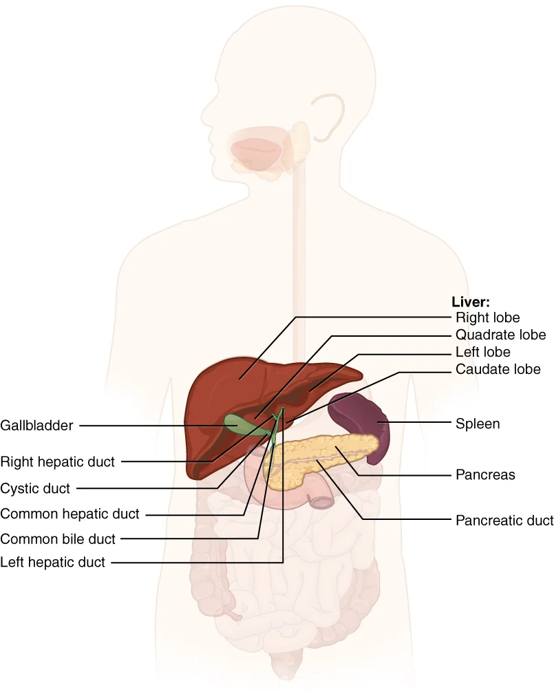
Long description
The liver, pancreas, and gallbladder are considered accessory digestive organs, but their roles in the digestive system are vital.

Figure 1: The liver, pancreas, and gallbladder are considered accessory digestive organs, but their roles in the digestive system are vital.
The hepatic lobule is organized around a central vein, with blood flowing from the periphery toward the center. At the periphery of each lobule lies the portal triad, consisting of three structures: a bile duct, a hepatic artery branch, and a hepatic portal vein branch. Blood enters the lobule through hepatic sinusoids, which are open, porous blood spaces formed by fenestrated capillaries from the hepatic portal vein and hepatic artery. The liver has three main components: hepatocytes, bile canaliculi, and hepatic sinusoids.
Between adjacent hepatocytes, bile canaliculi form small ducts that accumulate bile produced by hepatocytes. Bile flows from canaliculi into bile ductules and then into larger bile ducts, eventually forming the common hepatic duct. The gallbladder has a simple columnar epithelium organized in rugae, with no submucosa. Its muscular coat contracts to eject bile, and the mucosa absorbs water and ions, concentrating bile by up to 10-fold. [1]
The pancreas has both exocrine and endocrine components. The exocrine part consists of acini (grape-like cell clusters) that secrete enzyme-rich pancreatic juice into ducts. These ducts merge to form two dominant ducts, with the larger fusing with the common bile duct before entering the duodenum. Scattered among the exocrine acini are the islets of Langerhans, which contain endocrine cells producing hormones including insulin, glucagon, somatostatin, and pancreatic polypeptide.
Pathological conditions include cirrhosis (characterized by fibrosis and nodule formation), steatosis (fatty infiltration of hepatocytes), and pancreatitis (inflammation of the pancreatic tissue). The liver's stellate cells and pancreatic stellate cells play roles in fibrogenic processes in these conditions[1].
Under light microscopy, the liver and pancreas exhibit distinct histological features that reflect their specialized functions. The liver is organized into lobules, while the pancreas contains both exocrine and endocrine components.
| Structure | Appearance | Cell types | Key features |
|---|---|---|---|
| Liver lobule | Hexagonal arrangement of hepatocytes radiating from a central vein | Hepatocytes, Kupffer cells, stellate cells (Ito cells), sinusoidal endothelial cells | Hepatocytes form cords separated by sinusoids; central vein at the center; portal triads at corners |
| The pancreas has a unique structure that supports both exocrine and endocrine functions. This soft, oblong glandular organ lies transversely in the retroperitoneum behind the stomach. Its head is nestled into the "c-shaped" curvature of the duodenum, with the body extending to the left about 15.2 cm (6 inches) and ending as a tapering tail in the hilum of the spleen. The pancreas produces enzyme- and bicarbonate-rich pancreatic juice, which it delivers to the small intestine through ducts. This [1][2] | |||
| Pancreas endocrine | Islets of Langerhans scattered among exocrine tissue | Alpha cells, beta cells, delta cells, PP cells | Islets are paler staining than exocrine tissue; contain hormone-producing cells |
| Pancreatic stellate cells (PSCs) | Located adjacent to acinar cells and ducts | Quiescent PSCs with vitamin A droplets | Comprise 4–7% of total pancreatic cell mass; express desmin, GFAP, vimentin [3] |
The darker staining Kupffer cells are identifiable within the sinusoids. In the pancreas, the exocrine component appears as clusters of acinar cells with their characteristic pyramidal shape and basophilic cytoplasm. The lighter staining islets of Langerhans are scattered throughout the exocrine tissue.
The pancreatic stellate cells, discovered relatively recently (first described in 1982), play a critical role in pancreatic fibrosis in diseases like chronic pancreatitis and pancreatic cancer. In their quiescent state, these cells contain abundant vitamin A lipid droplets in their cytoplasm, distinguishing them from other cell types. [3]
The liver lobule represents the fundamental functional unit, organized around a central vein and demarcated by portal triads. Each portal triad contains three essential components: hepatic arterioles, portal venules, and bile ducts. This arrangement facilitates the liver's dual blood supply and its role in processing nutrients, toxins, and bile production. Hepatocytes constitute the primary cellular component of the liver, arranged in plates that radiate from the central vein. These cells are responsible for numerous metabolic functions, including protein synthesis, detoxification, and the production of bile. Between the hepatocyte plates, sinusoids allow blood to flow from the portal triads toward the central vein, creating an efficient exchange system between blood and liver cells. [3]
Due to their hydrophobic nature, fatty acids can only be exported from the liver after being packed into water-soluble VLDL particles alongside cholesterol, phospholipids, and apolipoproteins. VLDL particles are formed in the endoplasmic reticulum, where apolipoprotein B100 (apoB100) is lipidated in a process catalyzed by the enzyme microsomal triglyceride transfer protein (MTTP). The nascent VLDL particle is then transferred to the Golgi apparatus, and during this process, the particle is further lipidated until a mature VLDL particle is formed. [2]
The pancreas consists of both exocrine and endocrine components. The exocrine portion comprises acinar cells that produce digestive enzymes, which are transported through ducts to the duodenum. These acinar cells are organized into lobules separated by thin connective tissue septa. Embedded within the exocrine tissue are clusters of endocrine cells known as the islets of Langerhans. These islets contain alpha, beta, delta, and other specialized cells that produce hormones like insulin, glucagon, and somatostatin, which regulate blood glucose levels. [1]
These cells play a crucial role in maintaining the normal architecture of the pancreas and participate in tissue repair processes. However, when activated during injury or disease, PSCs undergo morphological changes, lose their lipid droplets, and transform into proliferative, contractile, and fibrogenic cells that produce excessive amounts of extracellular matrix components, particularly collagen. [3]
In non-alcoholic fatty liver disease (NAFLD), there is an accumulation of triglycerides within hepatocytes. Interestingly, ChREBP, a regulator of fatty acid metabolism, is downregulated in NAFLD patients compared to healthy controls. Instead, SREBP1c becomes a predominant regulator, upregulating genes involved in de novo lipogenesis. In high fat fed mice, adenovirus-mediated ChREBP overexpression resulted in hepatic steatosis and increased de novo lipogenesis, but insulin sensitivity and glucose tolerance were maintained, likely owing to an increased conversion of saturated fatty acids to monounsaturated fatty acids mediated by stearoyl-CoA desaturase-1 (SCD1). [2] [1]
The liver exports excess lipids through very-low-density lipoprotein (VLDL) particles. These particles require apolipoprotein B100 and are formed in the endoplasmic reticulum, with further lipidation occurring in the Golgi apparatus. While one molecule of apoB100 is associated with each VLDL particle, and is required for VLDL export, the triglyceride content of the VLDL particle can vary considerably. This export mechanism represents the only way to reduce hepatic lipid content beyond fatty acid oxidation.
In chronic pancreatitis, pancreatic stellate cells (PSCs) play a crucial role in fibrosis development. These cells are activated early in the course of injury through paracrine effects from injured acinar cells and inflammatory cells. Once activated, PSCs become the major source of collagen in fibrotic areas and can maintain their activated state through autocrine pathways, explaining disease progression even after the initial insult ceases. [3]
Special stains are essential for visualizing specific components of liver and pancreatic tissue that are not clearly visible with standard hematoxylin and eosin (H&E) staining. These stains highlight particular structures, cells, or pathological changes that are crucial for understanding the histology of these organs.
In liver histology, special stains help identify various components. The Masson's trichrome stain is particularly valuable for demonstrating connective tissue in the liver, highlighting fibrosis that may occur in chronic liver disease. Sirius Red is another stain used specifically for collagen detection, which is particularly useful in assessing fibrotic changes in both liver and pancreas. For lipid content, special stains like Oil Red O can be employed on frozen sections to visualize fat droplets within hepatocytes, which is particularly relevant in conditions like non-alcoholic fatty liver disease (NAFLD).
In pancreatic histology, special stains play a crucial role in evaluating chronic pancreatitis. Standard histological stains like H&E provide basic architecture, but special stains reveal more detailed pathological changes. Masson's trichrome and Sirius Red stains are used to identify and quantify collagen deposition in the fibrotic pancreas. These stains help distinguish between normal and fibrotic tissue by highlighting the increased extracellular matrix characteristic of chronic pancreatitis.
Immunohistochemical techniques are considered specialized staining methods that provide valuable information about cellular components in both organs. In the pancreas, immunostaining for alpha smooth muscle actin (αSMA) helps identify activated pancreatic stellate cells (PSCs), which play a key role in fibrogenesis. Studies have shown that areas staining positive for collagen also stain positive for αSMA, indicating the presence of activated PSCs in fibrotic areas of the pancreas. Furthermore, dual immunostaining techniques have established that it is predominantly the activated PSCs that produce the collagen in fibrotic areas of the pancreas. [3]
The liver section demonstrates the classic lobular architecture with central veins and portal tracts. The fibrotic areas appear as blue deposits (Masson's trichrome) or red deposits (Sirius Red), indicating increased collagen deposition. The immunohistochemical staining for αSMA reveals activated stellate cells within these fibrotic regions, confirming their role in the fibrotic process.
The ultrastructure of the liver and pancreas reveals intricate cellular components visible only through electron microscopy. In hepatocytes, the plasma membrane contains specialized transporters for fatty acids, including fatty acid transport proteins (FATP) and cluster of differentiation 36 (CD36). These proteins facilitate the uptake of fatty acids, with FATP2 and FATP5 being the primary isoforms found in the liver. The cytoplasm contains numerous organelles: rough endoplasmic reticulum for protein synthesis, smooth endoplasmic reticulum for lipid metabolism, and peroxisomes where fatty acid oxidation occurs. Hepatocytes also contain lipid droplets that store triglycerides, which become prominent in conditions like steatosis.
Pancreatic stellate cells (PSCs) exhibit distinctive ultrastructural features. On electron microscopic examination, PSCs reveal a prominent rough endoplasmic reticulum, collagen fibrils, and vacuoles (lipid droplets) surrounding a central nucleus. In their quiescent state, PSCs exhibit a flattened polygonal shape with prominent lipid droplets in the cytoplasm surrounding the central nucleus. These lipid droplets contain vitamin A, which can be visualized when the cells are exposed to UV light at 328 nm, eliciting a transient blue-green fluorescence. [3]
The endoplasmic reticulum in both liver and pancreatic cells plays crucial roles in cellular function. Hepatocytes contain both rough and smooth endoplasmic reticulum, with the rough form studded with ribosomes for protein synthesis. In pancreatic stellate cells, the rough endoplasmic reticulum is particularly prominent, reflecting their active synthetic capabilities. The smooth endoplasmic reticulum in hepatocytes is involved in lipid metabolism and detoxification processes.
The liver and pancreas are complex organs with specialized structures that enable their diverse functions. In the liver, the space of Disse is a crucial anatomical feature that facilitates the exchange of substances between hepatocytes and blood. This space lies between the plates of hepatocytes and the endothelial cells of the hepatic sinusoids. It contains a network of reticular fibers and is bathed in blood plasma, allowing for efficient transfer of nutrients, hormones, and other molecules be [1]
The pancreas has a unique structure that supports both exocrine and endocrine functions. This soft, oblong glandular organ lies transversely in the retroperitoneum behind the stomach. Its head is nestled into the "c-shaped" curvature of the duodenum, with the body extending to the left about 15.2 cm (6 inches) and ending as a tapering tail in the hilum of the spleen. The pancreas produces enzyme- and bicarbonate-rich pancreatic juice, which it delivers to the small intestine through ducts. This
The exocrine function of the pancreas involves the secretion of digestive enzymes. The enzymes that digest starch (amylase), fat (lipase), and nucleic acids (nuclease) are secreted in their active forms, as they do not attack the pancreas itself. This is in contrast to the protein-digesting enzymes, which must be secreted in inactive forms to prevent autodigestion of the pancreatic tissue. [1]
Within the liver, the space of Disse plays a vital role in lipid metabolism. Lipids are hydrophobic and do not dissolve in water, so before they can be digested in the small intestine, large lipid globules must be broken down into smaller ones through emulsification. Bile, secreted by the liver, accomplishes this emulsification process. The space of Disse allows for efficient exchange between hepatocytes and sinusoidal blood, facilitating the transport of bile components and other metabolites. [1]
The pancreas also contains specialized cells called pancreatic stellate cells (PSCs) that contribute to the organ's structure and function. These cells can function as progenitor cells, immune cells, or intermediary cells in cholecystokinin (CCK)-induced pancreatic digestive enzyme secretion. PSCs express Toll-like receptors (TLR2, 3, 4, 5 and 9) which recognize foreign pathogen-associated molecular patterns (PAMPs), suggesting a role in innate immunity. They also respond to various signaling molecules such as platelet-derived growth factor (PDGF), which mediates PSC proliferation through ERK and JAK/STAT pathways, while PI3K pathway regulates PDGF-induced migration. [3]
The liver is the largest internal organ and can be identified on a slide by its distinctive lobular structure. Each liver lobule is typically hexagonal and centered around a central vein, which runs from the center toward the periphery. Radiating outward from the central vein are plates of hepatocytes, one cell thick, which form the functional parenchyma of the liver. Between these hepatocyte plates are sinusoids, specialized vascular spaces where blood from the hepatic artery and portal vein mix before draining into the central vein. The lobule's periphery contains portal triads, each consisting of a branch of the hepatic artery, a portal venule, and a bile duct.
DefinitionLiver lobule
A hexagonal functional unit centered on a central vein, with portal triads at each corner.
The gallbladder, an 8-10 cm muscular sac, can be identified by its location nested in a shallow area on the posterior aspect of the right lobe of the liver. It has three distinct regions: the fundus (the widest portion), the body (tapering medially from the fundus), and the neck (which narrows and angles slightly superiorly to approach the hepatic duct). The cystic duct, a 1-2 cm tube, connects the gallbladder neck to the hepatic duct. [1]
The pancreas is identified by its distinctive exocrine and endocrine components. The exocrine portion consists of clusters of secretory cells called acini, which produce pancreatic enzymes. These acini drain into small ductules that eventually form larger ducts. The endocrine portion consists of islets of Langerhans, which are scattered throughout the pancreas and contain alpha, beta, delta, and other hormone-producing cells. The pancreas produces over a liter of pancreatic juice daily, which is clear and alkaline (pH 7.1 to 8.2) due to its sodium bicarbonate content. [1]
NotePancreatic juice composition
Clear fluid containing water, salts, sodium bicarbonate, and digestive enzymes active in the digestion of sugars, proteins, and fats.
When examining liver and pancreas slides, it's important to distinguish between normal tissue and pathological changes. In the liver, steatosis (fatty liver) appears as lipid droplets within hepatocytes, while fibrosis shows as increased collagen deposition. In the pancreas, chronic pancreatitis reveals fibrosis with activation of pancreatic stellate cells (PSCs), which exhibit a myofibroblastic phenotype and express alpha-smooth muscle actin. Normal pancreatic tissue should show minimal connective tissue between acini.
| Feature | Liver | Pancreas |
|---|---|---|
| Basic structure | Lobular with central vein and portal triads | Acinar with ducts and scattered islets |
| Main cell type | Hepatocytes | Acinar cells (exocrine) and islet cells (endocrine) |
| Distinguishing feature | Sinusoids between hepatocyte plates | Zymogen granules in acinar cells |
| Common pathology | Steatosis (fat droplets), fibrosis | Acinar atrophy, fibrosis, islet changes |
Radiating from the central vein are hepatocyte plates separated by sinusoids. Scattered among the acini are islets of Langerhans, which appear as lighter-staining clusters of cells.
The liver exhibits characteristic histological changes in various disease states. In non-alcoholic fatty liver disease (NAFLD), hepatic triglyceride content can be quantified using magnetic resonance spectroscopy, with classifications of no steatosis (< 5.56%), mild steatosis (5.56–15%), or severe steatosis (> 15%). Interestingly, the presence of NAFLD rather than the amount of hepatic triglycerides is associated with hepatic insulin resistance. Liver biopsies reveal that cytoplasmic diacylgl [2]
The hepatocytes demonstrate uniform eosinophilic cytoplasm and prominent nuclei. In steatotic liver, hepatocytes contain clear lipid vacuoles that displace the nucleus to the periphery of the cell. The portal tract contains bile ducts, hepatic artery branches, and portal vein tributaries. In NASH, additional features include hepatocyte ballooning, Mallory bodies (eosinophilic cytoplasmic inclusions), and mixed inflammatory infiltrate in the lobules and portal tracts.
Islets of Langerhans are visible as clusters of lighter-staining cells. In chronic pancreatitis, note the replacement of normal pancreatic tissue with fibrosis, characterized by dense collagen deposition. Activated pancreatic stellate cells (PSCs) are identified by their stellate shape and positive staining for alpha-smooth muscle actin. Inflammatory infiltrate is present around the ducts and within the fibrotic areas.
In NAFLD, antioxidant defense systems are compromised, with decreased levels of glutathione, glutathione peroxidase, and superoxide dismutase in liver biopsies. This creates a vicious cycle where oxidative stress depletes these antioxidants further. NASH patients show significantly increased oxidative DNA damage compared to healthy controls, despite similar rates of fatty acid oxidation. [2]
Hepatic stellate cells (HSCs) play a central role in liver fibrogenesis, being established as key producers of extracellular matrix proteins. Beyond fibrosis, HSCs contribute to liver development, regeneration, retinoid metabolism, and immunomodulation. In the pancreas, pancreatic stellate cells (PSCs) similarly drive fibrogenesis in chronic pancreatitis. These activated PSCs express senescence markers such as senescence associated β-galactosidase (SA-βGal) and become susceptible to immune-mediated cytotoxicity. The relative contributions of apoptosis, senescence, and reversion to quiescence in removing activated PSCs after pancreatic injury remain unclear. [3]
Clinical pearlLiver histology
The liver lobule is the functional unit, centered on a central vein and surrounded by portal triads containing hepatic arterioles, portal venules, and bile ducts. Hepatocytes account for approximately 80% of liver volume and perform over 500 metabolic functions.
Exam trapNAFLD pathophysiology
Instead, SREBP1c becomes the predominant regulator of de novo lipogenesis in this condition. [2]
Clinical pearlVLDL formation
VLDL particles require apolipoprotein B100 and are formed in the endoplasmic reticulum, with further lipidation occurring in the Golgi apparatus. Each VLDL particle contains exactly one molecule of apoB100, which is essential for VLDL export, while triglyceride content can vary considerably. [2]
Exam trapNAFLD treatment
There is currently no approved pharmacological treatment for NAFLD. A major limiting factor in developing new treatments is the lack of predictive pre-clinical models that accurately reflect human disease with regard to liver histology, pathophysiology, and metabolic abnormalities. [2]
Clinical pearlPancreatic stellate cells
Pancreatic stellate cells (PSCs) are the major source of collagen in fibrotic areas of chronic pancreatitis. These cells are activated early in the course of injury through paracrine effects of factors produced by injured acinar cells and inflammatory cells during the acute phase. [3]
Exam trapFibrosis mechanisms
Pancreatic fibrosis was historically considered an epiphenomenon rather than a key pathological process, despite being a predominant histological feature of two major pancreatic diseases: chronic pancreatitis and pancreatic cancer.
Clinical pearlPSC activation
PSCs are activated early in pancreatic injury through paracrine effects from injured acinar cells and inflammatory cells.
Clinical pearlPDGF role
Increased PDGF receptor expression on PSCs contributes to their increased numbers in fibrotic areas of chronic pancreatitis. PDGF is a potent mitogenic and chemotactic factor for PSCs, and its receptor expression is increased at both mRNA and protein levels in areas of fibrosis. [3]
Exam trapEthanol effects
This occurs through different mechanisms than the oxidative ethanol metabolite acetaldehyde, as non-oxidative ethanol metabolites (FAEEs) have not been reported to activate PSCs. [3]
Clinical pearlPSC perpetuation
Activated PSCs can maintain their state through autocrine pathways, explaining disease progression after initial insult cessation. This phenomenon may represent one of the mechanisms responsible for progression of chronic pancreatitis despite the cessation of the initial insult, for example alcohol and/or acute flare. [3]
NoteTreatment challenges
Any intervention targeting NAFLD pathways is likely to have consequences on multiple cellular signaling pathways, which should be taken into careful consideration when developing future treatment options. Inter-individual differences further complicate treatment approaches.
Exam trapConfusing hepatocytes with Kupffer cells
Hepatocytes are the primary functional cells of the liver, responsible for metabolism, detoxification, and bile production, while Kupffer cells are specialized macrophages that line the sinusoids and are responsible for immune surveillance and phagocytosis.
Exam trapMisunderstanding pancreatic stellate cell activation
Pancreatic stellate cells (PSCs) exist in a quiescent state in the normal pancreas, containing vitamin A lipid droplets, but become activated during injury or disease, losing these droplets and transforming into proliferative, fibrogenic cells.
Exam trapAssuming ethanol only affects PSCs after inflammation
Ethanol can activate PSCs directly from their quiescent state without requiring pre-activation or necroinflammation, and also inhibits PSC apoptosis, facilitating their survival and perpetuating fibrosis. [3]
Exam trapOverlooking the role of cytokines in PSC activation
Multiple cytokines play distinct roles in PSC activation: PDGF acts as a proliferative and chemotactic factor; TGFβ stimulates ECM protein synthesis and MMP2 production; and TNFα, MCP-1, IL1, IL6, and IL13 promote proliferation, αSMA expression, and collagen synthesis.
Exam trapIgnoring the paracrine effects in chronic pancreatitis
In chronic pancreatitis, pancreatic acinar cells adjacent to fibrosis express TGFβ, which may have a paracrine effect on PSCs, leading to increased collagen synthesis, while NGF expression is also increased in this condition.
Exam trapUnderestimating PSC involvement in pancreatic cancer
PSCs contribute to pancreatic cancer progression by enhancing tumor angiogenesis through VEGF secretion, promoting chemoresistance by sequestering drugs like gemcitabine in the stroma, and protecting cancer cells from radiation via β1-integrin dependent pathways. [3]
Exam trapNeglecting therapeutic targeting of PSCs
The pancreatic stroma, particularly PSCs, represents an important therapeutic target in pancreatic cancer, with approaches like inhibition of the Sonic hedgehog pathway in PSCs showing promise in pre-clinical models by depleting stroma and increasing drug accumulation in cancer cells.
NoteClinical assessment of liver function
Understanding hepatocyte function is crucial in clinical practice for assessing liver function, particularly the production and regulation of bile, which increases when fatty chyme enters the duodenum and stimulates cholecystokinin secretion.
NoteDiagnosis of pancreatic fibrosis
Knowledge of PSC activation mechanisms helps in understanding the pathogenesis of pancreatic fibrosis, where activated PSCs contribute to excessive extracellular matrix production, a hallmark of chronic pancreatitis.
| Cytokine | Effect on PSCs |
|---|---|
| PDGF | Potent proliferative and chemotactic factor |
| TGFβ | Stimulates ECM protein synthesis and MMP2 production |
| TNFα, MCP-1, IL1, IL6, IL13 | Promote proliferation, αSMA expression, and collagen synthesis |
| VEGF | Mediates PSC-induced angiogenesis in pancreatic cancer |
Pancreatic stellate cell activation in disease
- Quiescent statePSCs contain vitamin A lipid droplets
- Question: Injury or stimulus?
Yes
- ActivationLoss of lipid droplets, transformation to myofibroblasts
- ProliferationDriven by PDGF and other growth factors
- FibrogenesisECM production stimulated by TGFβ and CTGF
- Disease progressionFibrosis in chronic pancreatitis or contribution to pancreatic cancer
No
- MaintenanceQuiescent state preserved
Clinical pearlTherapeutic implications in pancreatic cancer
Understanding PSC involvement in pancreatic cancer has led to novel therapeutic strategies targeting the stroma, such as inhibiting the Sonic hedgehog pathway to deplete stroma and increase drug delivery to cancer cells, potentially overcoming chemoresistance.
Clinical pearlImaging considerations
Knowledge of PSC activation and fibrosis patterns helps in interpreting imaging studies of the pancreas, where fibrotic bands and increased stromal density are characteristic features of chronic pancreatitis and pancreatic cancer.
Clinical pearlSurgical planning
Understanding the histological changes in liver and pancreas, including bile production regulation and PSC activation patterns, is essential for surgical planning, particularly in procedures involving the hepatobiliary system or pancreatic resections.
- Hepatocytes continuously produce bile, which is conserved between meals by diverting to the gallbladder when the hepatopancreatic ampulla closes [1]
- Pancreatic stellate cells exist in a quiescent state with vitamin A lipid droplets and become activated during injury or disease
- Ethanol can directly activate PSCs from their quiescent state and inhibits their apoptosis, facilitating fibrosis development
- Multiple cytokines regulate PSC activation: PDGF promotes proliferation, TGFβ stimulates ECM production, and various inflammatory cytokines enhance fibrogenic activity
- In chronic pancreatitis, acinar cells may secrete TGFβ that acts on PSCs in a paracrine manner to promote fibrosis
- PSCs contribute to pancreatic cancer progression by enhancing angiogenesis via VEGF secretion and promoting chemoresistance by sequestering drugs
- Targeting pancreatic stellate cells represents a promising therapeutic approach to overcome treatment resistance in pancreatic cancer
Answer each one in your head first, then open the answer.
What is the primary function of hepatocytes in bile production?
Show answerHide answer
Hepatocytes continuously produce bile, which is conserved between meals by diverting to the gallbladder when the hepatopancreatic ampulla closesWhat is a key characteristic of quiescent pancreatic stellate cells?
Show answerHide answer
Quiescent pancreatic stellate cells contain vitamin A lipid droplets in their cytoplasmHow does ethanol affect pancreatic stellate cells?
Show answerHide answer
Ethanol can activate PSCs directly from their quiescent state and inhibits their apoptosis, facilitating their survival and perpetuating fibrosisWhich cytokine is known to stimulate extracellular matrix protein synthesis by pancreatic stellate cells?
Show answerHide answer
TGFβ stimulates the synthesis and secretion of ECM proteins (collagen, fibronectin, and laminin) by pancreatic stellate cellsHow do pancreatic stellate cells contribute to chemoresistance in pancreatic cancer?
Show answerHide answer
PSCs contribute to chemoresistance by sequestering chemotherapeutic agents like gemcitabine in the stromal area, limiting their availability to cancer cells
- Betts JG, Young KA, Wise JA, et al. Accessory Organs in Digestion: The Liver, Pancreas, and Gallbladder. OpenStax, Anatomy and Physiology 2e (CC BY-NC-SA 4.0). Accessed .
- Ipsen DH, Lykkesfeldt J, Tveden-Nyborg P. Molecular mechanisms of hepatic lipid accumulation in non-alcoholic fatty liver disease. Cellular and molecular life sciences : CMLS 2018;75(18):3313-3327. doi:10.1007/s00018-018-2860-6. Accessed .
- Apte MV, Pirola RC, Wilson JS. Pancreatic stellate cells: a starring role in normal and diseased pancreas. Frontiers in physiology 2012;3:344. doi:10.3389/fphys.2012.00344. Accessed .
Figure credits
Educational content for medical students and doctors. It is not a substitute for clinical judgement or local guidelines.