Renal histology
Renal histology examines kidney structure and function at microscopic level.
The kidney is divided into two main regions: the renal cortex and the renal medulla[1]. The renal cortex appears lighter in color compared to the medulla and contains all renal corpuscles, proximal convoluted tubules (PCTs), and distal convoluted tubules[1]. The renal medulla contains renal pyramids and renal papillae, which are bundles of collecting ducts that transport urine to the calyces[1].
The functional unit of the kidney is the nephron, consisting of the renal corpuscle, PCT, loop of Henle, and DCT. Each kidney contains about 1.3 million nephrons[1]. Nephrons are classified as cortical nephrons (with short loops of Henle) or juxtamedullary nephrons (with long loops extending into the medulla), with approximately 15 percent of nephrons being juxtamedullary[1]. [2]
The renal corpuscle consists of the glomerulus and Bowman's capsule, forming the initial filtering unit of the nephron. The glomerulus contains afferent and efferent arterioles that regulate blood flow and pressure. The afferent arteriole brings blood into the glomerulus, while the efferent arteriole carries filtered blood away. Between these arterioles lies the glomerular capillary network, which is specialized for filtration. Bowman's capsule surrounds the glomerulus and consists of a parietal [1]
Podocytes are visceral epithelial cells with large cell bodies that bulge into Bowman's space and do not touch the GBM directly. From each cell body, primary processes emerge and branch into secondary foot processes (pedicels) that interdigitate with those of neighbouring podocytes, covering the outer GBM surface. Between adjacent foot processes lies the filtration slit, about 25–30 nm wide, bridged by a slit diaphragm composed of nephrin, podocin and CD2AP. The slit diaphragm is the final size- [1]
Mesangial cells in the filtration membrane can contract to help regulate the rate of filtration. The juxtaglomerular apparatus contains specialized cells that produce paracrine signals to regulate blood flow and filtration rates of the glomerulus. After filtration, the blood flows through a second capillary bed (peritubular capillaries and vasa recta) surrounding the nephron tubule, forming a portal system[1].
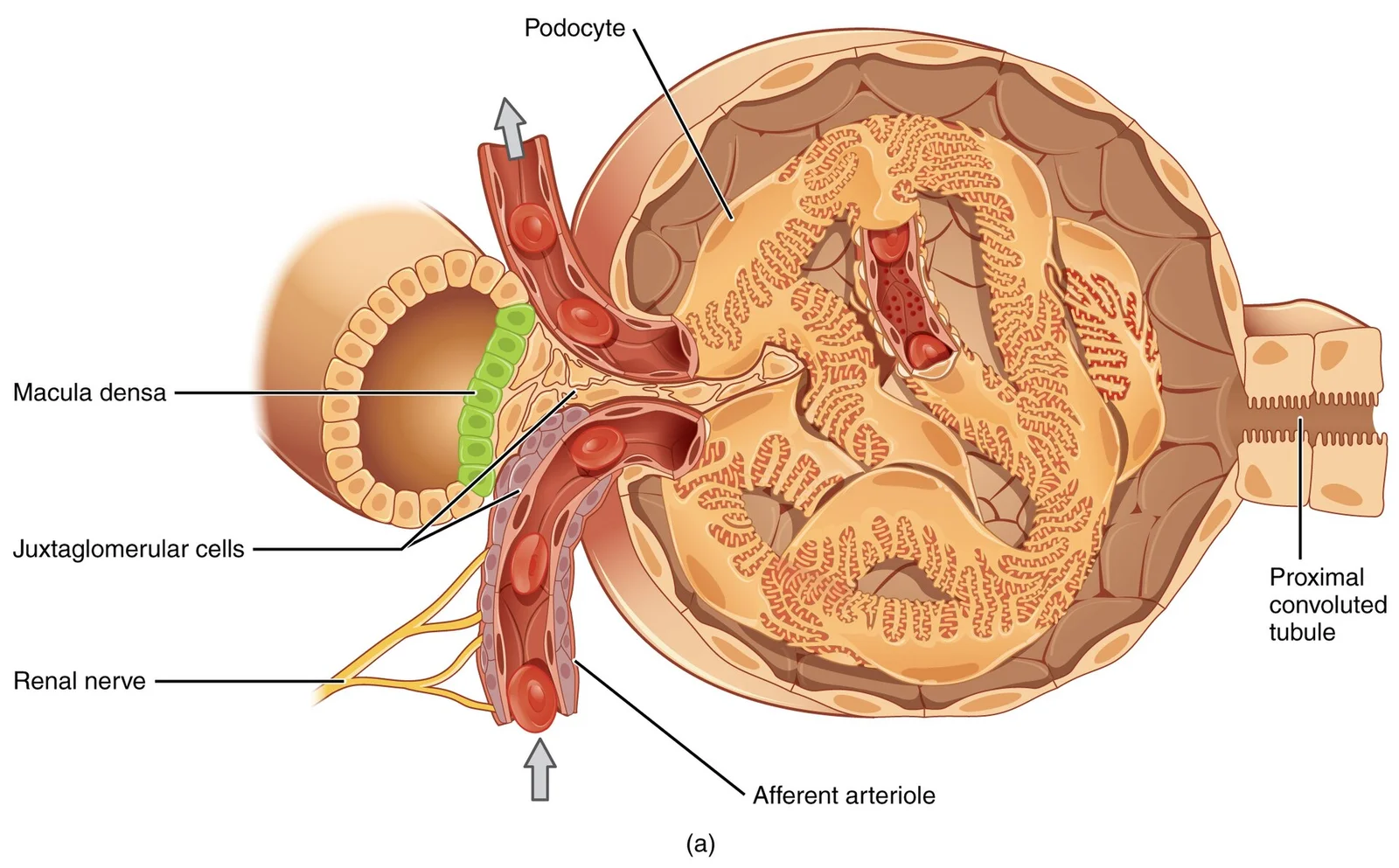
Figure 12615 Juxtaglomerular Apparatus
Long description
(a) The JGA allows specialized cells to monitor the composition of the fluid in the DCT and adjust the glomerular filtration rate. (b) This micrograph shows the glomerulus and surrounding structures. LM × 1540. (Micrograph provided by the Regents of University of Michigan Medical School © 2012)

Figure 1: (a) The JGA allows specialized cells to monitor the composition of the fluid in the DCT and adjust the glomerular filtration rate.
Under light microscopy, renal histology reveals the intricate architecture of the nephron and its associated structures. The H&E staining demonstrates distinct layers and cell types that work together to filter blood and form urine. Each kidney contains approximately 1.3 million nephrons, which are the functional units responsible for maintaining homeostasis through filtration, reabsorption, and secretion[1]. The renal structures cannot be seen by the naked eye and require microscopic examination to reveal their functional anatomy[1].
| Tubule Segment | Epithelial Type | Key Features |
|---|---|---|
| Proximal Convoluted Tubule (PCT) | Simple cuboidal | Numerous microvilli, highly active in reabsorption, eosinophilic cytoplasm |
| Loop of Henle - Descending thick | Simple cuboidal | Similar to PCT, continues from PCT |
| Loop of Henle - Descending thin | Simple squamous | Important for water permeability, forms hairpin turn |
| Loop of Henle - Ascending thin | Simple squamous | Different permeability properties, adjacent to descending limb |
| Loop of Henle - Ascending thick | Simple cuboidal | Similar to Distal Convoluted Tubule, contains ion pumps |
| Distal Convoluted Tubule (DCT) | Simple cuboidal | Fewer microvilli than PCT, many mitochondria for ion transport |
| Collecting Duct | Simple cuboidal or columnar | ADH-responsive, contains aquaporin channels for water reabsorption |
The renal corpuscle consists of the glomerulus and Bowman's capsule. The glomerulus is a tuft of capillaries where blood filtration occurs. Between 10 to 20 percent of plasma filters into Bowman's capsule[1]. The filtration membrane comprises the capillary endothelium with fenestrae, shared basement membrane, and podocyte "fingers" with filtration slits. These three features work together to create a selective barrier
The juxtaglomerular apparatus is a specialized structure where the distal tubule comes into contact with the afferent arteriole. It contains juxtaglomerular cells - modified smooth muscle cells that regulate glomerular filtration rate based on filtrate osmolarity[1]. When the osmolarity of the filtrate is too high (hyperosmotic), these cells contract, decreasing the glomerular filtration rate (GFR) so less plasma is filtered[1]. Conversely, if the osmolarity is too low, the j
The loop of Henle consists of descending and ascending portions that run adjacent and parallel after making a hairpin turn. The descending loop has an initial short, thick portion with simple cuboidal epithelium similar to the PCT, followed by a long, thin portion with simple squamous epithelium. The ascending loop begins with a thin portion (simple squamous) and transitions to a thick portion with simple cuboidal epithelium similar to the DCT. These differ [1]
The distal convoluted tubule (DCT) is tortuous like the PCT but shorter, formed by simple cuboidal epithelium with fewer microvilli on the apical surface[1]. Despite fewer microvilli, these cells contain large numbers of mitochondria (although fewer than in the PCT) to pump ions against concentration gradients[1].
Collecting ducts, which are continuous with the nephron but not technically part of it, merge as they descend deeper in the medulla to form about 30 terminal ducts[1]. They are lined with simple cuboidal or columnar epithelium containing receptors for ADH[1]. When stimulated by ADH, these cells insert aquaporin channel proteins into their membranes, allowing water to pass from the duct lumen through the cells and into the interstitial spaces[1]. These aquaporin channels are so small that only water can pass, enabling precise control of water reabsorption[1].
The glomerulus appears as a tuft of capillary loops surrounded by Bowman's capsule. The proximal convoluted tubule can be identified by its prominent brush border and eosinophilic cytoplasm. The loop of Henle demonstrates both thick and thin segments with different epithelial characteristics. The distal convoluted tubule has fewer microvilli but contains numerous mitochondria. Collecting ducts appear as larger tubules with cuboidal or columnar epithelium.
Special stains are essential techniques in renal histology that highlight specific components of the kidney tissue that are not visible with routine hematoxylin and eosin (H&E) staining. These stains allow for detailed examination of the glomerular basement membrane, tubular structures, and extracellular matrix components.
DefinitionPeriodic acid-Schiff (PAS) stain
The PAS stain is one of the most commonly used special stains in renal pathology. It stains glycogen, glycoproteins, and glycolipids, while the background appears blue. This stain is used for evaluating the glomerular basement membrane and mesangial matrix. [1]
| Stain | Target structures | Clinical application |
|---|---|---|
| Periodic acid-Schiff (PAS) | Glycogen, glycoproteins, glycolipids | Evaluation of glomerular basement membrane thickness, mesangial matrix expansion |
| Masson's trichrome | Collagen, fibrin | [1] |
| Silver stain (Jones, methenamine silver) | Basement membranes, reticular fibers | [1] |
| Congo red | Amyloid protein | [1] |
The silver stain, particularly the Jones methenamine silver technique, is crucial for examining the glomerular basement membrane. This stain blackens the basement membranes, making them clearly visible against a pale background. It is especially useful for assessing the thickness of the glomerular basement membrane. [1]
Masson's trichrome stain is valuable for identifying collagen and fibrous tissue within the kidney. With this stain, collagen appears blue or green, while cytoplasm is red and nuclei are black. This stain helps in evaluating the extent of interstitial fibrosis and tubular atrophy in chronic kidney disease.
NoteCongo red stain
The Congo red stain is specifically used to detect amyloid deposits in renal tissue. When positive, these deposits exhibit apple-green birefringence under polarized light, which is diagnostic for amyloidosis. [1]
The glomerular basement membrane appears as a magenta-colored structure surrounding the capillary loops.
Electron microscopy reveals the renal corpuscle as a highly specialised filtration apparatus built from three layers: the fenestrated endothelium, the glomerular basement membrane, and the podocyte layer with its slit diaphragms. Each layer contributes a distinct size and charge barrier, and together they produce the ultrafiltrate that enters Bowman's space.
The glomerular capillary endothelium is a thin, attenuated sheet of cytoplasm perforated by numerous circular fenestrae, each about 70 nm in diameter. These fenestrae lack diaphragms, unlike those of peritubular capillaries. The endothelial cell body lies near the mesangial aspect of the loop, while its fenestrated periphery faces the basement membrane. A thin glycocalyx coats the luminal surface and carries a negative charge from si [1]
The glomerular basement membrane (GBM) is a trilaminar structure, formed by fusion of the endothelial and podocyte basal laminae. Its three layers are the lamina rara interna (adjacent to endothelium), the thick central lamina densa, and the lamina rara externa (adjacent to podocytes). The lamina densa is a meshwork of type IV collagen, laminin, nidogen and heparan sulphate proteoglycans; the latter confer the anionic charge that impedes albumin passage. In Alport [1]
Podocytes are visceral epithelial cells with large cell bodies that bulge into Bowman's space and do not touch the GBM directly. From each cell body, primary processes emerge and branch into secondary foot processes (pedicels) that interdigitate with those of neighbouring podocytes, covering the outer GBM surface. Between adjacent foot processes lies the filtration slit, bridged by a slit diaphragm composed of nephrin, podocin and CD2AP. The slit diaphragm is the final size-
From top to bottom, the reader first sees the fenestrated endothelium lining the capillary, then the trilaminar GBM with its three laminae, then the interdigitating podocyte foot processes with the slit diaphragms between them, and finally the podocyte cell body in Bowman's space.
The mesangium consists of mesangial cells embedded in a mesangial matrix, lying between capillary loops and forming the stalk of the glomerular tuft. Mesangial cells are contractile, phagocytic and capable of proliferation; they contain actin filaments, dense bodies and occasional renin granules. The mesangial matrix is a fibrillar material similar to but less organised than the GBM, and it is continuous with the lamina rara interna at the capillary periphery. Mesangial cells are separated from the capillary lumen only by the fenestrated endothelium, with no basement membrane interposed, which allows them to take up immune complexes and macromolecules that escape the filtration barrier. [1]
The proximal tubule ultrastructure reflects its massive reabsorptive workload. Its cells have a tall brush border of microvilli, about 1–2 μm long and 0.1 μm wide, increasing the apical surface area roughly 20-fold. The apical cytoplasm contains numerous endocytic vesicles, lysosomes and a well-developed endosomal apparatus for protein reabsorption. The basolateral membrane is deeply infolded, with elongated mitochondria oriented perpendicular to the basement membrane between the folds, providin [1]
The kidney's microscopic structure is intricately designed to perform its essential functions of blood filtration, waste excretion, and homeostasis. Each component has a specific anatomical arrangement that directly supports its physiological role.
The nephron is the functional unit of the kidney, consisting of the renal corpuscle and renal tubule. The renal corpuscle, composed of Bowman's capsule and the glomerulus, is responsible for the initial filtration of blood. Bowman's capsule surrounds the glomerular capillaries and collects the filtrate. The glomerulus is a network of capillaries with fenestrated endothelium, a basement membrane, and podocytes with foot processes. This structure allows for the passage of water and small solutes while retaining blood cells and most proteins.
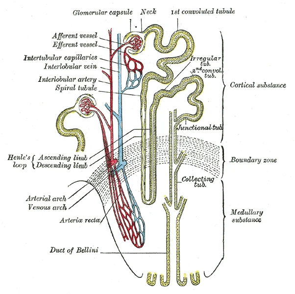
Figure 2Gray1128
Long description
Scheme of renal tubule and its vascular supply.

Figure 2: Scheme of renal tubule and its vascular supply.
The proximal convoluted tubule (PCT) follows Bowman's capsule and is characterized by its tortuous path. It is lined with simple cuboidal cells that possess prominent microvilli on their luminal surface, forming a brush border. These microvilli create a large surface area to maximize the absorption and secretion of solutes such as Na+, Cl-, and glucose. The cells are rich in mitochondria to provide the ATP needed for active ion transport. [1]
The macula densa is a specialized region of the distal tubule that lies adjacent to the juxtaglomerular cells of the afferent arteriole. It plays a crucial role in regulating filtration rate and renin release. When blood osmolarity increases, filtration and urine formation decrease, leading to water retention. Conversely, when blood osmolarity decreases, filtration and urine formation increase, resulting in water loss. The macula densa also regulates renin release from the juxtaglomerular cells. Renin, a protein of 304 amino acids, cleaves angiotensinogen to form angiotensin I, which is then converted to angiotensin II by angiotensin-converting enzyme (ACE) in the lungs. Angiotensin II acts as a systemic vasoconstrictor to help regulate blood pressure. [1]
Renal histology provides the foundation for understanding various clinical conditions. Damage to the glomerular filtration barrier can lead to proteinuria, as seen in glomerulonephritis. The fenestrated endothelium, basement membrane, and podocyte foot processes work together to prevent the loss of proteins into the urine. When this barrier is compromised, proteins such as albumin leak into the filtrate, resulting in proteinuria.
The proximal convoluted tubule's extensive brush border and high mitochondrial content make it particularly vulnerable to ischemic and toxic injuries. Conditions such as acute tubular necrosis often primarily affect the PCT due to its high metabolic activity and exposure to filtered toxins. Impaired function of the PCT can lead to defects in solute reabsorption, resulting in glycosuria or electrolyte imbalances.
The juxtaglomerular apparatus plays a critical role in blood pressure regulation through the renin-angiotensin-aldosterone system (RAAS). Dysregulation of this system can contribute to hypertension. ACE inhibitors, a class of antihypertensive medications, work by blocking the conversion of angiotensin I to angiotensin II, thereby reducing vasoconstriction and blood pressure. Understanding the histological basis of RAAS function helps explain the mechanism of action of these drugs.
Renal histology is characterised by the distinct organisation of the nephron and its associated vasculature. To identify kidney tissue, look for the presence of renal corpuscles, which consist of a globular cluster of capillaries called the glomerulus surrounded by a double-walled Bowman's capsule.
The glomerulus is a high-pressure capillary network that serves as the first capillary bed of a unique portal system. In a histological section, it appears as a dense, eosinophilic sphere of cells and capillaries. Surrounding this is Bowman's capsule, the space between which collects the glomerular filtrate.
The renal corpuscle is the most prominent feature. The glomerulus is the central capillary tuft, surrounded by the urinary space (Bowman's space), which is the clear gap between the tuft and the outer parietal layer of Bowman's capsule.
The JGA is a specialised structure located just outside Bowman's capsule and the glomerulus. It is found at the specific juncture where the afferent and efferent arterioles enter and leave the capsule. [1]
DefinitionMacula Densa
A cluster of cuboidal epithelial cells in the wall of the distal convoluted tubule (DCT) that monitors fluid composition.
On a slide, the macula densa is identified as a region of the DCT that comes into direct contact with the arterioles. These cells are more densely packed than those in other parts of the tubule. They possess a single, nonmotile cilium used to respond to the rate of fluid movement. [1]
The kidney is unique because it contains a portal system where an arteriole is positioned between two capillary beds. The first bed is the glomerulus, and the second consists of the peritubular capillary networks that surround the tubules.
| Structure | Key Histological Feature | Distinguishing Characteristic |
|---|---|---|
| Glomerulus | Capillary tuft | Located within Bowman's capsule |
| Macula Densa | Dense cuboidal cells | Direct contact with afferent/efferent arterioles [1] |
| DCT | Simple cuboidal epithelium | Lacks the prominent brush border seen in the proximal tubule |
| Peritubular Capillaries | Fine vessel networks | Recover solutes and water from the nephron |
The renal cortex can be confused with other glandular tissues. However, the presence of the renal corpuscle is pathognomonic. While other portal systems exist, such as those linking the hypothalamus to the anterior pituitary or the digestive viscera to the liver, the renal system is the only one with an intervening arteriole between the two capillary beds. [2]
The renal corpuscle consists of the glomerulus and Bowman's capsule, forming the initial filtering unit of the nephron. The glomerulus contains afferent and efferent arterioles that regulate blood flow and pressure. The afferent arteriole brings blood into the glomerulus, while the efferent arteriole carries filtered blood away. Between these arterioles lies the glomerular capillary network, which is specialized for filtration. Bowman's capsule surrounds the glomerulus and consists of a parietal
The proximal convoluted tubule (PCT) follows the renal corpuscle and is responsible for the reabsorption of approximately 65% of filtered sodium, water, and glucose. Histologically, the PCT has a simple cuboidal epithelium with a prominent brush border, which increases the surface area for reabsorption. The loop of Henle consists of a descending limb and an ascending limb, creating the countercurrent multiplier system. The descending limb is permeable to water but not solutes, while the ascending limb is impermeable to water but permeable to solutes. [1]
The distal convoluted tubule (DCT) connects to the collecting duct system and is responsible for fine-tuning sodium and potassium balance under hormonal control. The DCT has a simple cuboidal epithelium but lacks the prominent brush border seen in the PCT. The collecting duct system begins with the connecting tubule and continues through the cortical and medullary collecting ducts, ultimately draining into the renal pelvis. [1]
The glomerulus appears as a tuft of capillaries surrounded by Bowman's capsule. The PCT can be identified by its eosinophilic cytoplasm and brush border. The collecting ducts are larger in diameter and appear as empty spaces in the section.
DefinitionRenal Corpuscle Composition
The renal corpuscle is part of the functional unit of the kidney, the nephron, comprising the glomerulus and Bowman's capsule. It is the site where plasma is filtered to form the initial ultrafiltrate. [1]
Clinical pearlFiltration Barrier Layers
The glomerular filtration barrier consists of three distinct layers: fenestrated endothelium, the basement membrane, and podocyte foot processes. The basement membrane contains negatively charged glycoproteins that repel negatively charged plasma proteins, preventing their passage into the urine. [1]
Exam trapPodocyte Limitations
Podocytes are terminally differentiated cells that cannot undergo mitosis. Consequently, injury to podocytes leads to their permanent loss and cannot be replaced by cell division, making them highly susceptible to progressive damage. [1]
Clinical pearlJuxtaglomerular Apparatus
The juxtaglomerular apparatus is a specialized structure consisting of macula densa cells, juxtaglomerular cells, and extraglomerular mesangial cells. It regulates glomerular filtration rate via tubuloglomerular feedback, adjusting blood flow based on sodium chloride concentration.
NoteLoop of Henle Segments
The loop of Henle is divided into a thin descending limb, a thin ascending limb, and a thick ascending limb. The thick ascending limb is impermeable to water but actively transports sodium, chloride, and potassium, which is crucial for establishing the medullary gradient. [1]
Clinical pearlCollecting Duct Pathway
The collecting duct system begins at the medullary ray and extends to the renal papilla. It is responsible for final urine concentration under the influence of antidiuretic hormone (ADH), allowing for water reabsorption. [1]
Clinical pearlDaily Filtration Volume
The renal corpuscle filters about 180 liters of fluid per day in adults. This high volume ensures that all plasma is filtered multiple times, maximizing the efficiency of waste removal. [1]
NoteProximal Tubule Function
Proximal convoluted tubules feature a brush border with numerous microvilli to increase surface area, facilitating efficient reabsorption. [1]
Clinical pearlRenal Interstitium Role
The renal interstitium contains fibroblasts, macrophages, and extracellular matrix. It provides structural support and participates in fluid and electrolyte balance, acting as a reservoir for interstitial fluid. [1]
Exam trapRenal Innervation
The kidney is innervated by sympathetic nerves from the celiac plexus. Parasympathetic innervation is minimal and has no significant functional role, meaning renal blood flow is primarily regulated by sympathetic tone. [1]
Clinical pearlArterial Branching Pattern
The renal artery branches into segmental, interlobar, arcuate, and interlobular arteries. Afferent arterioles lead to glomeruli, and efferent arterioles exit to form peritubular capillaries, maintaining high hydrostatic pressure for filtration. [1]
NoteLymphatic Drainage
Lymphatic vessels follow the blood vessels and drain into lumbar lymph nodes. They help remove excess fluid and proteins from the renal interstitium, preventing edema and maintaining tissue homeostasis. [1]
Exam trapConfusing nephron segments
Students often mix up the functions of the proximal convoluted tubule (PCT) and distal convoluted tubule (DCT). The PCT handles most absorption and secretion, while the DCT does so to a lesser extent. This misunderstanding can lead to errors in predicting the effects of toxins or drugs that target specific segments. For example, proximal tubule damage impairs glucose reabsorption, causing glucosuria, while distal tubule issues affect potassium and hydrogen ion balance. [1]
Exam trapMisunderstanding JGA function
The juxtaglomerular apparatus (JGA) produces renin for blood pressure regulation, not for filtration itself. Students often incorrectly attribute filtration functions to the JGA. In reality, renin release is triggered by decreased renal perfusion or sympathetic stimulation, initiating the renin-angiotensin-aldosterone system. This misunderstanding can lead to confusion about the pathophysiology of hypertension and the mechanism of ACE inhibitors.
Exam trapOverestimating kidney size
Many students believe kidneys are larger than they actually are. They are roughly the size of a fist, with male kidneys typically only slightly larger than female ones. This misconception can lead to errors in interpreting imaging studies. In adults, the average kidney length is approximately 10-12 cm, and any significant deviation from this norm may indicate pathology such as polycystic kidney disease or atrophy. [2]
Exam trapIgnoring renal blood flow
Students often underestimate the kidneys' blood supply. They receive about 25% of cardiac output at rest, highlighting their high metabolic demand. This high perfusion is necessary for adequate filtration. Understanding this helps explain why renal ischemia can occur quickly in states of low cardiac output, and why contrast-induced nephropathy is a risk with intravenous contrast administration.
Exam trapConfusing collecting duct function
The collecting duct doesn't produce urine but collects forming urine from multiple nephrons and fine-tunes water recovery under ADH influence. Students often incorrectly believe that urine formation occurs primarily in the collecting ducts. In reality, the collecting duct's main function is to concentrate or dilute the filtrate based on the body's hydration status, with ADH being the key regulator of this process. [1]
Exam trapMisrepresenting loop of Henle structure
The loop of Henle has both descending and ascending limbs, each with thick and thin segments, which is crucial for the countercurrent multiplier system. Students frequently oversimplify this structure, failing to recognize the functional differences between the thick and thin segments. The thick ascending limb is impermeable to water but actively transports sodium and chloride, while the thin segments allow passive movement of ions and water, essential for creating the medullary osmotic gradient. [1]
Exam trapNeglecting retroperitoneal location
The kidneys lie in the retroperitoneal space between the parietal peritoneum and the posterior abdominal wall. This location explains why kidney pain is often referred to the flank and why peritoneal signs may be absent in early pyelonephritis. Understanding this anatomical relationship is crucial for differentiating renal from intra-abdominal pathology and for proper surgical approaches to the kidney. [2]
NoteClinical assessment
Knowledge of renal histology is essential when assessing patients with kidney disease. Understanding the nephron's segments helps localize where dysfunction might occur, such as proximal tubule defects in Fanconi syndrome. For example, isolated proximal tubule dysfunction presents with glucosuria, aminoaciduria, and phosphaturia despite normal glomerular filtration. In contrast, distal tubule disorders affect potassium and acid-base balance, presenting with hypokalemia and metabolic acidosis.
NotePharmacology
Many drugs target specific nephron segments. Loop diuretics act on the thick ascending limb, inhibiting the Na-K-2Cl cotransporter, while thiazides target the distal convoluted tubule, inhibiting the Na-Cl cotransporter. This segment-specific action explains their different efficacy profiles and side effects. Loop diuretics are more potent but have a shorter duration of action, while thiazides are less potent but have a longer duration, making them suitable for different clinical scenarios.
NoteImaging interpretation
Radiologists use knowledge of renal anatomy and vasculature when interpreting CT and MRI scans. The retroperitoneal location and relation to surrounding structures are crucial for identifying abnormalities. For example, understanding that the kidneys are surrounded by perirenal fat helps differentiate normal renal contours from pathological masses. Additionally, knowledge of the renal vasculature is essential for identifying renal artery stenosis or aneurysms on angiographic studies.
NoteSurgical planning
Surgeons must understand renal histology when performing nephrectomies or partial nephrectomies. The relationship between the kidney and surrounding structures like the parietal peritoneum is vital for safe surgical approaches. For example, during laparoscopic nephrectomy, the surgeon must navigate the retroperitoneal space to avoid injury to adjacent organs. Understanding the blood supply is crucial for vascular control during partial nephrectomy to preserve renal function.
Renal function assessment
- Patient presents with renal symptomse.g., abnormal urinalysis, elevated creatinine
- Question: Is there evidence of tubular dysfunction?
Yes
- Consider proximal vs distal tubule involvementBased on specific transport defects
- Evaluate for Fanconi syndrome or other tubulopathies
No
- Question: Is there evidence of glomerular disease?
Yes
- Consider biopsy for definitive diagnosis
No
- Evaluate vascular or interstitial causes
NoteExamination preparation
Medical students must understand renal histology to answer questions about kidney function, structure, and the effects of diseases on specific nephron segments. This knowledge forms the foundation for understanding renal physiology and pathophysiology. For example, questions may ask about the site of action of diuretics or the consequences of specific tubular defects. In clinical examinations, students must be able to correlate histological findings with clinical presentations and laboratory abnormalities.
- The kidneys receive about 25% of cardiac output at rest
- The juxtaglomerular apparatus produces renin for blood pressure regulation
- The proximal convoluted tubule handles most absorption and secretion
- The collecting duct fine-tunes water recovery under ADH influence
- Kidneys are roughly the size of a fist and lie in the retroperitoneal space
- The loop of Henle has both descending and ascending limbs with thick and thin segments
Answer each one in your head first, then open the answer.
What percentage of cardiac output do the kidneys receive at rest?
Show answerHide answer
25 percentWhich part of the nephron produces renin?
Show answerHide answer
Juxtaglomerular apparatusWhere does most absorption and secretion occur in the nephron?
Show answerHide answer
Proximal convoluted tubuleWhat hormone influences water recovery in the collecting duct?
Show answerHide answer
ADH (antidiuretic hormone)Where are the kidneys located in the body?
Show answerHide answer
Retroperitoneal space between the parietal peritoneum and the posterior abdominal wall
- Betts JG, Young KA, Wise JA, et al. Microscopic Anatomy of the Kidney. OpenStax, Anatomy and Physiology 2e (CC BY-NC-SA 4.0). Accessed .
- Betts JG, Young KA, Wise JA, et al. Gross Anatomy of the Kidney. OpenStax, Anatomy and Physiology 2e (CC BY-NC-SA 4.0). Accessed .
Figure credits
Educational content for medical students and doctors. It is not a substitute for clinical judgement or local guidelines.