Endocrine histology
Endocrine system glands and cells secreting hormones for body regulation.
The endocrine system consists of cells, tissues, and organs that secrete hormones as a primary or secondary function. These ductless glands secrete their hormones directly into the surrounding fluid, which then transports them throughout the body via interstitial fluid and blood vessels. The endocrine system includes the pituitary, thyroid, parathyroid, adrenal, and pineal glands. Some glands have both endocrine and non-endocrine functions; for example, the pancreas contains digestive cells as well as hormone-secreting cells that regulate blood glucose levels. [3]
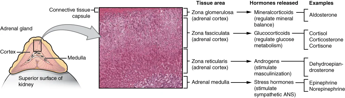
Figure 11818 The Adrenal Glands
Long description
Both adrenal glands sit atop the kidneys and are composed of an outer cortex and an inner medulla, all surrounded by a connective tissue capsule. The cortex can be subdivided into additional zones, all of which produce different types of hormones. LM × 204. (Micrograph provided by the Regents of Uni

Figure 1: Both adrenal glands sit atop the kidneys and are composed of an outer cortex and an inner medulla, all surrounded by a connective tissue capsule.
The endocrine system arises from all three embryonic germ layers. The pituitary gland has dual origins: the anterior pituitary arises from oral ectoderm, while the posterior pituitary arises from neural ectoderm at the base of the hypothalamus. The pineal gland also arises from ectoderm. The adrenal glands have different origins: the cortex from mesoderm and the medulla from ectoderm neural cells. The thyroid and parathyroid glands, along with the pancreas and thymus, arise from endoderm. [1]
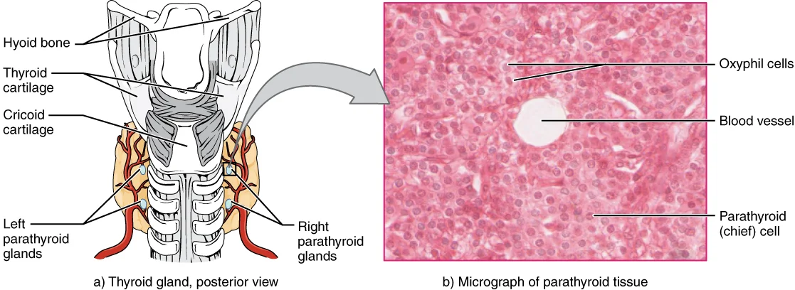
Figure 21814 The Parathyroid Glands
Long description
The small parathyroid glands are embedded in the posterior surface of the thyroid gland. LM × 760. (Micrograph provided by the Regents of University of Michigan Medical School © 2012)

Figure 2: The small parathyroid glands are embedded in the posterior surface of the thyroid gland (lm × 760).
The pineal gland is a small, pinecone-shaped endocrine gland located in the epithalamus that participates in biological rhythm regulation. It projects posteriorly and inferiorly into the quadrigeminal cistern, with boundaries including the back of the third ventricle wall, the splenium of the corpus callosum superiorly, and the thalamus on both sides. The pineal gland's major product is melatonin, a multifunctional endogenous indoleamine. Tumors of the pineal region account for approximately 3–11% of pediatric brain neoplasms but fewer than 1% of brain neoplasms in adults. [3]
The pineal gland contains two populations of cells: about 95% are pinealocytes with dendritic processes, and 5% are neuroglial supporting cells that resemble astrocytes. Both cell types can form neoplasms, along with residual germ cells and cells derived from nearby structures. Pineal parenchymal tumors are neuroepithelial neoplasms arising from pineocytes, accounting for fewer than 1% of all primitive central nervous system tumors and constituting 15% to 30% of pineal gland tumors. The World Health Organization recognizes four distinct categories: pineocytomas, pineoblastomas, papillary pineal tumors, and pineal parenchymal tumors of intermediate differentiation. [3]
Pineal papillary tumors are uncommon WHO grade II or III neuroepithelial neoplasms. Among them, 68% have been found to recur at a mean follow-up of 4.2 years, with overall survival rates of 73% at 5 years and 58% at 10 years. In a pediatric population, 47% of papillary tumors of the pineal gland recurred at a mean follow-up of 6.5 years. These tumors appear as partially cystic masses with obstructive hydrocephalus on MRI. The main symptom reported by patients with pineal papillary tumors is headaches, related to obstructive hydrocephalus. [3]
Under the light microscope, endocrine glands are recognised by their lack of ducts and their rich vascular network, as hormones are secreted directly into the extracellular fluid and then into the blood[1]. The classic H&E appearance shows cords, follicles or clusters of epithelial cells intimately associated with fenestrated capillaries or sinusoids. The connective tissue stroma is sparse, and the cells often contain cytoplasmic granules that represent stored hormone.
The pituitary gland (hypophysis) sits in the sella turcica and has two distinct parts. The anterior lobe (adenohypophysis) is derived from oral ectoderm and stains variably with H&E because it contains several cell types. The posterior lobe (neurohypophysis) is neural tissue, appearing paler and less cellular, with nerve fibres and specialised glial cells called pituicytes.
| Region | H&E appearance | Main components |
|---|---|---|
| Adenohypophysis (anterior lobe) | Cellular, with cords and acini of epithelial cells; variable cytoplasmic staining (acidophilic, basophilic, chromophobic) | Glandular epithelial cells, sinusoids |
| Neurohypophysis (posterior lobe) | Pale, fibrillar, relatively acellular; no glandular epithelium | Unmyelinated axons, pituicytes, Herring bodies |
| Pars intermedia | Thin, often cystic zone between the lobes | Basophilic cells, colloid-filled follicles |
The adenohypophysis contains five hormone-secreting cell types, classifiable by their staining and hormone product. Acidophils stain pink with eosin and secrete growth hormone (somatotrophs) or prolactin (lactotrophs). Basophils stain blue-purple with haematoxylin and secrete ACTH (corticotrophs), TSH (thyrotrophs) or FSH/LH (gonadotrophs). Chromophobes have few granules and appear pale; they are thought to be degranulated or reserve cells. With age, [2]
The neurohypophysis is not a true gland but a storage and release site for hormones made in the hypothalamus. Its axons run from the supraoptic and paraventricular nuclei down the infundibulum, and their dilated terminals (Herring bodies) store oxytocin and vasopressin (ADH). Pituicytes are modified astrocytes that support the axons. [2]
In the pituitary field, the adenohypophysis lies on the left, with dense clusters of epithelial cells around sinusoids; the neurohypophysis on the right is paler, with wavy nerve fibres and scattered pituicyte nuclei. In the pancreatic field, the islet of Langerhans appears as a pale, rounded cluster of small cells embedded in the darker exocrine acinar tissue.
The pancreas has both exocrine and endocrine functions[1]. The endocrine portion consists of the islets of Langerhans, which are pale-staining, rounded clusters of cells scattered among the darker, enzyme-secreting acini. Each islet contains four main cell types, best distinguished by special stains or immunohistochemistry, though they can be approximated on H&E by granule colour and position.
| Cell type | Approximate proportion | Location within islet | Secretion | H&E appearance |
|---|---|---|---|---|
| Beta (β) cells | ~70% | Central | Insulin | Pale, granular cytoplasm |
| Alpha (α) cells | ~20% | Peripheral | Glucagon | Slightly eosinophilic granules |
| Delta (δ) cells | ~5% | Peripheral | Somatostatin | Sparse, small granules |
| PP (F) cells | ~1–2% | Peripheral, especially in head | Pancreatic polypeptide | Small, inconspicuous |
On H&E, beta cells dominate the islet centre, giving it a pale, uniform look; alpha cells rim the periphery. The islets are richly capillarised, with fenestrated vessels allowing direct hormone entry into the blood. The exocrine acini around them are strongly eosinophilic due to zymogen granules, providing a sharp contrast that makes islets easy to identify at low power.
Special stains are essential tools in endocrine histology, allowing us to identify and characterize specific hormone-producing cells and tissues. These stains utilize chemical properties of cellular components to highlight structures that would otherwise be difficult to distinguish in routine hematoxylin and eosin (H&E) preparations.
DefinitionHormone-producing cells
Cells that synthesize and secrete hormones directly into the bloodstream to regulate physiological processes.
One of the most commonly used special stains in endocrine pathology is the argentaffin stain, which identifies cells containing silver-reducing substances. Another important stain is the chromaffin stain, which uses chromium salts to stain cells that produce catecholamines, such as those in the adrenal medulla. The chromaffin reaction is based on the oxidation of catecholamines to fo [1]
For pituitary gland examination, special stains like periodic acid-Schiff (PAS) can highlight glycoprotein hormones in acidophils and basophils. The PAS reaction stains carbohydrates and is particularly useful for identifying the glycoprotein hormones produced by the anterior pituitary, such as growth hormone and thyroid-stimulating hormone. [1]
In pancreatic islets, the aldehyde fuchsin stain is valuable for identifying beta cells, which produce insulin. This stain selectively stains beta cells purple, allowing for their quantification and study in conditions like diabetes mellitus. [1]
| Stain | Target cells/tissues | Application |
|---|---|---|
| Argentaffin | Serotonin-producing cells | Identification of carcinoid tumors |
| Chromaffin | Catecholamine-producing cells | Study of adrenal medulla and pheochromocytomas |
| PAS | Glycoprotein hormones | Pituitary gland examination |
| Aldehyde fuchsin | Insulin-producing beta cells | Pancreatic islet studies in diabetes |
Under the electron microscope, endocrine cells reveal features that directly reflect their function of synthesising and secreting hormones. The most striking common feature is an abundance of rough endoplasmic reticulum (RER) and a prominent Golgi apparatus, reflecting high rates of protein synthesis and packaging. Mitochondria are numerous and often elongated, providing the ATP needed for hormone production and exocytosis. The nuclei are typically euchromatic, indicating active transcription, and often have a rounded or oval profile with one or two prominent nucleoli.
The defining ultrastructural feature of most endocrine cells is the presence of membrane-bound secretory granules. These granules store the finished hormone before release. Their appearance varies by hormone type: peptide hormone granules are usually dense and homogeneous, while those storing glycoprotein hormones may appear less electron-dense with a flocculent content. The granules are distributed throughout the cytoplasm but often accumulate near the cell me
In the centre of the field lies a large, round nucleus with dispersed chromatin and a prominent nucleolus. To one side of the nucleus, the Golgi apparatus appears as a curved stack of smooth, flattened sacs with associated small vesicles.
The capillary endothelium in endocrine glands is characteristically fenestrated, with pores often closed by a thin diaphragm. This structural adaptation greatly facilitates the passage of secreted hormones from the interstitial space into the blood. The basal lamina of the endothelial cell and that of the endocrine cell may fuse, reducing the diffusion distance to less than 1 µm in some glands. This intimate vascular relationship is a key diagnostic feature of endo [1]
Lipid-soluble hormone-producing cells, such as those of the adrenal cortex and gonads, show a different ultrastructure. Their hallmark is abundant smooth endoplasmic reticulum (SER), appearing as a network of branching tubules, and numerous mitochondria with tubular or vesicular cristae rather than the lamellar cristae of most other cells. These cells also contain many lipid droplets, which serve as the cholesterol substrate for steroid synthesis. Secretory granules are sparse or absent because steroid hormones diffuse out of the cell immediately after synthesis rather than being stored.
The thyroid follicular cell provides a special ultrastructural case. Its apical surface bears microvilli projecting into the colloid, and the cytoplasm contains large endocytotic vesicles and lysosomes. The electron micrograph shows reabsorption of thyroglobulin from the colloid, its fusion with lysosomes, and the release of thyroid hormones at the basolateral surface. This polarised arrangement, with synthetic machinery basally and endocytotic activity apically, is unique among endocrine cells.
The pineal gland is a small endocrine structure located in the epithalamus, near the center of the brain. It sits beneath the posterior end of the corpus callosum and above the quadrigeminal cistern, surrounded by the cerebral aqueduct. The gland is attached to the roof of the third ventricle by a stalk that connects it to the habenular commissure.
Histologically, the pineal gland consists of pinealocytes (the primary secretory cells), glial cells, and calcified structures called brain sand or corpora arenacea. The pinealocytes are responsible for producing and secreting melatonin, a hormone that regulates circadian rhythms and sleep-wake cycles. These cells are arranged in cords and clusters, surrounded by a rich capillary network that facilitates hormone release into the bloodstream.
The functional significance of the pineal gland is primarily related to its role in melatonin secretion. Melatonin production follows a circadian pattern, with higher levels during darkness and lower levels during light exposure. This rhythm helps regulate various physiological processes, including sleep patterns, seasonal reproduction, and mood regulation.
Pathological conditions affecting the pineal gland can disrupt its normal function. Pineal region masses may produce nonspecific signs and symptoms, often causing syndromes of mass effect including headaches, aqueductal stenosis, and hydrocephalus. These masses can also compress the hypothalamus, leading to conditions such as diabetes insipidus and slowed growth. The close anatomical relationship between the pineal gland and surrounding structures explains why pineal tumors can have such diverse clinical manifestations. [3]
When examining endocrine histology slides, the key features to identify include the presence of specialized secretory cells arranged in specific patterns. Endocrine glands are characterized by cells that produce and secrete hormones directly into the bloodstream, rather than through ducts. These cells typically have abundant granules containing hormones, which may be visible with special stains. [1]
The pineal gland, an endocrine structure, can be identified by its unique histological features. Pineal parenchymal cells (pineocytes) are arranged in cords and clusters, surrounded by a rich capillary network. These cells have large, round nuclei and eosinophilic cytoplasm. Pinealocytes often form rosette-like structures called pinealocytomatous rosettes, which are characteristic of pineocytomas[1].
Papillary tumors of the pineal gland show distinctive papillary morphological features with ependymal rosettes and pseudorosettes. The rosetted cells have thick processes resting on collagen from adjacent blood vessels. These cells display small round/oval nuclei, stippled chromatin, small nucleoli, and eosinophilic cytoplasm with distinct cell borders[1]. Mitotic figures are uncommon, while necrosis is often present[1]. [3]
Pineal parenchymal tumors of intermediate differentiation present features between those of pineocytomas and pineoblastomas[1]. These tumors show wide morphological variability and can occur at all ages, though they appear more commonly among women, teenagers, and middle-aged patients[1]. [3]
When differentiating pineal papillary tumors from other pineal tumors, immunohistochemistry is helpful. Pineal papillary tumors typically show neuron-specific enolase reactivity but do not express neurofilament proteins[1]. This pattern helps differentiate them from pineal parenchymal tumors with intermediate differentiation, while MAP-2 expression aids in distinguishing them from choroid plexus papillomas[1]. [3]
DefinitionPinealocyte
Specialized neuroendocrine cells in the pineal gland that produce melatonin and are arranged in cords and clusters.
| Tumor Type | Histological Features | Distinguishing Characteristics |
|---|---|---|
| Pineocytoma | Pinealocytomatous rosettes, uniform cells, rare mitoses | Well-differentiated, calcifications common |
| Pineoblastoma | Small blue cells, high mitotic rate, necrosis | Primitive neuroectodermal tumor, aggressive |
| Pineal papillary tumor | Papillary structures, ependymal rosettes, NSE positive | No neurofilament expression, MAP-2 positive |
| Pineal parenchymal tumor (intermediate) | Features between pineocytoma and pineoblastoma | Variable morphology, occurs at all ages |
| Germinoma | Large cells, lymphocytic infiltration, calcifications | Heterogeneous on imaging, solid/cystic masses |
The tumor cells exhibit small round nuclei with stippled chromatin and eosinophilic cytoplasm. The rosettes are formed by tumor cell processes extending toward the central blood vessels, which is a typical feature of pineal parenchymal tumors.
The endocrine glands share a common histologic theme: epithelial cells arranged in cords, follicles, or clusters, intimately associated with a rich capillary network. This reflects their function—secreting hormones directly into the bloodstream. Unlike exocrine glands, they have no ducts; their secretory products pass through the basement membrane into fenestrated capillaries.
The adenohypophysis contains five hormone-secreting cell types, classifiable by their staining and hormone product. Acidophils stain pink with eosin and secrete growth hormone (somatotrophs) or prolactin (lactotrophs). Basophils stain blue-purple with haematoxylin and secrete ACTH (corticotrophs), TSH (thyrotrophs) or FSH/LH (gonadotrophs). Chromophobes have few granules and appear pale; they are thought to be degranulated or reserve cells. With age,
The thyroid gland is unique: its functional unit is the follicle, a spherical sac lined by simple cuboidal epithelium surrounding colloid. The colloid is a homogeneous eosinophilic mass of thyroglobulin. When the gland is active, the epithelium becomes tall cuboidal to columnar, and the colloid shows scalloped edges (resorption vacuoles). When inactive, the epithelium flattens to squamous-like, and colloid is abundant and smooth. Parafollicular C cells (clear cells) lie singly or in small clusters within the follicular basement membrane, secreting calcitonin; they are best seen with silver stains or immunohistochemistry.
The parathyroid glands are four small ovoid structures embedded on the posterior surface of the thyroid. They contain two cell types: chief cells (small, round nuclei, pale cytoplasm) arranged in cords or nests, and oxyphil cells (larger, intensely eosinophilic cytoplasm) appearing after puberty. Chief cells secrete parathyroid hormone; oxyphil cells are thought to be degenerate chief cells with no known secretory function.
The adrenal gland has a cortex and medulla. The cortex, derived from mesoderm, shows three zones: zona glomerulosa (outermost, cells in rounded clusters, secreting aldosterone), zona fasciculata (thickest, cells in straight cords with foamy lipid-rich cytoplasm, secreting cortisol), and zona reticularis (innermost, cells in anastomosing cords with lipofuscin pigment, secreting androgens). The medulla, derived from neural crest, contains large polyhedral chromaffin cells arranged in nests or cords, secreting catecholamines; they stain brown with dichromate (chromaffin reaction).
The pancreatic islets (islets of Langerhans) are pale-staining spherical clusters scattered among the exocrine acini, most numerous in the tail. They contain five cell types: beta cells (about 70%, central, secrete insulin), alpha cells (about 20%, peripheral, secrete glucagon), delta cells (about 5%, secrete somatostatin), PP cells (secrete pancreatic polypeptide), and epsilon cells (secrete ghrelin). On H&E, islet cells appear as pale cords with fine granules; immunostaining is required to distinguish the types.
Clinical pearlThyroid aging
The thyroid gland produces less thyroid hormones with age, causing decreased basal metabolic rate and increased body fat. This age-related reduction in thyroid hormone production is a gradual process that affects thermoregulation and metabolism. [2]
Exam trapParathyroid paradox
Parathyroid hormone increases with age, potentially due to reduced dietary calcium, but this combined with decreased calcitonin can lead to osteoporosis in elderly people regardless of sex. The compensatory increase in PTH attempts to maintain calcium homeostasis but ultimately contributes to bone demineralization. [2]
Their incidence varies with age, sex, and ethnicity, making them relatively rare in the general population.
These tumors are characterized by cellular sheets or lobules of uniform cells with large round nuclei, prominent nucleoli, and clear cytoplasm. [3]
This specific presentation requires treatment approaches that address both locations. [3]
They contain significant amounts of lipids and macromolecules compared to other pineal gland tumors. [3]
Clinical pearlTeratoma characteristics
Teratomas are characterized by multipotential cells producing tissues from two or more embryological layers (ectoderm, mesoderm, endoderm). They can be partially or totally encapsulated, but may also be unencapsulated and locally invasive. [3]
DefinitionTeratoma imaging
On MRI, pineal teratomas appear as lobular, multiloculated, and heterogeneously wide masses with foci of fat, calcification, and cystic regions. These imaging characteristics help distinguish teratomas from other pineal region tumors. [3]
These aggressive tumors often present with hydrocephalus due to obstruction of CSF flow. [3]
These metastatic lesions may appear histologically similar to primary pineal tumors but require different treatment approaches.
Exam trapMisunderstanding paracrine signaling
Paracrine factors act only on neighboring cells, not distant tissues. Histamine in asthma is an example that affects nearby bronchial smooth muscle cells. [1]
Exam trapOverestimating testosterone decline in aging
Andropause involves a gradual decline in testosterone levels, but this is much less dramatic than estrogen decline in females and rarely affects sperm production until very old age.
Exam trapConfusing pineal gland function
The pineal gland's primary purpose is producing and releasing melatonin, a pleiotropic indoleamine hormone, not regulating calcium or other functions sometimes attributed to it.
Exam trapOver-reliance on imaging for pineal tumors
Imaging alone cannot distinguish between germinomas, nongerminomatous germ cell tumors, and pineal parenchymal tumors. Complete evaluation including serum and CSF markers is essential.
Exam trapMisinterpreting choriocarcinoma characteristics
Choriocarcinomas show stromal vascular ducts forming blood lakes and intratumoral hemorrhagic necrosis, which correlate with poor prognosis. They appear as ovoid, heterogeneous, slightly hyperdense masses on imaging.
Exam trapOverlooking tumor vasculogenic mimicry
Some tumors, like choriocarcinomas, may have sinusoids expressing laminin but not CD34, allowing blood flow from CD34-positive vessels to sinusoids, potentially leading to clotting and hemorrhagic necrosis [Q39].
Exam trapConfusing pineocytoma and pineoblastoma
Pineocytomas are slow-growing grade I/II tumors with well-differentiated mature cells, while pineoblastomas are composed of undifferentiated or immature pineal cells. [3]
Exam trapMisjudging pineoblastoma treatment
Aggressive surgical resection is first-line therapy for pineoblastoma, with radiotherapy after resection increasing patient survival. Combined therapy appears more effective than single approaches.
Exam trapOveremphasizing tumor size as prognostic factor
For pineal tumors, tissue invasion near the pineal gland increases death risk about threefold, while tumor size may not be considered a prognostic factor (risk of death decreases by 4% per 1mm increase).
NoteClinical diagnosis of pineal tumors
Diagnosis of pineal region neoplasms requires clinical presentation, imaging, and pathology results. Serum and CSF biomarkers provide additional data before invasive procedures like open surgical resection or stereotactic/endoscopic biopsy.
NoteGerminoma identification
Germinomas are diagnosed using imaging combined with serum and CSF markers, showing high expression of oncoproteins such as alpha-fetoprotein, beta human chorionic gonadotropin, lactate dehydrogenase, and placental alkaline phosphatase.
NoteChoriocarcinoma markers
Choriocarcinomas are associated with elevated levels of both CSF and plasma human chorionic gonadotropin, similar to germinomas which also show elevated beta human chorionic gonadotropin expression.
NotePineocytoma characteristics
Pineocytomas are slow-growing grade I/II tumors that may remain locally confined. They occur at all ages but are more frequent in adults aged 30 to 60 years old.
NotePineoblastoma treatment approach
Aggressive surgical resection is first-line therapy for pineoblastoma, with radiotherapy after resection improving survival rates. The International Gamma Knife Research Foundation reported 27% local control and 48% survival rates at 5 years following stereotactic radiosurgery. [3]
NotePineal papillary tumor management
Pineal papillary tumors often recur, and radiotherapy is frequently effective. Both radiotherapy and chemotherapy are used in initial tumor management and for recurrences after gross total resection.
NoteMetastatic cancer to pineal gland
Metastatic cancer spreading to the pineal gland is exceedingly uncommon, originating most commonly from lung carcinoma, with documented cases also from pancreatic, esophageal, and bladder neoplasms.
NotePineal cyst progression
Surgically treated pineal cysts may represent a progressive disease with acute or progressive hydrocephalus at the final stage. Young women with active sexual hormone status (aged >10 years) are at highest risk for pineal cyst progression. [3]
Pineal Tumor Diagnosis Pathway
- Clinical PresentationHeadaches, visual disturbances, hydrocephalus
- Question: Imaging performed?
Yes
- Question: Characteristic findings?
Yes
- Consider germinoma, choriocarcinoma, pineocytoma, or pineoblastoma
No
- Proceed to biopsy or CSF/blood markers
No
- Perform imaging (MRI preferred)
- Paracrine factors act only on neighboring cells, not distant tissues
- Andropause involves a gradual testosterone decline that rarely affects sperm production until very old age [2]
- The pineal gland produces and releases melatonin as its primary function [3]
- Imaging alone cannot distinguish between different pineal tumors; complete evaluation including markers is essential [3]
- Choriocarcinomas show characteristic blood lakes and necrosis on imaging with poor prognosis [3]
- Pineocytomas are slow-growing with well-differentiated cells, while pineoblastomas are undifferentiated
- Aggressive surgical resection followed by radiotherapy is first-line for pineoblastoma [3]
- Tissue invasion near pineal gland increases death risk threefold, while tumor size has minimal prognostic value [3]
- Metastatic cancer to pineal gland is uncommon, most often from lung carcinoma [3]
- Young women with active sexual hormone status are at highest risk for pineal cyst progression
Answer each one in your head first, then open the answer.
What is the primary function of the pineal gland?
Show answerHide answer
Production and release of melatoninWhich imaging technique cannot distinguish between different types of pineal tumors?
Show answerHide answer
All imaging techniques alone cannot distinguish between pineal tumor typesWhat is the first-line therapy for pineoblastoma?
Show answerHide answer
Aggressive surgical resectionWhich patient group is at highest risk for pineal cyst progression?
Show answerHide answer
Young women with active sexual hormone status (aged >10 years)What is the most common origin of metastatic cancer spreading to the pineal gland?
Show answerHide answer
Lung carcinoma
- Betts JG, Young KA, Wise JA, et al. An Overview of the Endocrine System. OpenStax, Anatomy and Physiology 2e (CC BY-NC-SA 4.0). Accessed .
- Betts JG, Young KA, Wise JA, et al. Development and Aging of the Endocrine System. OpenStax, Anatomy and Physiology 2e (CC BY-NC-SA 4.0). Accessed .
- Favero G, Bonomini F, Rezzani R. Pineal Gland Tumors: A Review. Cancers 2021;13(7):1547. doi:10.3390/cancers13071547. Accessed .
Figure credits
Educational content for medical students and doctors. It is not a substitute for clinical judgement or local guidelines.