Reproductive histology
Histological structure and functional cycles of the male and female reproductive systems
The ovary is enclosed by an outer covering of cuboidal epithelium known as the ovarian surface epithelium, which sits superficial to a dense connective tissue layer called the tunica albuginea. The organ is divided into an outer cortex and an inner medulla. The cortex consists of the ovarian stroma, where oocytes develop surrounded by supporting cells to form follicles. The medulla contains the nerves, lymph vessels, and blood vessels. [1]
Folliculogenesis is the process of growth and development of these follicles. A female infant is born with one to two million oocytes, a number that declines until menopause. Follicles progress through four stages: primordial, primary, secondary, and tertiary. The oocyte remains a primary oocyte until just before ovulation. [1]
DefinitionAtresia
The death of ovarian follicles, which can occur at any point during their development.
During the follicular phase, tertiary follicles secrete estrogen. Usually, only one dominant follicle survives the reduction of FSH and LH to induce ovulation. After ovulation, the granulosa cells of the collapsed follicle luteinize to form the corpus luteum, which produces progesterone.
The uterine tube uses beating cilia to transport the ovulated oocyte toward the uterus. Fertilization occurs within the uterine tube.
The uterus consists of the fundus, body, and cervix. Its wall comprises the outer perimetrium, the muscular myometrium, and the inner endometrium. The endometrium undergoes a menstrual cycle, which typically lasts 28 days, though it can vary from 21 to 32 days. [1]
| Phase | Primary Hormone | Histological Change |
|---|---|---|
| Proliferative | Estrogen | Thickening of the stratum functionalis |
| Secretory | Progesterone | Endometrial glands secrete glycogen-rich fluid |
| Menstrual | Progesterone decline | Sloughing of the inner endometrium (menses) |
The cervix is the lower region of the uterus. The vagina's acidity is slightly decreased by high estrogen levels to facilitate sperm hospitality.
The testis contains tightly coiled seminiferous tubules, which form the bulk of the organ. These tubules contain developing sperm cells surrounding a central lumen.
Sperm Transport Path
- Seminiferous TubulesSperm produced in the lumen
- Straight TubulesAlso known as tubuli recti
- Rete TestesFine meshwork of tubules
- Efferent Ductules15 to 20 ductules crossing the tunica albuginea
The epididymis, prostate, placenta, and mammary glands are essential accessory and supporting structures of the reproductive system.
Under light microscopy, the reproductive organs are examined in haematoxylin and eosin (H&E) stained sections, which distinguish basophilic (blue) nuclei and cytoplasm from eosinophilic (pink) extracellular material and cytoplasm. The key structures differ markedly between male and female systems, and each shows a characteristic layered organisation.
Spermatids and mature sperm are found near the lumen. [2]
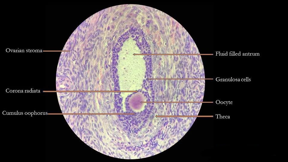
Tertiary (antral) follicles contain a fluid-filled antrum and cumulus oophorus surrounding the oocyte. [1]
| Structure | Layer / region | Cell types | H&E appearance |
|---|---|---|---|
| Seminiferous tubule (testis) | Basement membrane to lumen | Spermatogonia, primary spermatocytes, secondary spermatocytes, spermatids, spermatozoa, sustentacular cells | Progressive maturation from dark basal cells to elongated luminal sperm; sustentacular cells pale with irregular nuclei |
| Interstitium (testis) | Between tubules | Leydig cells | Eosinophilic, polygonal, clustered near capillaries |
| Ovary – cortex | Outer zone | Primordial, primary, secondary, antral follicles; corpus luteum | Follicles at varying sizes; oocyte with large nucleus; lutein cells pale and vacuolated |
| Ovary – medulla | Inner zone | Connective tissue, blood vessels, hilus cells | Loose stroma, prominent vessels |
Between the tubules, small clusters of Leydig cells appear as pink islands.
The uterine tube (fallopian tube) shows a folded mucosa with ciliated columnar cells and secretory peg cells; the muscularis has inner circular and outer longitudinal smooth muscle. The uterus has three layers: endometrium (surface epithelium, glands, stroma), myometrium (thick smooth muscle) and perimetrium (serosa). The endometrium varies with the menstrual cycle: proliferative phase shows straight glands and mitoses; secretory phase shows coiled, vacuolated glands and oedematous strom [1]
In reproductive histology, routine haematoxylin and eosin (H&E) staining is the backbone for identifying general architecture, but special stains are essential for resolving specific components that H&E cannot distinguish clearly. The choice of stain depends on the tissue and the question asked: connective tissue framework, elastic fibres, mucins, glycogen, or the presence of specific secretory products.
For the ovary, a Masson's trichrome stain is particularly useful because it differentiates collagen (stained blue or green) from smooth muscle and cytoplasm (stained red). In the ovarian cortex, the tunica albuginea appears as a dense blue band of collagen beneath the surface epithelium. In the corpus luteum, the granulosa-lutein cells stain red, while the central coagulated blood clot and the surrounding theca-lutein layer show a delicate blue reticular network of collagen. This contrast allows the student to appreciate the luteinisation process, where granulosa cells enlarge and acquire lipid droplets that are lost during routine processing.
Periodic acid–Schiff (PAS) staining is invaluable for the female tract because it highlights neutral mucopolysaccharides and glycogen. In the cervix, the endocervical glands secrete mucus that is strongly PAS-positive, appearing magenta. The endometrial glands in the proliferative phase show PAS-positive glycogen at the basal aspect of the cells; in the secretory phase, the glycogen moves to the supranuclear region and then is discharged into the gland lumen, a classic teaching point for dating the endometrium. The zona pellucida of the ovarian follicle is also PAS-positive, as it is composed of glycoproteins.
Alcian blue at pH 2.5 stains acidic mucins blue and is used to distinguish cervical mucus from endometrial secretions. The endocervical glands are strongly Alcian blue-positive, whereas the endometrial glands are negative or only weakly positive. This distinction is clinically relevant because the cervical mucus changes its viscosity and staining intensity under oestrogen and progesterone influence, and the presence of acidic mucins correlates with the ferning pattern seen in dried smears at mid-cycle. [1] [2]
For the male reproductive tract, Verhoeff–van Gieson elastic stain is used on the penis and the ductus deferens. The tunica albuginea of the penis contains abundant elastic fibres that appear black, allowing the student to see how this fibrous sheath permits expansion during erection while maintaining rigidity. In the ductus deferens, the thick muscular coat shows a characteristic three-layer arrangement (inner and outer longitudinal, middle circular), and the elastic fibres are concentrated
Oil Red O (on frozen sections) or Sudan black demonstrates lipids in the steroid-secreting cells of the testis and ovary. The Leydig cells of the testis, the theca interna cells, and the luteinised granulosa cells all contain lipid droplets that stain red or black. This confirms their role in steroidogenesis, as cholesterol esters are stored in these droplets. In the prostate, Congo red is not used routinely, but immunohistochemistry for prostate-specific antigen (PSA) is the special stain of choice to confirm prostatic origin in metastatic deposits, as the normal prostatic acinar cells show strong cytoplasmic positivity.
A Gomori reticulin stain is helpful in the testis to outline the seminiferous tubule basement membrane and the peritubular myoid cell layer. The reticulin fibres appear black, forming a delicate network around each tubule and around individual Leydig cell clusters. In the ovary, the same stain outlines the theca externa and the stromal cell network, helping to distinguish a theca-fibroma from a granulosa cell tumour, as the former shows abundant reticulin surrounding individual cells, whereas the latter shows reticulin only around groups of cells. [2]
Finally, Mucicarmine stains epithelial mucins a deep red and is used to identify mucinous differentiation in ovarian tumours. In the normal fallopian tube, the ciliated cells are mucicarmine-negative, but the secretory (non-ciliated) cells show apical positivity. This stain is also used to confirm the presence of mucin-secreting cells in a suspected mucinous cystadenoma of the ovary, where the lining epithelium shows intense red apical granules.
Under the electron microscope, the reproductive organs reveal specialised cellular machinery that light microscopy cannot resolve. In the testis, the seminiferous epithelium shows Sertoli cells with extensive, branching cytoplasm that envelops developing germ cells. Their nuclei are characteristically indented and contain a prominent nucleolus. The tight junctions between adjacent Sertoli cells form the blood–testis barrier, visible as a continuous belt of fusion points between lateral membranes. Spermatogonia lie external to this barrier, while primary spermatocytes, secondary spermatocytes and spermatids are internal. The acrosome of the spermatid appears as a dense, membrane-bound vesicle capping the nucleus; its formation from Golgi-derived proacrosomal granules can be traced stepwise. The sperm flagellum shows the classic 9+2 microtubule arrangement in the axoneme, surrounded by nine outer dense fibres and a fibrous sheath, all visible in cross-section. [1]
In the ovary, the granulosa cells of the tertiary follicle show microvilli on their apical surfaces and gap junctions between adjacent cells, allowing metabolic coupling. The oocyte itself has a thick extracellular coat, the zona pellucida, which under EM appears as a finely granular, moderately electron-dense layer penetrated by microvilli from both the oocyte and the granulosa cells. The oocyte cytoplasm contains cortical granules, small membrane-bound vesicles lying just beneath the plasma membrane; these are released at fertilisation to prevent polyspermy. The theca interna cells show features of steroid synthesis: abundant smooth endoplasmic reticulum, mitochondria with tubular cristae, and lipid droplets. The corpus luteum contains large lutein cells with the same steroidogenic ultrastructure, plus smaller theca-lutein cells.
Figure 1Stages of folliculogenesis
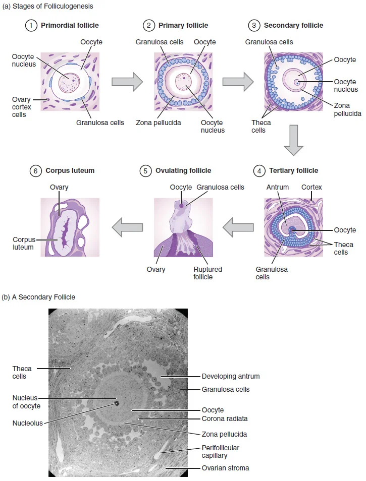
Long description
(a) The maturation of a follicle is shown in a clockwise direction proceeding from the primordial follicles. FSH stimulates the growth of a tertiary follicle, and LH stimulates the production of estrogen by granulosa and theca cells. Once the follicle is mature, it ruptures and releases the oocyte.

Figure 1: (a) The maturation of a follicle is shown in a clockwise direction proceeding from the primordial follicles.
The endometrium of the uterus shows surface epithelial cells with microvilli and cilia. The ciliated cells beat toward the uterine cavity, helping to distribute secretions. The underlying stromal cells are fibroblast-like, but in the secretory phase they enlarge and show dilated rough endoplasmic reticulum, reflecting glycogen and glycoprotein secretion. The spiral arteries have a thick tunica media of smooth muscle; their endothelial cells show fenestrations in the late secretory phase, facilitating the plasma protein leakage that prepares the endometrium for implantation. The uterine glands are simple tubular glands whose cells show apical secretory granules and basal nuclei; under progesterone influence, their lumina become distended with glycogen-rich secretion. [1]
The reproductive system exhibits remarkable structural adaptations that directly support its functions in gamete production, fertilization, and hormone secretion. The vagina, a muscular canal approximately 10 cm long, serves as both the entrance to the reproductive tract and the exit from the uterus during menses and childbirth. Its walls consist of three layers: an outer fibrous adventitia, a middle layer of smooth muscle, and an inner layer of stratified squamous epithelium that rests on a lamina propria. The vaginal epithelium forms transverse folds called rugae, which increase surface area and allow for stretching, enabling the vagina to accommodate intercourse and childbirth. [1]
In the ovaries, oogenesis begins with oogonia formed during fetal development, which divide via mitosis to form primary oocytes arrested in meiosis I. The number of primary oocytes declines from one to two million in an infant to approximately 400,000 at puberty, reaching zero by menopause. This structural arrangement ensures a limited supply of oocytes throughout a female's reproductive life. [1]
Sperm cells demonstrate specialized structures directly related to their function. With a volume 85,000 times less than that of the female gamete, sperm are produced at a rate of 100 to 300 million per day, compared to the single oocyte typically ovulated monthly by females. The sperm consists of three distinct regions: a head containing a compact haploid nucleus, a mid-piece filled with tightly packed mitochondria, and a tail. The head is covered by an acrosome, a cap filled with lysosomal enzymes essential for fertilization. These structural adaptations enable sperm to efficiently navigate and penetrate the female reproductive tract. [1] [2]
The testicular microenvironment maintains testosterone concentrations 100 times higher than in circulation, with Leydig cells producing approximately 6 to 7 mg of testosterone daily. This high intratesticular concentration is critical for promoting spermatogenesis, as low levels can lead to infertility. The hypothalamic-pituitary axis regulates testosterone production through pulsatile release of gonadotropin-releasing hormone (GnRH), which stimulates the anterior pituitary to release luteinizing hormone (LH) and follicle-stimulating hormone (FSH). FSH primarily binds to Sertoli cells to promote spermatogenesis, while LH stimulates Leydig cells to produce testosterone. [2]
Figure 2Graafian follicle
Long description
Histology of Graafian follicle with labelled parts.

Figure 2: Histology of Graafian follicle with labelled parts.
Secondary follicles exhibit multiple layers of granulosa cells with the theca layer beginning to form. [1]
Spermatogonia lie near the basement membrane, while primary and secondary spermatocytes occupy the middle layers. [2]
DefinitionDominant Follicle
The single tertiary follicle that survives FSH decline and will ovulate, producing extremely high estrogen levels that trigger positive feedback. [1]
Look-alikes can be challenging. Ovarian cortical stroma may resemble primordial follicles but lacks the oocyte. The rete ovarii appears as irregular tubules lined with cuboidal epithelium and should not be confused with developing follicles. In the testis, Leydig cells in the interstitium can be mistaken from a distance but contain characteristic lipid droplets and are located between tubules.
The antrum appears as a large fluid-filled cavity surrounded by multiple layers of granulosa cells. The theca interna and externa layers surround the follicle, with the interna containing more vascular tissue. The basal lamina separates the granulosa cells from the theca layer.
The epididymis, when visible, shows pseudostratified columnar epithelium with stereocilia and numerous sperm in the lumen. [2]
In reproductive histology, the histopathologist examines tissue architecture to distinguish normal cyclical variation from disease. The uterus provides the clearest example: its wall has three layers, and each shows characteristic changes. The most superficial is the perimetrium, a serous membrane of epithelial tissue covering the exterior. Beneath it lies the myometrium, a thick smooth-muscle layer whose fibres run horizontally, vertically and diagonally, permitting the powerful contractions of labour and the weaker cramps that expel menstrual blood. Anteriorly directed myometrial contractions near ovulation may facilitate sperm transport. The deepest layer, the endometrium, is not described in the passages but is the site of cyclical shedding and implantation; its histology is graded by dating criteria (e.g., proliferative versus secretory phase) in routine practice. [1]
At the top lies the perimetrium, a thin serosal covering. Below it, the myometrium occupies most of the field, with interlacing bundles of smooth muscle cut in various planes. The deepest layer, the endometrium, lines the lumen at the bottom; its glands and stroma vary with the menstrual cycle.
Histopathological correlates in the male tract focus on the testis. In normal seminiferous tubules, spermatogenesis proceeds from the basement membrane to the lumen: spermatogonia lie peripherally, then primary and secondary spermatocytes, then spermatids, then mature spermatozoa. The intervening sustentacular (Sertoli) cells support and nourish developing germ cells. In disease, this orderly sequence is lost. Although this is a parasite, the principle—that apoptosis of germ cells and phagocytosis by supporting cells is a histopathological correlate of reproductive injury—applies to mammalian testes. [2] [3]
Histopathological correlates include atretic follicles (degenerating oocytes with pyknotic nuclei and folded granulosa layers) and, in polycystic ovary syndrome, multiple subcortical cysts with a thickened, luteinised theca interna. The fallopian tube's mucosa shows ciliated and secretory cells; salpingitis is marked by plasma-cell infiltration and plicae blunting.
Clinical pearlOogenesis Efficiency
While the process of oogenesis can produce up to four cells, only one of these survives. [1]
Exam trapFollicle Selection
A follicle does not become dominant by chance; its selection depends on its size, the total number of granulosa cells present, and the specific number of FSH receptors located on those granulosa cells. [1]
DefinitionEndometrial Zonation
The endometrium is divided into the stratum basalis (basal layer) and the stratum functionalis (functional layer). The stratum basalis is adjacent to the myometrium and does not shed during menses, whereas the thicker stratum functionalis contains the glandular portion and is the layer that grows and thickens under the influence of estrogen and progesterone. [1]
Clinical pearlUterine Vasculature
During the luteal phase of the menstrual cycle, the thickened stratum functionalis is specifically supplied by spiral arteries, which are specialized branches of the uterine artery. [1]
Clinical pearlThermoregulation of Testes
The dartos muscle (subcutaneous) and cremaster muscles (descending from the internal oblique) contract simultaneously in cold weather to elevate the testes closer to the body core. This mechanism decreases the surface area of the scrotum to retain heat. Conversely, warmth causes the scrotum to relax, increasing surface area to promote heat loss. [2]
Exam trapScrotal Anatomy
The scrotal septum, which divides the scrotum into two separate compartments for each testis, is a continuation of the dartos muscle. Externally, the raphae is the raised medial thickening on the scrotal surface. [2]
DefinitionCryptorchidism
Cryptorchidism refers to the clinical condition where one or both testes fail to descend into the scrotum before birth. [2]
Clinical pearlSpermatogenesis Yield
A single primary spermatocyte undergoes meiosis I to produce two secondary spermatocytes, which then undergo meiosis II to produce a total of four haploid spermatids. [2]
Exam trapSpermiogenesis vs Spermatogenesis
Spermatogenesis is the overall process of producing haploid cells, but spermiogenesis is the specific final stage where spermatids are transformed into spermatozoa (formed sperm). [2]
Clinical pearlSperm Transit Path
During ejaculation, they travel through the ductus deferens, meet fructose and proteins from the seminal vesicle at the ejaculatory duct, and receive prostatic secretions in the prostatic urethra to form semen. [2]
Exam trapProstate Pathology
Prostate cancer is the second most common cancer in males. While its symptoms may mimic benign prostatic hyperplasia (BPH), there is no link between BPH and the development of prostate cancer. [2]
Clinical pearlOogonial Morphology
They are characterized by heterochromatic nuclei (sometimes in mitosis with one or two nucleoli) and a thin basophilic layer of cytoplasm. [3]
Exam trapConfusing the origin of the clitoris
The clitoris originates from the same cells as the glans penis, not from the labia minora. The superior, anterior portions of the labia minora come together to encircle the clitoris. [1]
Exam trapMisinterpreting the hymen as proof of virginity
The hymen is a thin membrane that sometimes partially covers the entrance to the vagina, but an intact hymen cannot be used as an indication of "virginity." Even at birth, this is only a partial membrane, as menstrual fluid and other secretions must be able to exit the body, regardless of penile-vaginal intercourse. [1]
Exam trapAssuming all follicles reach ovulation
Most follicles don't make it to ovulation. Several follicles reach the tertiary stage at the same time, and most of these will undergo atresia. The one that does not die will continue to grow and develop until ovulation. [1]
Exam trapMisunderstanding the LH surge
The LH surge (called the LH surge) leads to ovulation of the dominant follicle. It induces many changes in the dominant follicle, including stimulating the resumption of meiosis of the primary oocyte to a secondary oocyte. [1]
Exam trapConfusing the function of the corpus luteum
The corpus luteum is formed after ovulation and produces large amounts of progesterone, not estrogen. The luteinized granulosa and theca cells of the corpus luteum begin to produce large amounts of the sex steroid hormone progesterone, a hormone that is critical for the establishment and maintenance of pregnancy. [1]
Exam trapMisunderstanding fertilization location
Fertilization often occurs in the ampulla of the uterine tube, not in the ovary. The middle region of the tube, called the ampulla, is where fertilization often occurs. [1]
Exam trapConfusing menstrual phase timing
The menses phase occurs during the early days of the follicular phase of the ovarian cycle, when progesterone, FSH, and LH levels are low. The menses phase can last from 2 to 7 days, or longer, averaging approximately five days. [1]
NoteClinical examination
Knowledge of reproductive histology is essential for clinical examination. Understanding the normal anatomy of the external genitalia, including the relationship between the vaginal opening, urethra, and anus, is fundamental for gynecological examinations.
NoteInfertility evaluation
Understanding folliculogenesis is crucial in evaluating infertility. In the 28-day ovarian cycle, early tertiary follicles with their fluid-filled antrum will be stimulated by an increase in FSH to grow. Supporting granulosa and theca cells in the growing follicles produce estrogens, until the level of estrogen in the bloodstream is high enough that it triggers negative feedback at the hypothalamus and pituitary. [1]
NoteOvulation timing
Knowledge of the LH surge is critical for timing ovulation in fertility treatments. The LH surge induces many changes in the dominant follicle, including stimulating the resumption of meiosis of the primary oocyte to a secondary oocyte.
NoteMenstrual disorders
Understanding the menstrual cycle phases helps diagnose and treat menstrual disorders. The menses phase occurs during the early days of the follicular phase of the ovarian cycle, when progesterone, FSH, and LH levels are low. This decline in progesterone triggers the shedding of the stratum functionalis of the endometrium.
Ovulation Process
- Dominant follicle reaches tertiary stageFluid-filled antrum becomes large and fully formed
- Question: LH surge occurs?
Yes
- Resumption of meiosisPrimary oocyte becomes secondary oocyte
- Breakdown of ovarian wallProteases cleave structural proteins
- OvulationExpulsion of oocyte into peritoneal cavity
- Formation of corpus luteumProduction of progesterone begins
No
- Follicle undergoes atresiaMost follicles don't make it to ovulation
NoteSurgical procedures
Knowledge of uterine tube anatomy is essential for tubal surgeries and tubal ligation procedures. The uterine tubes are divided into sections: the isthmus (narrow medial end connected to the uterus), the infundibulum (wide distal end with fimbriae), and the ampulla (middle region where fertilization often occurs).
NotePathology interpretation
Understanding normal histology is essential for interpreting pathological specimens. The corpus luteum is a temporary endocrine structure that forms after ovulation and produces progesterone, which is critical for pregnancy maintenance.
- The clitoris originates from the same cells as the glans penis
- The hymen is not a reliable indicator of virginity
- Most follicles undergo atresia and never reach ovulation
- The LH surge triggers ovulation and the resumption of meiosis
- The corpus luteum produces progesterone, not estrogen
- Fertilization typically occurs in the ampulla of the uterine tube
- The menses phase lasts 2-7 days on average [1]
Answer each one in your head first, then open the answer.
What structure originates from the same cells as the glans penis?
Show answerHide answer
clitorisWhat hormone triggers ovulation and the resumption of meiosis?
Show answerHide answer
LH (luteinizing hormone)Where does fertilization typically occur in the female reproductive tract?
Show answerHide answer
ampulla of the uterine tubeWhat hormone does the corpus luteum produce in large amounts?
Show answerHide answer
progesteroneHow long does the menses phase typically last?
Show answerHide answer
2 to 7 days, averaging approximately five days
- Betts JG, Young KA, Wise JA, et al. Anatomy and Physiology of the Ovarian Reproductive System. OpenStax, Anatomy and Physiology 2e (CC BY-NC-SA 4.0). Accessed .
- Betts JG, Young KA, Wise JA, et al. Anatomy and Physiology of the Testicular Reproductive System. OpenStax, Anatomy and Physiology 2e (CC BY-NC-SA 4.0). Accessed .
- Hanna R. Fasciola hepatica: Histology of the Reproductive Organs and Differential Effects of Triclabendazole on Drug-Sensitive and Drug-Resistant Fluke Isolates and on Flukes from Selected Field Cases. Pathogens (Basel, Switzerland) 2015;4(3):431-456. doi:10.3390/pathogens4030431. Accessed .
Figure credits
Educational content for medical students and doctors. It is not a substitute for clinical judgement or local guidelines.