Skin and its appendages
Histology of skin and appendages for medical students, from epidermis to glands.
The skin is the largest organ, composed of an outer epidermis (keratinised stratified squamous epithelium) and an underlying connective-tissue dermis. The dermis projects into the epidermis as dermal papillae, which interdigitate with epidermal rete ridges; this increases the surface area for nutrient exchange and mechanical anchoring. Beneath the dermis lies the hypodermis (subcutaneous layer), containing adipose tissue and larger blood vessels; accessory structures such as hair follicles and s [1]
Figure 1Gray942
Long description
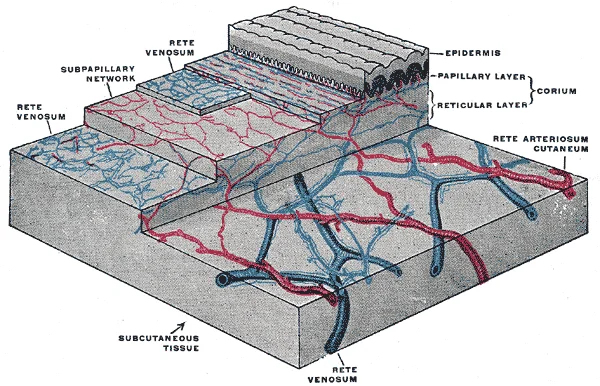
The distribution of the bloodvessels in the skin of the sole of the foot. (Spalteholz.)

Figure 1: The distribution of the bloodvessels in the skin of the sole of the foot (Spalteholz.).
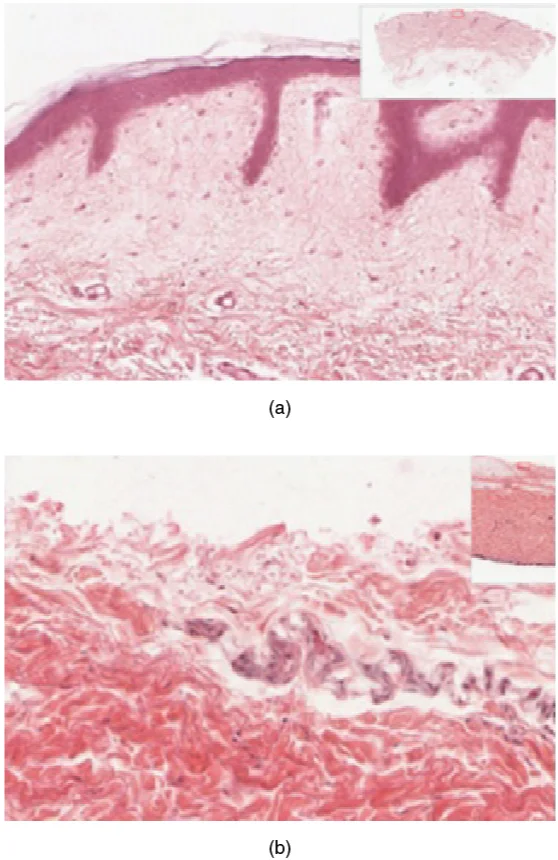
Thick skin (palms and soles) has a prominent stratum corneum and stratum lucidum but lacks hair follicles and sebaceous glands. Thin skin covers the rest of the body, has a thinner keratin layer, and contains hair follicles, sebaceous glands, and arrector pili muscles. The dermis has two layers: the papillary dermis (loose connective tissue forming dermal papillae) and the reticular dermis (dense irregular connective tissue with collagen and elastin). [1]
Figure 2Gray945
Long description
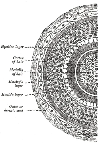
Transverse section of hair follicle. Labelled illustration from Wikimedia Commons, used under its open licence.

Figure 2: Transverse section of hair follicle.
Figure 3Gray944
Long description
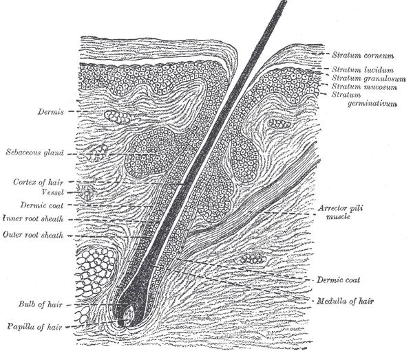
Section of skin, showing the epidermis and dermis; a hair in its follicle; the Arrector pili muscle; sebaceous glands.

Figure 3: Section of skin, showing the epidermis and dermis; a hair in its follicle; the Arrector pili muscle; sebaceous glands.
The hair follicle is an epidermal downgrowth into the dermis. The hair shaft is the exposed dead keratinised part; the hair root is anchored in the follicle and ends at the hair bulb, which contains the hair matrix of mitotically active basal cells. The hair papilla is a connective-tissue core with capillaries and nerves. The seba [2]
Sensory receptors include Meissner's corpuscles (light touch in dermal papillae), Pacinia [1]
Melanocytes (in the basal layer) produce melanin for pigmentation and UV protection. Langerhans cells (in the suprabasal epidermis) are antigen-presenting dendritic cells. Merkel cells (in the basal layer) are mechanoreceptors associated with nerve endings.
DefinitionDyskeratosis
Premature keratinisation of individual keratinocytes within the epidermis, seen as eosinophilic cells.
DefinitionHyperkeratosis
Thickening of the stratum corneum, often with retained nuclei (parakeratosis) or without (orthokeratosis).
DefinitionParakeratosis
Retention of nuclei in the stratum corneum, indicating rapid epidermal turnover.
DefinitionHypergranulosis
Increase in the thickness of the stratum granulosum, with prominent keratohyalin granules.
DefinitionSpongiosis
Intercellular oedema of the epidermis, widening the intercellular spaces.
DefinitionAcantholysis
Loss of cohesion between keratinocytes, leading to intraepidermal clefts or blisters.
DefinitionAcanthosis
Thickening of the stratum spinosum (prickle cell layer), often due to increased proliferation.
These terms are used in histopathological reporting. Dyskeratosis is seen in actinic keratosis and squamous cell carcinoma. Hyperkeratosis with parakeratosis occurs in psoriasis; hypergranulosis is typical of lichen planus. Spongiosis is the hallmark of eczematous dermatitis. Acantholysis is found in pemphigus vulgaris. Acanthosis accompanies chronic irritation or psoriasis.
| Structure | Origin | Location | Secretion/Product | Function |
|---|---|---|---|---|
| Hair follicle | Epidermal downgrowth | Dermis to hypodermis | Keratinised hair shaft | Protection, sensation, thermoregulation |
| Sebaceous gland | Epidermal bud | Dermis, opens into follicle | Sebum (holocrine) | Moisturises skin, keeps keratin layer healthy |
| Eccrine sweat gland | Epidermal projection | Dermis, duct to surface | Watery sweat (merocrine) | Cooling, excretion |
| Apocrine sweat gland | Epidermal projection | Deep dermis/hypodermis, duct to follicle | Thick organic sweat | Pheromone response, smell |
| Nail | Epidermal keratinisation | Dorsal digits | Hard keratin plate | Protection of fingertips/toes |
Figure 4501 Structure of the skin
Long description
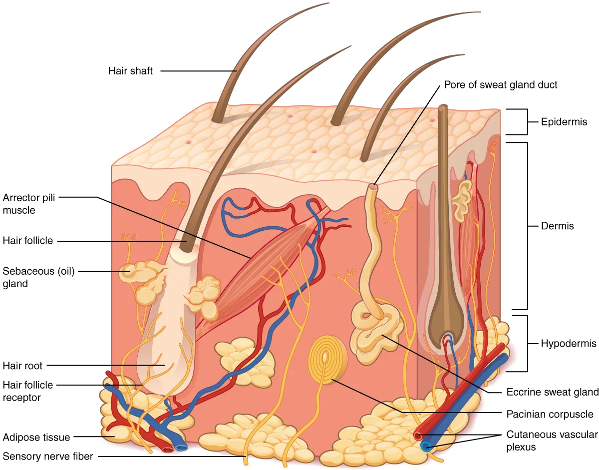
The skin is composed of two main layers: the epidermis, made of closely packed epithelial cells, and the dermis, made of dense, irregular connective tissue that houses blood vessels, hair follicles, sweat glands, and other structures. Beneath the dermis lies the hypodermis, which is composed mainly

Figure 4: The skin is composed of two main layers: the epidermis, made of closely packed epithelial cells, and the dermis, made of dense, irregular connective tissue that houses blood vessels, hair follicles, sweat glands, and other structures.
Light microscopy reveals the complex structure of skin and its appendages. The epidermis appears as the outermost layer, composed of keratinized, stratified squamous epithelium without any blood vessels (avascular)[1]. The dermis lies beneath the epidermis and contains blood vessels, lymph vessels, nerves, hair follicles, and sweat glands[1].
Figure 5502ab Thin Skin versus Thick Skin
Long description
These slides show cross-sections of the epidermis and dermis of (a) thin and (b) thick skin. Note the significant difference in the thickness of the epithelial layer of the thick skin. From top, LM × 40, LM × 40. (Micrographs provided by the Regents of University of Michigan Medical School © 2012)

Figure 5: These slides show cross-sections of the epidermis and dermis of (a) thin and (b) thick skin.
| Layer | Location | Cell Types | Characteristics |
|---|---|---|---|
| Stratum basale | Deepest layer | Basal cells, Merkel cells, melanocytes | Single layer of cuboidal stem cells; attaches to basal lamina[1] |
| Stratum spinosum | Above stratum basale | Keratinocytes, Langerhans cells | 8-10 layers with spiny appearance due to desmosomes; contains Langerhans cells[1] |
| Stratum granulosum | Above stratum spinosum | Keratinocytes | Contains keratohyalin granules |
| Stratum lucidum | Above stratum granulosum (only in thick skin) | Dead keratinocytes | Thin, translucent layer[1] |
| Stratum corneum | Most superficial layer | Dead keratinocytes | 15-30 layers; provides protection against microbes and dehydration[1] |
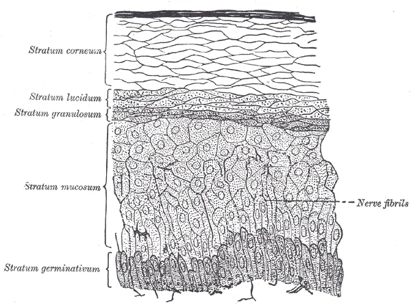
The epidermis consists of four or five layers depending on body location. Most skin has four layers (thin skin), while thick skin found on palms and soles has a fifth layer called the stratum lucidum[1]. The stratum basale (also called stratum germinativum) is the deepest epidermal layer and attaches the epidermis to the basal lamina via basement membrane[1]. This layer contains basal cells, which are cuboidal-shaped stem cells that produce all keratinocytes through mitosis[1]. Merkel cells in this layer function as touch receptors, while melanocytes produce melanin pigment[1].
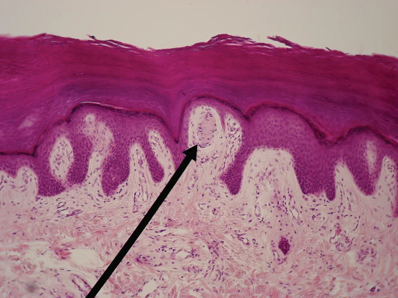
Figure 6514 Light Micrograph of a Meissner Corpuscle
Long description
In this micrograph of a skin cross-section, you can see a Meissner corpuscle (arrow), a type of touch receptor located in a dermal papilla adjacent to the basement membrane and stratum basale of the overlying epidermis. LM × 100. (credit: “Wbensmith”/Wikimedia Commons)

Figure 6: In this micrograph of a skin cross-section, you can see a Meissner corpuscle (arrow), a type of touch receptor located in a dermal papilla adjacent to the basement membrane and stratum basale of the overlying epidermis (lm × 100).
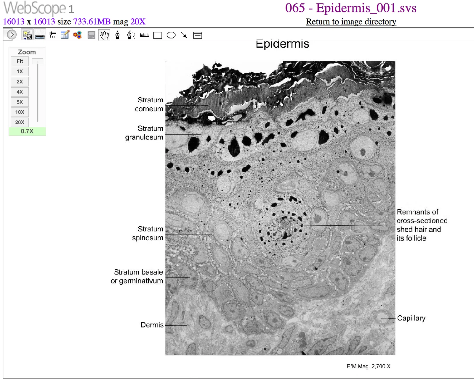
Figure 7505 Cells of the Epidermis
Long description
The cells in the different layers of the epidermis originate from basal cells located in the stratum basale, yet the cells of each layer are distinctively different. EM × 2700. (Micrograph provided by the Regents of University of Michigan Medical School © 2012)

Figure 7: The cells in the different layers of the epidermis originate from basal cells located in the stratum basale, yet the cells of each layer are distinctively different (em × 2700).
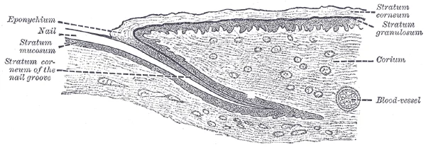
Figure 8Gray943
Long description
Longitudinal section through nail and its nail groove (sulcus).

Figure 8: Longitudinal section through nail and its nail groove (sulcus).
Figure 9Gray941
Long description
Section of epidermis. (Ranvier.). Labelled illustration from Wikimedia Commons, used under its open licence.

Figure 9: Section of epidermis (Ranvier.).
This layer contains 8-10 layers of keratinocytes and Langerhans cells that function as macrophages[1]. These cells are shed and replaced over approximately 4 weeks[1].
The dermis consists of two layers of connective tissue with elastin and collagenous fibers produced by fibroblasts[1]. It forms the "core" of the integumentary system and contains various structures including hair follicles and sweat glands[1].
Special stains are histochemical techniques that highlight specific components of skin and its appendages that are not visible with standard hematoxylin and eosin staining. These stains allow for the identification and study of various structures and substances within the skin.
The stain reveals the distribution of melanin in the epidermis, particularly in the stratum basale where melanocytes are located. Special stains can also demonstrate the presence of sebum within sebaceous glands, which are found all over the body and help to lubricate and waterproof the skin and hair. These stains are essential for understanding the microscopic anatomy of the skin and its appendages. [1] [2]
DefinitionSpecial stains
Histochemical techniques that highlight specific components of skin and its appendages not visible with standard staining.
Special stains are particularly useful for identifying melanin, which gives hair and skin its color and helps protect the living cells of the epidermis from ultraviolet radiation damage. Melanin occurs in two primary forms: eumelanin (black and brown) and pheomelanin (red). Special stains can demonstrate how melanin is transferred from melanocytes into keratinocytes via a cellular vesicle called a melanosome. [1]
For studying the dermis, special stains can highlight collagen and elastin fibers. The reticular layer of the dermis is composed of dense, irregular connective tissue and appears reticulated (net-like) due to a tight meshwork of fibers. Elastin fibers provide elasticity to the skin, enabling movement, while collagen fibers provide structure and tensile strength. Special stains can also demonstrate how collagen binds water to keep the skin hydrated. [1]
In the study of skin appendages, special stains can identify the components of eccrine sweat glands, which produce hypotonic sweat for thermoregulation. These glands are found all over the skin's surface but are especially abundant on the palms of the hand, the soles of the feet, and the forehead. Special stains can also highlight the lipid components of sebum produced by sebaceous glands, which help to lubricate and waterproof the skin and hair. [2]
The ultrastructure of skin, as revealed by electron microscopy, begins with the epidermis–dermis junction. The basal surface of the stratum basale rests on a basement membrane, a thin sheet of extracellular matrix that separates the avascular epidermis from the vascular dermis. Beneath this membrane lies the papillary layer of the dermis, which is made of loose, areolar connective tissue. In this layer, collagen and elastin fibers form a loose mesh rather than the dense parallel bundles seen deeper in the reticular layer. This meshwork is visible under the electron microscope as fine, wavy fibrils with intervening clear spaces filled with ground substance. [1]
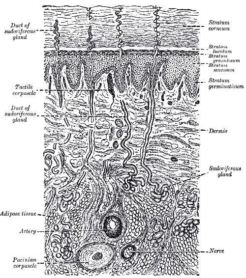
Figure 10Gray940
Long description
A diagrammatic sectional view of the skin (magnified).

Figure 10: A diagrammatic sectional view of the skin (magnified).
The papillary layer projects into the stratum basale of the epidermis to form finger-like dermal papillae. Each papilla is a small, conical elevation of connective tissue that increases the contact surface area between the two layers. On electron micrographs, these papillae appear as regular undulations of the dermal–epidermal interface, with the basement membrane following every contour. The height and density of papillae vary by body region: they are tall and numerous in thick skin of the palms and soles, but shorter and sparser in thin skin of the eyelids. [1]
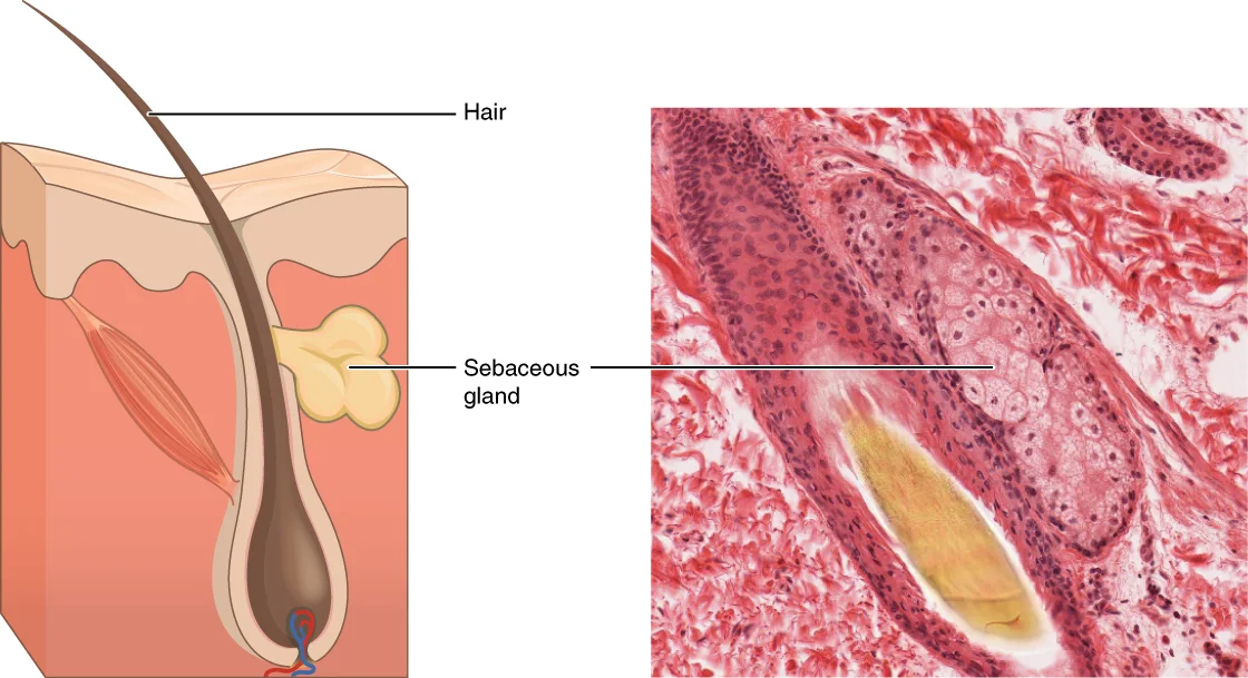
Figure 11407 Sebaceous Glands
Long description
These glands secrete oils that lubricate and protect the skin. They are holocrine glands and they are destroyed after releasing their contents. New glandular cells form to replace the cells that are lost. LM × 400. (Micrograph provided by the Regents of University of Michigan Medical School © 2012)

Figure 11: These glands secrete oils that lubricate and protect the skin.
Within the papillary layer, the principal resident cell is the fibroblast, a spindle-shaped cell with abundant rough endoplasmic reticulum and a prominent Golgi apparatus, reflecting its role in synthesising collagen, elastin and proteoglycans. A small number of fat cells (adipocytes) are also present, each appearing as a large, empty-looking vacuole with a thin rim of cytoplasm and a peripherally displaced nucleus. These adipocytes are scattered singly rather than clustered, unlike the fat lobules of the hypodermis. [1]
The papillary layer contains an abundance of small blood vessels, chiefly capillaries and post-capillary venules. Under the electron microscope, these vessels show a continuous endothelium with tight junctions, a basal lamina, and occasional pericytes. The capillaries loop up into the dermal papillae, bringing oxygen and nutrients close to the basement membrane, from which diffusion supplies the living layers of the epidermis. Lymphatic capillaries are also present, distinguished by their irregular, thin-walled lumen, absent basal lamina, and anchoring filaments that tether them to the surrounding collagen. [1]
Nerve fibres traverse the papillary layer, both myelinated and unmyelinated. Their terminals end in specialised touch receptors called the Meissner corpuscles, which lie within the dermal papillae, particularly in the fingertips, palms and soles. On electron microscopy, a Meissner corpuscle appears as an ovoid structure composed of flattened, stacked Schwann-like cells (lamellar cells) with the nerve terminal winding between them. The whole corpuscle is surrounded by a thin connective tissue capsule. These corpuscles are rapidly adapting mechanoreceptors, responding to light touch and low-frequency vibration. [1]
Phagocytes, defensive cells that help fight bacteria or other infections that have breached the skin, are scattered in the papillary layer. Ultrastructurally, these are macrophages with irregular surface folds, numerous lysosomes, and phagocytic vacuoles containing ingested debris. They sit close to blood vessels and lymphatic capillaries, ready to engulf pathogens that cross the epidermis. Together with the resident fibroblasts, adipocytes, capillaries, lymphatics, nerve fibres and Meissner corpuscles, the papillary layer forms a highly cellular, well-vascularised zone that supports the overlying epidermis and mediates its nutrition and sensation. [1]
The stratum basale appears as a single row of columnar cells resting on the basement membrane. Adjacent to the capillary lies a Meissner corpuscle, its lamellar cells arranged in concentric layers around the central nerve fibre.
The skin is the body's largest organ, with a surface area of approximately 1.5-2 m² in adults. It consists of three main layers: the epidermis, dermis, and hypodermis (subcutaneous tissue). The epidermis is the outermost layer, composed primarily of keratinocytes that undergo constant renewal, with a turnover time of about 28 days. This layer provides a waterproof barrier and protection against mechanical injury, pathogens, and ultraviolet radiation. [1]
The dermis lies beneath the epidermis and contains connective tissue, blood vessels, nerve endings, and appendages. It is approximately 1-2 mm thick in most areas but can be thicker in regions like the palms and soles. The dermis provides structural support, elasticity, and contains sensory receptors that detect touch, pressure, temperature, and pain. [1]
The hypodermis is the deepest layer, consisting mainly of adipose tissue that insulates the body and serves as an energy reserve. It also anchors the skin to underlying structures like muscle and bone. [1]
The skin appendages include hair follicles and sweat glands. Hair follicles are invaginations of the epidermis that extend into the dermis, with each follicle containing a hair shaft and root. The average person has about 5 million hair follicles, with 100,000 on the scalp alone. [1] [2]
Sebaceous glands are associated with hair follicles and secrete sebum, an oily substance that lubricates the skin and hair. Eccrine sweat glands are distributed throughout the body and produce sweat for thermoregulation, with each gland capable of secreting up to 10 liters of sweat per day during maximal activity. [2] [1]
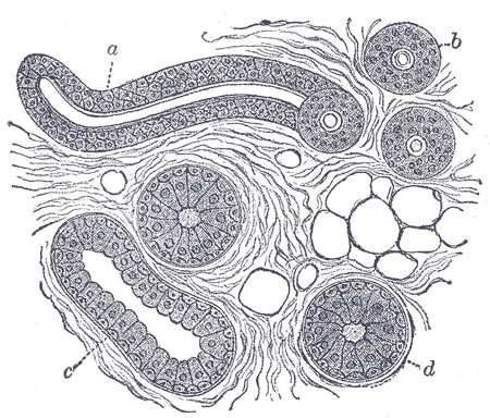
Figure 12Gray946
Long description
Body of a sudoriferous-gland cut in various deirections. a. Longitudinal section of the proximal part of the coiled tube. b. Transverse section of the same. c. Longitudinal section of the distal part of the coiled tube. d. Transverse section of the same. (Klein and Noble Smith.)

Figure 12: Body of a sudoriferous-gland cut in various deirections (a).
The epidermis appears as the stratified epithelial layer at the surface, the dermis as the connective tissue layer beneath it, and the hypodermis as the fatty layer at the bottom.
The skin is composed of two main layers: the epidermis and the dermis. The epidermis is the outermost layer and consists of stratified squamous epithelium. It has five distinct layers from deepest to most superficial: stratum basale, stratum spinosum, stratum granulosum, stratum lucidum, and stratum corneum. The dermis lies beneath the epidermis and contains connective tissue, blood vessels, nerve endings, and appendages like hair follicles and sweat glands. [1]
When examining a slide of skin, look for the characteristic layered appearance of the epidermis. The stratum corneum appears as the outermost layer of dead, keratinized cells. The stratum basale contains cuboidal to columnar cells and melanocytes. Melanin granules may be visible within melanocytes and transferred to keratinocytes. [1]
The nail apparatus can be identified by several key features. The nail bed appears pink due to its rich blood supply, except at the base where the lunula ("little moon") is visible as a crescent-shaped region formed by a thick layer of epithelium over the nail matrix.
Look-alikes include mucous membranes, which lack the stratified layers of the epidermis and have no stratum corneum. Unlike skin, mucous membranes contain goblet cells and often have underlying glands. Another potential confusion is with stratified squamous epithelium from other body sites, but skin is distinguished by the presence of hair follicles, sweat glands, and the specific organization of its epidermal layers.
The dermis contains blood vessels and nerve endings.
Histopathology of common skin diseases reveals characteristic changes that help establish diagnoses.
The dermis lies beneath, containing collagen fibers, blood vessels, and hair follicles.
In the psoriasis example, the epidermis is thickened with elongated rete ridges. Munro microabscesses are visible as collections of neutrophils within the stratum corneum. The dermis shows dilated capillaries and a perivascular lymphocytic infiltrate. [1]
The eczema section demonstrates spongiosis with widening of intercellular spaces in the epidermis. Microvesicles are forming due to this intercellular edema. Parakeratosis is present, with nuclei retained in the stratum corneum. The dermis shows a mixed inflammatory infiltrate around blood vessels. [1]
In basal cell carcinoma, the epidermis shows nests of basaloid cells with peripheral palisading. The tumor cells exhibit retraction artifact from the surrounding stroma. The dermis contains these tumor islands with areas of cystic degeneration.
Squamous cell carcinoma demonstrates atypical keratinocytes invading the dermis. Individual cell keratinization (dyskeratosis) and keratin pearl formation are characteristic features. The tumor shows invasive fronts with irregular borders.
Melanoma reveals atypical melanocytes with pleomorphic nuclei and prominent nucleoli. The epidermis shows pagetoid spread of melanocytes. The dermal component demonstrates nests and single cells of varying sizes and shapes, with dermal mitotic figures.
Clinical pearlStratum granulosum structure
The stratum granulosum is a critical layer of the epidermis that is specifically 3-5 cell layers thick. This layer represents a transitional zone where keratinocytes undergo significant changes as they move from the viable stratum spinosum toward the dead stratum corneum. [1]
Exam trapKeratohyalin vs lamellar bodies
A common misconception is confusing keratohyalin granules with lamellar bodies. Keratohyalin granules contain keratin precursors that aggregate and cross-link to form keratin bundles, while lamellar bodies contain glycolipids that are secreted to form the waterproof barrier. These are distinct structures with different functions. [1]
Clinical pearlKeratin production mechanism
Keratinocytes in the stratum granulosum generate large amounts of fibrous keratin protein, which constitutes the primary structural component of the epidermis. This process begins in the stratum granulosum and continues as cells migrate upward. [1]
Clinical pearlBarrier function development
The waterproof barrier function of the skin is established in the stratum granulosum through lamellar bodies that secrete glycolipids to the cell surface. This barrier is essential for preventing fluid loss from the body and protecting against external environmental factors. [1]
Exam trapCell morphology changes
As keratinocytes move to the stratum granulosum, they become flatter with thickened cell membranes, not larger as some students mistakenly believe. This flattening is part of the progressive transformation from living cells to dead, flattened corneocytes. [1]
Clinical pearlMicroscopic appearance
The characteristic "grainy" appearance of the stratum granulosum under microscopy is due to the accumulation of keratohyalin granules within the keratinocytes. This distinctive histological feature helps identify this layer in tissue sections. [1]
Clinical pearlProtein composition significance
Keratin and keratohyalin make up the bulk of the keratinocyte mass in the stratum granulosum, accounting for its distinctive appearance and function. These proteins are essential for the structural integrity and barrier properties of the epidermis. [1]
Exam trapKeratin precursor processing
While keratin precursors in keratohyalin granules aggregate, cross-link, and form bundles, this process is distinct from the function of lamellar bodies which produce the waterproof barrier. Confusing these two pathways is a common error in examinations. [1]
Clinical pearlBarrier formation process
The glycolipids from lamellar bodies are secreted to the cell surface where they help form the waterproof barrier that prevents transepidermal water loss. This barrier function is critical for maintaining homeostasis in the body. [1]
Clinical pearlMembrane thickening
Keratinocyte cell membranes thicken as they pass through the stratum granulosum, contributing to the formation of the cornified envelope. This thickening is part of the terminal differentiation process that creates the protective outer layer of the skin. [1]
Exam trapLayer thickness variation
Remember that the stratum granulosum is specifically 3-5 cell layers deep in normal skin, though this can vary in different body regions or pathological conditions. This precise measurement is important for histological identification. [1]
Clinical pearlKeratohyalin function
Keratohyalin contains keratin precursors that aggregate and cross-link to form keratin bundles, which provide strength to the epidermis. This protein processing is essential for the mechanical properties of the skin. [1]
A common mistake is confusing the layers of the epidermis and their functions. The epidermis consists of five layers: stratum basale, stratum spinosum, stratum granulosum, stratum lucidum, and stratum corneum. The stratum basale is the deepest layer where cells divide and push upward, while the stratum corneum is the outermost layer composed of dead cells filled with keratin. Understanding this progression from living to dead cells is essential for comprehending skin turnover and barrier function.
Students often misunderstand the formation of fingerprints. Fingerprints form where the cells of the stratum basale meet the papillae of the underlying dermal layer (papillary layer), resulting in the formation of the ridges on fingers. These patterns are unique to each individual and do not change with growth and aging, which is why they are used for forensic identification. [1]
Another error is overlooking the function of the arrector pili muscle. Each hair root is connected to a smooth muscle called the arrector pili that contracts in response to nerve signals from the sympathetic nervous system, making the external hair shaft "stand up". The primary purpose of this response is to trap a layer of air to add insulation, visible in humans as goose bumps. [2]
Many students incorrectly believe that hair loss only occurs due to aging. While aging can cause hair loss, it can also result from hormonal or dietary changes, or when more hair is shed than what is replaced. Understanding these multiple etiologies is crucial for proper assessment of patients with hair loss. [2]
Exam trapKeratin confusion
Keratin is a structural protein found in the stratum corneum, but it's not the only component. The stratum lucidum also contains keratohyalin and cell membranes.
NoteGoose bumps purpose
The visible goose bumps in humans represent a vestigial reflex that serves to trap air for insulation, a more obvious function in animals with heavier coats.
At the bedside, knowledge of skin histology is essential for understanding wound healing and skin graft procedures. When assessing a wound, clinicians consider the layers involved - epidermis, dermis, and possibly subcutaneous tissue - to determine the appropriate management approach. Understanding the stratum basal's regenerative capacity helps explain why superficial wounds heal without scarring, while deeper injuries may require intervention.
In the operating theatre, this knowledge guides surgical techniques. For example, during skin grafting, surgeons must preserve the dermal papillae and rete ridges to ensure proper integration and vascularization of the graft. The arrector pili muscles' relationship to hair follicles is important in procedures involving hair transplantation, where preserving these structures can improve graft survival and natural appearance.
Assessing Hair Loss
- HistoryDuration, pattern, associated symptoms
- Question: Recent hormonal or dietary changes?
Yes
- Address underlying cause
No
- Consider age-related changes
- Question: Is hair shedding greater than replacement?
Yes
- Investigate for pathological causes
No
- Monitor for progression
On imaging, understanding skin layers helps interpret findings. MRI and ultrasound can visualize the epidermis, dermis, and subcutaneous tissue, allowing assessment of thickness and abnormalities. The characteristic pattern of fingerprints can sometimes be visualized in high-resolution imaging, though this is primarily a forensic application.
In examinations, students are tested on their understanding of skin histology through various question formats. They may be asked to identify layers on histological slides, explain the process of keratinization, or describe the relationship between skin structures and their functions. Questions often focus on common pathological conditions and how they relate to normal skin structure and function.
Clinical pearlForensic application
The uniqueness and permanence of fingerprint patterns make them invaluable in forensic identification, as these patterns do not change with growth and aging.
The epidermis consists of five layers: stratum basale, stratum spinosum, stratum granulosum, stratum lucidum, and stratum corneum. Fingerprints form at the junction between the stratum basale and dermal papillae and remain unique throughout life. The arrector pili muscle contracts to cause goose bumps, primarily serving to trap air for insulation. Hair loss occurs when shedding exceeds replacement and can result from hormonal changes, dietary factors, or aging. Understanding skin histology is crucial for clinical assessment, surgical procedures, and forensic applications.
Answer each one in your head first, then open the answer.
What layer of the epidermis contains cells that divide and push upward?
Show answerHide answer
Stratum basaleWhat is the primary function of the arrector pili muscle?
Show answerHide answer
To trap a layer of air for insulationWhat causes visible goose bumps in humans?
Show answerHide answer
Contraction of arrector pili musclesBesides aging, what are two other causes of hair loss?
Show answerHide answer
Hormonal changes and dietary factorsWhat unique feature forms at the junction between the stratum basale and dermal papillae?
Show answerHide answer
Fingerprints
- Betts JG, Young KA, Wise JA, et al. Layers of the Skin. OpenStax, Anatomy and Physiology 2e (CC BY-NC-SA 4.0). Accessed .
- Betts JG, Young KA, Wise JA, et al. Accessory Structures of the Skin. OpenStax, Anatomy and Physiology 2e (CC BY-NC-SA 4.0). Accessed .
Figure credits
- Figure 1: Henry Vandyke Carter, via Wikimedia Commons, Public domain, Public domain.
- Figure 2: Henry Vandyke Carter, via Wikimedia Commons, Public domain, Public domain.
- Figure 3: Henry Vandyke Carter, via Wikimedia Commons, Public domain, Public domain.
- Figure 4: OpenStax College : J. Gordon Betts, Peter Desaix, Eddie Johnson., via Wikimedia Commons, CC BY 3.0, CC BY 3.0.
- Figure 5: OpenStax College, via Wikimedia Commons, CC BY 3.0, CC BY 3.0.
- Figure 6: OpenStax Anatomy and Physiology, via Wikimedia Commons, CC BY 4.0, CC BY 4.0.
- Figure 7: OpenStax College, via Wikimedia Commons, CC BY 3.0, CC BY 3.0. resized.
- Figure 8: Henry Vandyke Carter, via Wikimedia Commons, Public domain, Public domain.
- Figure 9: Henry Vandyke Carter, via Wikimedia Commons, Public domain, Public domain.
- Figure 10: Henry Vandyke Carter, via Wikimedia Commons, Public domain, Public domain.
- Figure 11: OpenStax College, via Wikimedia Commons, CC BY 3.0, CC BY 3.0.
- Figure 12: Henry Vandyke Carter, via Wikimedia Commons, Public domain, Public domain.
Educational content for medical students and doctors. It is not a substitute for clinical judgement or local guidelines.