Lymphoid organs
Lymphoid organs are specialized tissues for immune cell development and response.
Lymphoid organs are specialized tissues that play crucial roles in immune cell development, activation, and response to pathogens. The primary lymphoid organs include the thymus and bone marrow, where lymphocytes develop and mature. Secondary lymphoid organs, such as lymph nodes, spleen, tonsils, and mucosa-associated lymphoid tissue (MALT), provide environments for immune cells to encounter antigens and mount adaptive immune responses. [2] [1]
The lymph node is organized into three distinct regions: the cortex, paracortex, and medulla. The cortex contains densely packed B lymphocytes organized into follicles, while the paracortex is rich in T lymphocytes and contains high endothelial venules (HEVs) specialized for lymphocyte entry. The medulla contains plasma cells and lymphocytes that have migrated from the cortex and paracortex. [2]
The spleen is the largest secondary lymphoid organ and filters blood rather than lymph. It consists of red pulp, which contains red blood cells and macrophages for removing aged blood cells, and white pulp, which is organized around arteries and contains T and B cell zones for immune responses. [2]
The thymus is a primary lymphoid organ where T cells mature. It has a cortex, where immature T cells proliferate, and a medulla, where mature T cells undergo selection processes to ensure they can distinguish between self and non-self antigens.
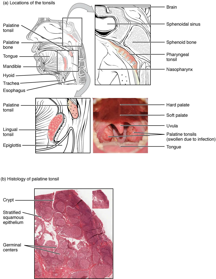
Tonsils are aggregates of lymphoid tissue located at the entrance of the respiratory and digestive tracts. The pharyngeal tonsils, also known as adenoids, are located in the nasopharynx and contain a rich supply of lymphocytes. They are particularly large in children but tend to regress with age. [1]
Figure 12209 Location and Histology of Tonsils
Long description
(a) The pharyngeal tonsil is located on the roof of the posterior superior wall of the nasopharynx. The palatine tonsils lay on each side of the pharynx. (b) A micrograph shows the palatine tonsil tissue. LM × 40. (Micrograph provided by the Regents of the University of Michigan Medical School © 201

Figure 1: (a) The pharyngeal tonsil is located on the roof of the posterior superior wall of the nasopharynx.
Mucosa-associated lymphoid tissue (MALT) includes lymphoid tissues in various mucosal surfaces throughout the body, such as the gut-associated lymphoid tissue (GALT) and bronchus-associated lymphoid tissue (BALT). These tissues provide immune surveillance at sites where pathogens commonly enter the body.
High endothelial venules (HEVs) are specialized blood vessels found in lymph nodes and other lymphoid tissues that facilitate the extravasation of lymphocytes from the bloodstream into the lymphoid organs. These vessels have plump endothelial cells that allow for efficient lymphocyte trafficking, which is essential for immune surveillance and response.
Under the light microscope, a lymphoid organ is first surveyed at low power to appreciate its overall architecture, then at high power to identify individual cells. The routine stain is haematoxylin and eosin (H&E): haematoxylin stains nucleic acids blue (hence nuclei and ribosome-rich cytoplasm), while eosin stains cytoplasmic and extracellular proteins pink. The key to interpreting any lymphoid organ is to recognise that it is a meshwork of reticular fibres and stromal cells, packed with lymphocytes at various stages of differentiation, interspersed with macrophages, dendritic cells and plasma cells.
The general layering seen in a lymph node is a useful template for all encapsulated lymphoid organs. From outside inwards, the capsule of dense connective tissue sends trabeculae into the parenchyma. Beneath the capsule lies the subcapsular sinus, a channel of lymph. The cortex contains lymphoid follicles, which are spherical aggregates of B cells. The paracortex (deep cortex) lies deep to the follicles and is a T-cell zone. The medulla contains medullary cords (plasma cells and macrophages) separated by medullary sinuses. The hilum is where blood vessels and efferent lymphatics enter and leave.
| Layer | H&E appearance | Main cell types |
|---|---|---|
| Capsule & trabeculae | Dense pink fibrous tissue | Fibroblasts, collagen |
| Subcapsular sinus | Pale, open space lined by endothelium | Macrophages, lymphocytes |
| Cortex (follicles) | Dark blue spherical nodules; pale germinal centre if active | B cells, follicular dendritic cells, tingible-body macrophages |
| Paracortex | Diffuse blue area, no nodules; high endothelial venules (HEVs) visible | T cells, interdigitating dendritic cells |
| Medulla | Pink cords separated by pale sinuses | Plasma cells, macrophages, B and T cells |
The B-cell zones are the follicles. A primary follicle is a uniform dense cluster of small, mature B cells. After antigenic stimulation, a secondary follicle develops with a pale central germinal centre surrounded by a dark mantle zone of small B cells. The germinal centre contains large, proliferating centroblasts and smaller centrocytes, plus tingible-body macrophages with engulfed apoptotic nuclei. The T-cell zones are the paracortex (in lymph nodes) and the periarteriolar lymphoid sheath (PALS) in the spleen. These areas contain small T cells, interdigitating dendritic cells, and the specialised post-capillary venules (HEVs) through which lymphocytes enter from blood.
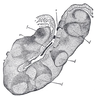
Figure 2Gray1027
Long description
Section through one of the crypts of the tonsil. (Stöhr.) Magnified. e. Stratified epithelium of general surface, continued into crypt. f, f. Nodules of lymphoid tissue—opposite each nodule numbers of lymph cells are passing into or through the epithelium. s, s. Cells which have thus escaped to mix

Figure 2: Section through one of the crypts of the tonsil ((stöhr.) magnified).
) magnified). The subcapsular sinus is the pale slit just beneath the capsule. Deep to the follicles, the paracortex is a diffuse, slightly paler blue region without nodules. The B-cell zones are the follicles; the T-cell zone is the paracortex, lying between the follicles and the medulla.
In the spleen, the white pulp is organised around central arterioles: the PALS is a T-cell zone, and B-cell follicles are attached to the PALS. The red pulp consists of splenic cords and venous sinuses. In the thymus, the cortex is densely packed with immature T cells (thymocytes) and the medulla is paler with fewer cells, Hassall's corpuscles and mature T cells. In mucosa-associated lymphoid tissue (MALT), such as Peyer's patches, B-cell follicles are present with intervening T-cell zones, but there is no capsule.
In routine haematoxylin and eosin (H&E) sections, the cellular architecture of lymphoid organs is evident, but many key components—reticular fibres, plasma cells, mast cells, and microorganisms—require special stains for confident identification. The most important special stains for lymphoid tissue are the silver impregnation methods for reticulin, the methyl green-pyronin (MGP) stain for plasma cells and activated lymphocytes, the periodic acid-Schiff (PAS) reaction for basement membranes and glycogen, and the Giemsa stain for mast cells and granulocytes.
The reticulin stain (e.g., Gordon and Sweet's silver method) is the classic stain for demonstrating the supporting framework of lymphoid organs. Reticulin fibres are fine, branching fibres of type III collagen that form a delicate meshwork around individual cells and small vessels. In a lymph node, the reticulin stain outlines the cortical follicles as relatively cell-dense areas with sparse fibres, whereas the paracortex and medullary cords show a denser, more regular network. In the spleen, reticulin fibres are prominent in the periarteriolar lymphoid sheath (PALS) and around the central arteriole, but are sparse within the germinal centres.
The methyl green-pyronin (MGP) stain is used to identify cells with high RNA content, which appear bright red (pyroninophilic). Plasma cells, with their abundant rough endoplasmic reticulum, stain intensely red, as do large activated lymphocytes (immunoblasts) in germinal centres. In a lymph node with reactive hyperplasia, MGP highlights the germinal centre as a red zone surrounded by a darker mantle of small, resting B cells that stain only faintly. In the medulla, plasma cells are seen as clusters of bright red cells with an eccentric nucleus and a pale perinuclear hof (the Golgi zone). This stain is particularly useful for distinguishing plasma cells from other round cells in infiltrates.
The periodic acid-Schiff (PAS) reaction stains neutral mucopolysaccharides, glycogen, and basement membranes a magenta colour. In lymphoid organs, PAS is useful for demonstrating the basement membrane of high endothelial venules (HEVs) in the paracortex of lymph nodes and in Peyer's patches. The HEV basement membrane appears as a thin, continuous magenta line surrounding the cuboidal endothelial cells. PAS also stains the basement membranes of the sinus-lining cells in the spleen and the reticular basement membrane of the thymic epithelium. In the thymus, PAS highlights the epithelial reticular cells, which form a delicate meshwork that is not visible on routine H&E.
The Giemsa stain is valuable for identifying mast cells and eosinophils in lymphoid tissue. Mast cells appear as oval cells filled with large, purple-red metachromatic granules that obscure the nucleus; they are found in small numbers in the capsule, trabeculae, and around blood vessels of lymph nodes and spleen. Eosinophils, with their bright red-orange granules and bilobed nucleus, are seen in the medulla of lymph nodes draining sites of allergic or parasitic inflammation. Giemsa also demonstrates plasma cells, whose cytoplasm stains a deep blue, and is useful for assessing the chromatin pattern of lymphocytes.
For the demonstration of immunoglobulin in plasma cells and follicular dendritic cells, the immunoperoxidase technique (using antibodies against kappa and lambda light chains) is the method of choice, though this is an immunohistochemical rather than a classical histochemical stain. In reactive follicles, the follicular dendritic cell network stains positively for CD21 and CD35, and the germinal centre B cells show surface immunoglobulin. In the bone marrow, the presence of a monoclonal population of plasma cells (all staining for one light chain only) is a key diagnostic feature of multiple myeloma, whereas a reactive plasmacytosis shows a mixture of kappa- and lambda-positive cells.
Finally, the Perls' Prussian blue stain for iron is occasionally used in the spleen and bone marrow to demonstrate haemosiderin-laden macrophages, which appear blue. This is relevant in conditions of haemolysis or repeated transfusion, where iron is deposited in the red pulp of the spleen and in the medullary cords of lymph nodes. The stain is simple: sections are treated with acid ferrocyanide, and ferric iron is converted to a blue pigment. In the normal spleen, small amounts of haemosiderin are seen in the red pulp macrophages; in pathological states, the blue granules become abundant and may be visible macroscopically.
Under the electron microscope, the lymphoid organs reveal a highly organised architecture that supports their function in immune surveillance and response. The fundamental unit is the reticular network, a three-dimensional meshwork of fibroblastic reticular cells (FRCs) and their associated reticular fibres. These FRCs are stellate cells with long cytoplasmic processes that wrap around bundles of collagen fibrils, creating a conduit system. This conduit system channels small soluble antigens and chemokines from the afferent lymphatics directly to the high endothelial venules (HEVs) and the T-cell zones, allowing rapid sampling of peripheral antigens without the need for cellular migration.
In the centre of the field lies a high endothelial venule (HEV), cut in cross-section. Its lumen is lined by plump, cuboidal endothelial cells with prominent nuclei and abundant cytoplasm containing numerous vesicles, reflecting active transcytosis of lymphocytes. Surrounding the HEV is the paracortical T-cell zone, where small lymphocytes with dense, heterochromatic nuclei and scant cytoplasm are packed tightly among the reticular fibres. The reticular fibres appear as electron-dense bundles of collagen fibrils, ensheathed by the thin cytoplasmic extensions of FRCs.
The high endothelial venules are a defining ultrastructural feature of secondary lymphoid organs, except the spleen. Their endothelial cells are tall and cuboidal, unlike the flat endothelium of most other vessels. The luminal surface bears microvilli, and the cells are joined by complex intercellular junctions that are transiently opened to allow lymphocyte passage. The basal lamina is often multilayered and incomplete, facilitating lymphocyte egress. The HEV endothelium expresses specific addressins, such as peripheral node addressin (PNAd) in lymph nodes, which bind to L-selectin on naïve lymphocytes, initiating the rolling and firm adhesion that precedes transmigration.
The reticular fibre network is composed of type III collagen (reticulin), which is visualised by silver staining in light microscopy but appears as thin, non-banded fibrils under the electron microscope. These fibres are produced by the FRCs, which also secrete the chemokines CCL19 and CCL21 that attract CCR7-expressing T cells and dendritic cells. The FRCs form a physical scaffold that defines the T-cell zone, and their processes are connected by desmosome-like junctions, creating a continuous cellular barrier that separates the conduit lumen from the surrounding lymphocytes. This barrier is not absolute, as dendritic cells can intercalate their processes into the conduits to sample antigen.
In the spleen, the ultrastructure differs because there are no afferent lymphatics or HEVs. The white pulp is organised around central arterioles, which are surrounded by a periarteriolar lymphoid sheath (PALS) of T cells. The marginal zone, at the border between white and red pulp, contains specialised macrophages and marginal zone B cells, which are positioned to capture blood-borne antigens. The red pulp consists of splenic cords (of Billroth) separated by venous sinuses; the sinusoidal endothelium has a discontinuous basal lamina and intercellular slits through which blood cells must squeeze to return to the circulation, allowing the spleen to filter aged or damaged erythrocytes.
The ultrastructure of the thymus is distinct, reflecting its role in T-cell development. The cortex contains densely packed immature thymocytes, which are large cells with abundant cytoplasm and prominent nucleoli, undergoing proliferation. These are supported by a network of cortical epithelial cells, which have long, branching processes that form a meshwork and create a microenvironment for positive selection. The epithelial cells are joined by desmosomes and contain characteristic tonofilaments. The medulla is less densely packed and contains mature thymocytes, medullary epithelial cells, and Hassall's corpuscles — concentric whorls of flattened, keratinised epithelial cells that are a hallmark of the thymic medulla and are thought to be involved in the negative selection of autoreactive T cells. The blood-thymus barrier in the cortex is formed by the continuous basal lamina of capillary endothelial cells, pericytes, and the processes of epithelial cells, preventing blood-borne antigens from reaching the developing thymocytes.
Lymphoid organs are organized structures that facilitate immune responses through specialized architecture and cellular components. The primary lymphoid organs, including the bone marrow and thymus, are responsible for the production and maturation of lymphocytes. The bone marrow serves as the site of hematopoiesis, giving rise to all blood cells including lymphocytes. The thymus provides the microenvironment for T cell maturation, where developing T cells undergo positive and negative selection to ensure self-tolerance.
Secondary lymphoid organs, such as lymph nodes, spleen, and mucosa-associated lymphoid tissue (MALT), are strategically positioned to encounter antigens. These organs contain specialized stromal cells that create a structured environment for immune cell interactions. Fibroblastic reticular cells (FRCs) form a network that provides chemokine cues and extracellular matrix, facilitating lymphocyte migration and activation. This organization allows for efficient antigen encounter and lymphocyte inte [2]
Tertiary lymphoid structures (TLOs) develop in sites of chronic inflammation, infection, or autoimmune conditions. These structures follow similar organizational principles as secondary lymphoid organs, containing organized T and B cell zones with antigen-presenting cells. The formation of TLOs involves the same cell types that control the development of secondary lymphoid organs, including lymphoid tissue inducer (LTi) cells and stromal cells that produce lymphotoxins. [2]
The function of lymphoid organs is closely tied to their structure. In lymph nodes, the cortex contains B cell follicles with germinal centers for antibody production, while the paracortex houses T cells. The medulla contains plasma cells and macrophages. This compartmentalization allows for efficient immune responses. Similarly, the spleen has white pulp with lymphoid follicles surrounding central arteries, and red pulp for filtration and blood storage.
The lymph node is surrounded by a capsule with afferent lymphatic vessels entering and efferent vessels exiting. Within the lymph node, high endothelial venules (HEVs) in the paracortex allow lymphocyte entry, while lymphatic sinuses facilitate antigen drainage. This organized structure enables efficient antigen presentation, lymphocyte activation, and immune response generation.
Key features to look for systematically:
- Capsule and trabeculae: A connective tissue capsule surrounds the organ. From it, trabeculae (septa) extend inward, dividing the organ into incomplete compartments. In the spleen, the capsule is relatively thick and contains smooth muscle; in lymph nodes it is thin. In the thymus, the cortex is very dark because of massive numbers of developing T cells; the medulla is paler and contains distinctive Hassall's corpuscles – concentric whorls of flattened, keratinised epithelial cells, often eosinophilic. These are pathognomonic for thymus.
- Lymphoid follicles: In lymph nodes and spleen (but not thymus), look for round, pale-staining aggregates of B cells called follicles. A primary follicle is uniform; a secondary follicle has a pale germinal centre (proliferating B cells) surrounded by a darker mantle zone of small resting B cells. Follicles are absent in the thymus.
- Sinuses: In lymph nodes, subcapsular, cortical and medullary sinuses appear as clear, slit-like spaces lined by endothelial cells, containing scattered macrophages and lymphocytes. In the spleen, the red pulp is a maze of venous sinuses (splenic sinuses) filled with red blood cells, separated by cords of Billroth (cellular cords).
- High endothelial venules (HEVs): In the paracortex of lymph nodes and in tonsils, look for venules lined by plump, cuboidal endothelial cells. These are the sites where lymphocytes leave the blood.
Look-alikes and how to tell them apart:
- Lymph node vs. spleen: Both have follicles and sinuses. The lymph node has a distinct hilum with efferent lymphatics and a subcapsular sinus; the spleen has no subcapsular sinus but has red pulp with abundant erythrocytes. If you see red blood cells in sinuses, it is spleen; if you see clear lymph, it is lymph node.
- Thymus vs. lymph node: The thymus has no follicles and no sinuses; it has a lobulated architecture with a very dark cortex and Hassall's corpuscles in the medulla. A lymph node has follicles and sinuses but no Hassall's corpuscles.
- Tonsil vs. lymph node: The tonsil has no capsule (or only a partial one), no subcapsular sinus, and its follicles lie directly beneath the epithelium. The crypts (invaginations of surface epithelium) are a hallmark of tonsils; lymph nodes never show crypts.
- Spleen white pulp vs. lymph node cortex: White pulp of the spleen forms periarteriolar lymphoid sheaths (PALS) – lymphocytes wrapped around a central arteriole. In a lymph node, lymphocytes are not arranged around arterioles. If you see a lymphoid cuff around a muscular artery, it is spleen.
Clinical pearlPearl
The fastest discriminator: Hassall's corpuscles = thymus; red pulp with sinuses = spleen; subcapsular sinus = lymph node; crypts with follicles = tonsil.
The capsule is the outer connective tissue rim; beneath it lies the subcapsular sinus, a clear space. The cortex contains follicles (B-cell zones) with pale germinal centres. Deep to the cortex is the paracortex (T-cell zone) containing HEVs. The hilum is at the bottom, where blood vessels enter and efferent lymphatics leave. Trabeculae extend from the capsule into the parenchyma.
The histopathology of lymphoid organs reveals distinct structural adaptations to their immune functions. The oropharynx contains two types of tonsils: palatine and lingual tonsils. The palatine tonsils are located laterally in the oropharynx in the area of the fauces, while the lingual tonsil is situated at the base of the tongue. These tonsils, along with the pharyngeal tonsil, are composed of lymphoid tissue that traps and destroys pathogens entering through the oral or nasal cavities. [1]
The surrounding connective tissue capsule with trabeculae extending inward provides structural support while allowing lymphocyte migration.
In pathological conditions, tertiary lymphoid organs (TLOs) can form in non-lymphoid tissues. These TLOs contribute to immunopathology via humoral mechanisms, as treatment with LTβR immunoglobulin or intravenous immunoglobulins (IVIGs) to saturate Fc receptors on effector cells prevented the disease. [2]
This demonstrates how local tissue environments shape immune cell function through metabolic pathways. [3]
A common trap is misclassifying the respiratory bronchioles as purely conducting – they contain occasional alveoli and thus participate in gas exchange. [1]
Traps: students forget that the meatuses are the spaces beneath each concha, not the conchae themselves. [1]
Trap: cilia beat toward the pharynx, not toward the lungs. [1]
The respiratory membrane is formed by alveolar epithelium plus capillary endothelium – a blood-air barrier for simple diffusion. [1]
Trap: alveolar ducts are not lined by continuous squamous epithelium – they are the entry tubes, not the sacs themselves. [1]
Trap: the epiglottis covers the glottis when closed, but it is not part of the glottis. [1]
Males tend to have larger folds, producing a deeper voice. [1]
Trap: odor sensing occurs in the nasal epithelium, not in the trachea or bronchi.
Exam trapConfusing lymphoid organ types
Students often mix up primary and secondary lymphoid organs. Primary lymphoid organs (bone marrow and thymus) are where lymphocytes develop and mature, while secondary lymphoid organs (lymph nodes, spleen, mucosa-associated lymphoid tissue) are where mature lymphocytes encounter antigens and mount immune responses.
Exam trapMisunderstanding TLO formation
Many students incorrectly assume that tertiary lymphoid organs (TLOs) form spontaneously. In reality, TLOs are induced by specific signals, particularly from dendritic cells (DCs) which are sufficient for TLO induction and can directly instruct stromal cells. [2]
Exam trapOverlooking tissue-specific TRM development
A common mistake is believing that tissue-resident memory T (TRM) cells form through identical pathways across all tissues. In fact, the signals driving TRM formation differ between tissue types - TGFβ signaling is required for lung TRM but not nasal cavity TRM, while IL-15 signaling is tissue-specific. [3]
Exam trapConfusing TRM retention mechanisms
Students often incorrectly assume that TRM cells use the same retention mechanisms in all tissues. Actually, TRM cells show reduced expression of S1PR1 and CCR7 that promote tissue egress, while expressing CD69 and CD103 (αE integrin) that promote tissue retention, with specific variations depending on the tissue location. [3]
Exam trapMisinterpreting P2RX7 in TRM formation
Many students incorrectly conclude that P2RX7 is essential for all TRM cell development. While P2RX7 deficiency affects TRM cell pool size in the small intestine, it doesn't impact TRM cell-forming capacity, suggesting alternative mechanisms exist for TGFβ receptor re-expression.
NoteClinical examination
Knowledge of lymphoid organ histology is essential for physical examination. Palpating lymph nodes helps assess for infection, malignancy, or autoimmune disease. Normal lymph nodes are usually less than 1 cm in diameter, mobile, and non-tender, while pathological nodes may be enlarged, fixed, or matted.
NoteSurgical pathology
In the operating theatre, surgeons must recognize lymphoid tissue during procedures. For example, during tonsillectomy, understanding the anatomy of palatine tonsils (mucosa-associated lymphoid tissue) is crucial to avoid damaging surrounding structures. Similarly, splenectomy requires knowledge of splenic architecture to control bleeding.
NoteImaging interpretation
Radiologists use lymphoid organ anatomy in interpreting imaging studies. The spleen's normal size is approximately 11 cm in craniocaudal length on ultrasound. Lymph nodes are considered pathological when short-axis diameter exceeds 1 cm on CT. Understanding the location of secondary lymphoid organs helps identify abnormal findings.
NoteExamination preparation
Medical students must understand lymphoid organ histology for board examinations. Common questions include identifying specific cell types in different lymphoid organs, describing the structure of lymph nodes, and explaining the functional differences between primary and secondary lymphoid tissues. The respiratory membrane thickness of approximately 0.5 μm is a frequently tested fact. [1]
Tertiary Lymphoid Organ Formation
- Antigen encounterPersistent antigen exposure in tissues
- Question: Dendritic cell activation?
Yes
- DC accumulation at siteCD11c+ cells expressing LTαβ
- Stromal instructionDirect or T cell-mediated
- LymphangiogenesisIn some tissues like thymus
- TLO formationInduction of myofibroblast differentiation
No
- No TLO formation
Lymphoid organs are categorized as primary (bone marrow, thymus) where lymphocytes mature, and secondary (lymph nodes, spleen, MALT) where immune responses occur. Tertiary lymphoid organs form in response to chronic inflammation or infection, induced primarily by dendritic cells. Tissue-resident memory T cells develop through tissue-specific pathways, with different requirements for TGFβ and IL-15 signaling across various tissues. The respiratory membrane, formed by alveolar and capillary endothelial cells, is approximately 0.5 μm thick, allowing efficient gas exchange. TRM cells exhibit unique molecular characteristics including reduced S1PR1/CCR7 expression and increased CD69/CD103 expression for tissue retention.
Answer each one in your head first, then open the answer.
What is the approximate thickness of the respiratory membrane?
Show answerHide answer
0.5 μmWhich cells are primarily responsible for inducing tertiary lymphoid organs?
Show answerHide answer
Dendritic cellsWhich signaling pathway is required for TRM formation in the lung but not the nasal cavity?
Show answerHide answer
TGFβ signalingWhich molecules promote tissue retention in TRM cells?
Show answerHide answer
CD69 and CD103What is the normal size of a non-pathological lymph node?
Show answerHide answer
Less than 1 cm in diameter
- Betts JG, Young KA, Wise JA, et al. Organs and Structures of the Respiratory System. OpenStax, Anatomy and Physiology 2e (CC BY-NC-SA 4.0). Accessed .
- Neyt K, Perros F, GeurtsvanKessel CH, et al. Tertiary lymphoid organs in infection and autoimmunity. Trends in immunology 2012;33(6):297-305. doi:10.1016/j.it.2012.04.006. Accessed .
- Kok L, Masopust D, Schumacher TN. The precursors of CD8<sup>+</sup> tissue resident memory T cells: from lymphoid organs to infected tissues. Nature reviews. Immunology 2022;22(5):283-293. doi:10.1038/s41577-021-00590-3. Accessed .
Figure credits
Educational content for medical students and doctors. It is not a substitute for clinical judgement or local guidelines.