Eye and ear histology
Microscopic structure of eye and ear tissues for medical students.
The eye and ear contain specialized sensory tissues that detect light and mechanical stimuli, respectively. The eye focuses light through the lens, which is suspended by ligaments attached to the ciliary body. The ciliary body contains smooth muscle that changes the shape of the lens, allowing accommodation for near and far vision. The iris, the colored part of the eye, is a smooth muscle that controls the size of the pupil to regulate light entry. The trabecular meshwork is a drainage structure in the anterior chamber that allows aqueous humor to exit the eye. [1]
The optic nerve head is where retinal ganglion cell axons exit the eye to form the optic nerve. The eyelid contains skin, muscle, and glands that protect the eye and distribute tears. The lacrimal gland produces tears that lubricate the eye and provide nutrients to the corneal surface. [1]
Figure 11427 Cochlea Micrograph
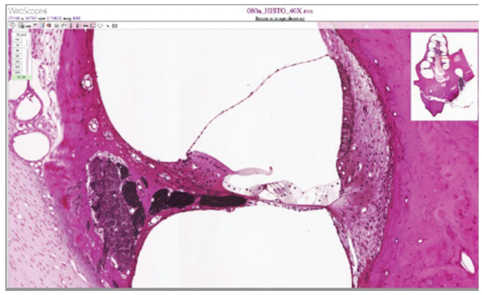
Long description
LM × 412. (Micrograph provided by the Regents of University of Michigan Medical School © 2012)

Figure 1: LM × 412.
The tympanic membrane, or eardrum, is a thin, cone-shaped membrane that vibrates in response to sound waves. It has a diameter of 9mm and thickness of 0.1mm. The organ of Corti in the cochlea contains hair cells with stereocilia that convert mechanical vibrations into electrical signals. The tectorial membrane slides across the stereocilia during sound stimulation, causing depolarization of hair cells. [3] [1]
Vestibular receptors in the inner ear detect head position and movement. The utricle and saccule contain maculae composed of hair cells surrounded by support cells. The stereocilia extend into an otolithic membrane containing calcium carbonate crystals that make it top-heavy. When the head tilts, the otolithic membrane slides over the macula, bending the stereocilia and depolarizing hair cells. The vestibular system is responsible for spatial orientation, motor coordination, and autonomic regulation. [1] [2]
DefinitionHair cell function
Hair cells convert mechanical stimuli into electrical signals through the bending of stereocilia, which opens ion channels and generates action potentials.
Under the light microscope, the eye and ear reveal highly ordered, layered epithelia and connective tissues. The eye is a hollow sphere of three tunics: the outer fibrous tunic (sclera and cornea), the middle vascular tunic (choroid, ciliary body, iris), and the inner neural tunic (retina). The ear shows the external canal, the tympanic membrane, the ossicles of the middle ear, and the sensory epithelia of the cochlea and vestibule. [1]
| Structure | H&E appearance | Key layers / cell types |
|---|---|---|
| Cornea | Anterior clear, avascular, convex; stains pink with H&E due to collagen | 5 layers: corneal epithelium (stratified squamous, non-keratinised), Bowman's membrane, stroma (regular collagen lamellae, keratocytes), Descemet's membrane, corneal endothelium (simple cuboidal) |
| Lens | Biconvex, avascular, eosinophilic; no nuclei in mature fibres | Lens capsule (basement membrane), lens epithelium (anterior, simple cuboidal), lens fibres (elongated, crystallin-filled) |
| Retina | Layered neural tissue; inner layers cellular, outer layers fibrous | 10 layers; photoreceptors (rods, cones) outermost; bipolar cells, amacrine cells, retinal ganglion cells (RGCs) inner; RGC axons form optic nerve |
| Organ of Corti | Spiral epithelial ridge on basilar membrane within cochlear duct | Inner hair cells (one row), outer hair cells (three rows), supporting cells (pillar, Deiters), tectorial membrane |
| Vestibular receptors | Maculae (utricle, saccule) and cristae (ampullae) with hair cells | Hair cells with stereocilia and kinocilium, supporting cells, otolithic membrane (maculae) or cupula (cristae) |
The cornea is the transparent anterior part of the fibrous tunic, continuous with the sclera, which covers five sixths of the eye surface. Its five layers are best seen in cross-section: the outermost stratified squamous epithelium rests on Bowman's membrane; the thick stroma of regular collagen lamellae gives strength and transparency; Descemet's membrane lies deep to the stroma; and the innermost simple cuboidal endothelium faces the anterior chamber. The cornea is avascular, so it appears clear; its nutrition comes from tears and aqueous humour. [1]
The retina is a multilayered neural sheet lining the posterior eye. From outside (choroid) to inside (vitreous), the layers are: retinal pigment epithelium, photoreceptor outer and inner segments, outer limiting membrane, outer nuclear layer (photoreceptor nuclei), outer plexiform layer (synapses with bipolar cells), inner nuclear layer (bipolar, amacrine, horizontal cells), inner plexiform layer (synapses with RGCs), ganglion cell layer, nerve fibre layer (RGC axons), and inner limiting membrane. At the fovea, the central retina, these inner layers are absent, leaving only photoreceptors, which maximises visual acuity. [1]
Figure 21413 Structure of the Eye
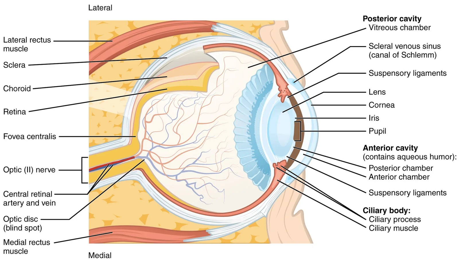
Long description
The sphere of the eye can be divided into anterior and posterior chambers. The wall of the eye is composed of three layers: the fibrous tunic, vascular tunic, and neural tunic. Within the neural tunic is the retina, with three layers of cells and two synaptic layers in between. The center of the ret

Figure 2: The sphere of the eye can be divided into anterior and posterior chambers.
Posterior to the cornea lies the lens, biconvex and avascular. In the ear, the organ of Corti sits on the basilar membrane within the cochlear duct; the tectorial membrane overlies the hair cells.
The organ of Corti is the sensory epithelium for hearing, located on the basilar membrane of the cochlear duct. It contains one row of inner hair cells and three rows of outer hair cells, with supporting pillar cells forming the tunnel of Corti. The tectorial membrane, a gelatinous shelf, contacts the stereocilia of the outer hair cells; sound-induced basilar membrane vibration bends the stereocilia, transducing mechanical stimuli into receptor potentials. The vestibular receptors—the maculae of the utricle and saccule, and the cristae of the ampullae—are similar: hair cells with stereocilia embedded in an otolithic membrane (maculae) or a cupula (cristae). Head movement displaces the otoliths or endolymph, bending the stereocilia and altering the membrane potential.
In routine haematoxylin and eosin (H&E) preparations, the eye and ear show their general architecture, but several specialised stains are needed to demonstrate key components that are otherwise poorly visible. These stains exploit the physical and chemical properties of specific extracellular matrices, cells and pigments.
For the eye, the Periodic acid-Schiff (PAS) stain is indispensable. It oxidises vicinal glycol groups in polysaccharides to aldehydes, which then react with Schiff reagent to produce a magenta colour. PAS strongly highlights the basal lamina of the corneal epithelium, the Descemet membrane (the thick basal lamina of the corneal endothelium), and the Bruch membrane of the choroid. It also stains the zonular fibres (suspensory ligaments of the lens) and the inner limiting membrane of the retina. In the sclera and cornea, PAS demonstrates the glycosaminoglycan-rich ground substance between collagen lamellae.
Masson trichrome is used to distinguish collagen from muscle and cytoplasm. In the eye, it stains scleral and corneal collagen blue or green, while the extraocular muscles (superior rectus, medial rectus, inferior rectus, lateral rectus, superior oblique, inferior oblique) appear red. This stain is particularly useful for assessing the fibrous tunic of the eye and for identifying fibrosis in the trabecular meshwork. The Verhoeff-Van Gieson stain demonstrates elastic fibres as black, and is used to show the elastic laminae of the choroidal vessels and the lamina cribrosa of the optic nerve head.
For the retina, silver impregnation methods (e.g., Bodian or Bielschowsky) stain neurofibrils and axons, allowing the three nuclear layers and the nerve fibre layer to be traced. Oil Red O or Sudan black can be used on frozen sections to demonstrate lipid droplets in the retinal pigment epithelium (RPE) in pathological states, though not in normal histology. The Fontana-Masson stain (melanin) is used to identify melanin granules in the RPE and in uveal melanocytes; it reduces silver nitrate to metallic silver, producing black granules.
In the ear, the Movat pentachrome stain is valuable for the temporal bone. It stains collagen yellow, mucin and ground substance blue, fibrin red, muscle red, and elastic fibres black. This is particularly useful for the basilar membrane of the cochlea, which contains collagenous and elastic components, and for the tectorial membrane, an acellular gelatinous structure overlying the organ of Corti. The osmic acid (osmium tetroxide) stain fixes and blackens lipids, and is used to demonstrate the myelin sheaths of the cochlear and vestibular nerve fibres within the modiolus.
Alcian blue at pH 2.5 stains acidic mucopolysaccharides and glycosaminoglycans. In the ear, it highlights the cupula of the crista ampullaris and the otolithic membrane of the maculae, which are rich in glycosaminoglycans. It also stains the endolymph within the membranous labyrinth, which is high in potassium and low in sodium, though this is not directly visualised by Alcian blue. The von Kossa stain for calcium is used to demonstrate the otoconia (otoliths) within the utricle and saccule, which are calcium carbonate crystals; they appear black.
The ultrastructure of the eye and ear reveals specialized cellular components essential for vision and hearing. Electron microscopy demonstrates the intricate organization of sensory cells and supporting structures.
In the retina, photoreceptors consist of rods and cones, each with distinct ultrastructural features. Rods contain rhodopsin within stacked membranous discs in their outer segments, while cones have invaginated membrane folds. These photopigments undergo conformational changes upon light absorption, initiating the visual transduction cascade. The inner segments of photoreceptors are packed with mitochondria to meet high energy demands, and synaptic terminals connect to bipolar cells via ribbon synapses specialized for rapid neurotransmitter release. [1]
The inner ear's sensory epithelia contain hair cells with characteristic stereocilia bundles. These mechanoreceptors have graded stereocilia heights arranged in a staircase pattern, connected by tip links that open mechanotransduction channels upon deflection. In the organ of Corti, inner hair cells are arranged in a single row and provide the primary afferent input to the cochlear nerve, while outer hair cells form three rows and amplify sound through electromotility. Hair cells rest on the basilar membrane, which contains specialized fibrocytes that contribute to the endocochlear potential essential for hearing. [1]
The middle ear ossicles—malleus, incus, and stapes—have distinctive ultrastructural features for efficient sound transmission. The malleus attaches to the tympanic membrane and articulates with the incus via a saddle-shaped joint, while the incus connects to the stapes through a similar articulation. The stapes footplate fits into the oval window, with its ultrastructure designed to minimize impedance mismatch between air-conducted sound and fluid movement in the cochlea. [1]
DefinitionHair Cell
A specialized mechanoreceptor with stereocilia that converts mechanical stimuli into neural signals in both the auditory and vestibular systems. [1]
The eye and ear are specialized sensory organs with highly specialized structures that enable their specific functions. The eye is responsible for vision, while the ear is responsible for both hearing and equilibrium. These organs contain specialized receptor cells that convert physical stimuli into neural signals that can be interpreted by the brain. [1]
DefinitionPhotoreceptors
Specialized receptor cells in the retina that convert light energy into neural signals.
The lacrimal gland produces tears that flow through lacrimal ducts, over the surface of the eyeball and conjunctiva to the medial corner of the eye, washing away foreign particles. Excess tears eventually flow into the nasal cavity, which is why the nose runs when a person cries. [1]
In the eye, the cornea is the transparent outer layer that allows light to enter. The iris controls the size of the pupil, regulating the amount of light that reaches the retina. The lens focuses light onto the retina, which contains photoreceptor cells that convert light into neural signals. [3]
In the ear, the outer ear collects sound waves and directs them toward the tympanic membrane (eardrum). The middle ear contains three small bones (ossicles) that transmit vibrations from the tympanic membrane to the oval window of the inner ear. The inner ear contains the cochlea, which is responsible for hearing, and the vestibular system, which is responsible for balance. The cochlea contains specialized hair cells that convert mechanical vibrations into neural signals, similar to how photoreceptors in the eye convert light into neural signals. [1]
Both the eye and ear demonstrate the principle of structure–function correlation, where specialized structures are adapted to perform specific functions efficiently. The photoreceptors in the eye and the hair cells in the ear are examples of specialized receptor cells that convert different forms of energy (light and mechanical vibrations, respectively) into neural signals that can be interpreted by the brain.
When examining histological slides of the eye and ear, specific key features allow for accurate identification of different structures. The eye is divided into two main cavities: the anterior cavity and the posterior cavity. The anterior cavity is located between the cornea and lens, including the iris and ciliary body, and is filled with aqueous humor. The posterior cavity extends behind the lens to the retina and contains vitreous humor. [1]
In the ear, the tympanic membrane (eardrum) is a key structure to identify. When viewed histologically, the tympanic membrane shows three layers: outer skin, middle fibrous layer, and inner mucosal layer.
NoteCommon Look-alikes
The cornea can be mistaken for the sclera, but the cornea is avascular and has a Bowman's layer, while the sclera is thicker and contains more collagen fibers.
Exam trapPitfall
The tympanic membrane may appear damaged if the probe was placed too close (<1mm) during measurement, potentially causing artifacts in the histological preparation.
| Structure | Key Features | Identification Tips |
|---|---|---|
| Cornea | Avascular, transparent | Presence of Bowman's layer |
| Lens | Biconvex structure | Epithelial cells on anterior surface |
| Tympanic membrane | Three-layered structure | Look for malleus attachment point |
| Taste buds | Oval clusters of cells | Located in tongue epithelium |
The histopathology of the eye and ear reveals important structural relationships that correlate with function.
These differences reflect the thicker tissue and higher concentration of blood vessels near the malleus. [3]
The color gradient represents temperature variations, with warmer colors (red) indicating higher temperatures near the malleus and cooler colors (blue) in more peripheral areas, reflecting the underlying vascular architecture and tissue thickness variations.
Clinical pearlOlfactory pathway
Olfactory neurons extend through the cribriform plate to the olfactory bulb, bypassing the thalamus and connecting directly to the primary olfactory cortex in the temporal lobe. This direct connection explains why smell can trigger emotional memories and responses without thalamic processing. This intimate connection between the olfactory system and the cerebral cortex is one reason why smell can be a potent trigger of memories and emotion. For example, the smell of food associated with one's bi [1]
Exam trapTaste nerve distribution
Facial nerve supplies anterior 2/3 of the tongue, glossopharyngeal nerve supplies posterior 1/3, and vagus nerve supplies extreme posterior tongue. The vagus nerve connects specifically to taste buds in the extreme posterior of the tongue, verging on the pharynx, which are more sensitive to noxious stimuli such as bitterness. Once the gustatory cells are activated by taste molecules, they release neurotransmitters onto the dendrites of sensory neurons from these three cranial nerves. This precise innervation pattern is crucial for understanding taste testing and neurological examinations of cranial nerve function. [1]
The anterior and posterior vertical canals are oriented at approximately 45 degrees relative to the sagittal plane. The base of each semicircular canal connects to an enlarged region known as the ampulla, which contains the hair cells that respond to rotational movement. The stereocilia of these hair cells extend into the cupula, a membrane that attaches to the top of the ampulla. When the head rotates in a plane parallel to the semicircular canal, the fluid lags, deflecting the cupula in the direction opposite to the head movement. This mechanism allows detection of angular acceleration during activities such as turning the head while saying "no". [1]
Mechanical, chemical, or thermal stimuli beyond a set threshold will elicit painful sensations. Stressed or damaged tissues release chemicals that activate receptor proteins in the nociceptors. For example, the sensation of heat associated with spicy foods involves capsaicin, the active molecule in hot peppers, which binds to a transmembrane ion channel in nociceptors that is sensitive to temperatures above 37°C. [1]
Photoreceptor cells have two parts: the inner segment containing the nucleus and other organelles, and the outer segment where photoreception takes place. The rod-shaped outer segments contain a stack of membrane-bound discs with the photosensitive pigment rhodopsin. In contrast, cone-shaped outer segments have their photosensitive pigments in infoldings of the cell membrane. There are three cone photopigments called opsins, each sensitive to a particular wavelength of light, enabling color vision. Light falling on the retina causes chemical changes to these pigment molecules, ultimately leading to a change in the activity of the retinal ganglion cells. [1]
This knowledge is crucial for accurate clinical temperature measurement, as improper probe positioning can lead to significant errors in readings. [3]
Clinical pearlEmotional memory connection
Olfactory system connects directly to limbic system and hypothalamus, explaining smell's potent trigger for memories and emotions. Unlike other sensory modalities, smell does not synapse in the thalamus before connecting to the cerebral cortex. This direct pathway allows smells to become associated with long-term memory and emotional responses through connections to the limbic system and hypothalamus. The intimate connection between the olfactory system and the cerebral cortex is one reason why smell can be a potent trigger of memories and emotion. This explains why certain smells can instantly evoke vivid memories and emotional responses that other sensory stimuli cannot match. [1]
Exam trapThermoreceptors
Some thermoreceptors respond only to cold, others only to heat; temperature detection occurs when local temperatures differ from body temperature. Two different types of somatosensory signals transduced by free nerve endings are pain and temperature, using thermoreceptors and nociceptors respectively. Temperature receptors are stimulated when local temperatures differ from body temperature, with some receptors sensitive to just cold and others to just heat. This specialized detection system allo [1]
Clinical pearlGustatory transduction
Taste cells release neurotransmitters onto facial, glossopharyngeal, and vagus nerve dendrites after activation by taste molecules. These neurons are part of the facial and glossopharyngeal cranial nerves, as well as a component within the vagus nerve dedicated to the gag reflex. The facial nerve connects specifically to taste buds in the anterior two thirds of the tongue, while the glossopharyngeal nerve connects to taste buds in the posterior one third. The vagus nerve connects to taste buds in the extreme posterior of the tongue, verging on the pharynx, which are more sensitive to noxious stimuli such as bitterness. This precise innervation pattern is essential for the complete gustatory function and neurological assessment of taste sensation. [1]
Exam trapFacial cooling effect
Facial cooling affects tympanic membrane temperature, but this sensitivity can be reduced by proper probe positioning. This knowledge is important for accurate clinical temperature monitoring, especially in environments where facial cooling might occur. [3]
Clinical pearlPhototransduction
Light causes chemical changes in photoreceptor pigments, ultimately changing retinal ganglion cell activity. Photoreceptor cells have specialized outer segments where photoreception takes place. In rods, this involves rhodopsin in stacked membrane-bound discs, while cones have three different opsins in membrane infoldings, each sensitive to specific wavelengths. The chemical changes in these pigments initiate a cascade that ultimately leads to changes in the activity of retinal ganglion cells. This process converts light energy into neural signals that can be interpreted by the brain as visual images, forming the basis of vision. [1]
This effect must be accounted for in surgical and critical care environments where temperature monitoring is crucial. [3]
Exam trapReceptor classification confusion
Students often confuse the three types of sensory receptors: free nerve endings, encapsulated endings, and specialized receptor cells. The pain and temperature receptors in the skin are examples of neurons with free nerve endings, while lamellated corpuscles in the skin are neurons with encapsulated endings that respond to pressure and touch. The photoreceptors in the retina represent specialized receptor cells. [1]
Exam trapMisunderstanding receptor locations
A common mistake is failing to distinguish between exteroceptors, interoceptors, and proprioceptors based on their locations. Exteroceptors are located near external stimuli, like somatosensory receptors in the skin. Interoceptors interpret stimuli from internal organs, such as blood pressure receptors in the aorta. Proprioceptors are located near moving body parts like muscles to interpret tissue positions during movement.
Exam trapPeripheral vs. foveal vision confusion
Students often underestimate the difference in visual acuity between the fovea and peripheral retina. In the fovea, each retinal ganglion cell (RGC) receives input from a single photoreceptor, maximizing accuracy. Toward the edges of the retina, up to 50 photoreceptors can converge on a single RGC, significantly reducing visual acuity. [1]
Exam trapColor perception in darkness
Many students believe they can see colors in complete darkness, but this is incorrect. If you think you can see colors in the dark, it is most likely because your brain is relying on memory of what color something should be, not actual visual perception.
Exam trapTympanic membrane temperature measurement
A common error in tympanic temperature measurement is assuming the probe should physically engage the tympanic membrane. While this would ensure all measurements are taken at the same proximity, it is unrealistic and unsafe in conscious individuals.
Exam trapIgnoring environmental temperature effects
Students often overlook how environmental temperature affects temperature measurement sites. When environment temperature is lower than core body temperature, we seek the hottest spot. However, if the environment is hotter than core body temperature, heat flow reverses, and we should seek the coolest point instead.
Exam trapGeneral anesthesia effects on temperature
Many students fail to recognize that general anesthesia lowers core body temperature. This effect is observed at the start of all experiments when general anesthesia is administered, with temperature measurements at all sites falling below the healthy range. The body's thermoregulation function gradually returns after anesthesia is halted.
NoteClinical assessment of sensory function
Knowledge of sensory receptors is fundamental in clinical practice. When assessing a patient's sensory system, clinicians must understand that free nerve endings detect pain and temperature, encapsulated endings like lamellated corpuscles respond to pressure and touch, and specialized receptors like photoreceptors in the retina detect light. This understanding guides neurological examinations and helps localize lesions in the nervous system.
NoteFlavor perception in nutrition
Understanding taste receptors has practical applications in nutrition and food science. The umami taste, based on G protein-coupled receptors activated by L-glutamate, explains why protein-rich foods like meat are described as savory. This knowledge helps dietitians design nutrition plans and food scientists develop flavor-enhancing products. [1]
NoteCapsaicin as a topical analgesic
The unusual binding dynamics of capsaicin with its transmembrane ion channel has practical therapeutic applications. This property makes capsaicin useful as a topical analgesic in products like Icy Hot™. [3]
NoteVisual field testing
Knowledge of retinal anatomy is crucial in ophthalmology and neurology. The difference in visual acuity between the fovea and peripheral retina explains why visual field testing is performed by having patients fixate on a central point while identifying objects in their periphery. This principle guides the interpretation of visual field defects in conditions like glaucoma or optic nerve damage.
Tympanic Temperature Measurement
- Position probe in ear canalEnsure proper placement without touching tympanic membrane
- Question: Environment temperature < core body temperature?
Yes
- Locate hottest spot in tympanic membraneTypically lower anterior quadrant or at malleus bone
No
- Locate coolest spot in tympanic membraneHeat flow direction reverses when environment is hotter
- Record temperatureNote that general anesthesia lowers readings
NoteIntraoperative temperature monitoring
Knowledge of tympanic membrane temperature measurement has applications in the operating theater. During general anesthesia, which lowers core body temperature, monitoring tympanic temperature provides insight into the patient's thermoregulation status. The rate of change in tympanic membrane temperature can indicate when the body's thermoregulation function is being restored after anesthesia is halted.
NoteExamination preparation
Understanding eye and ear histology is essential for medical examinations. Students must recognize the structural differences between various sensory receptors, understand the principles of visual transduction, and be able to explain how environmental factors affect temperature measurements. These concepts frequently appear in questions about sensory systems, ophthalmology, and clinical measurement techniques.
- The eye contains specialized structures such as the lens, ciliary body, iris, and trabecular meshwork that work together to focus light and regulate aqueous humor drainage.
- The retina is a multilayered neural tissue with photoreceptors (rods and cones) that convert light into neural signals, with the fovea having the highest visual acuity due to minimal convergence of photoreceptors to ganglion cells. [1]
- The ear consists of the tympanic membrane that vibrates in response to sound waves, and the organ of Corti in the cochlea that contains hair cells with stereocilia converting mechanical vibrations into electrical signals.
- Vestibular receptors in the inner ear, including the utricle and saccule, detect head position and movement through hair cells with stereocilia that depolarize when the otolithic membrane slides over them during head tilts.
- Hair cells in both the auditory and vestibular systems convert mechanical stimuli into electrical signals through the bending of stereocilia, which opens ion channels and generates action potentials.
- The cornea is transparent and avascular, consisting of five layers: corneal epithelium, Bowman's membrane, stroma, Descemet's membrane, and corneal endothelium.
- Special stains like PAS highlight basal lamina structures in the eye, while Movat pentachrome stain is valuable for demonstrating collagen, elastic fibers, and glycosaminoglycans in the ear.
- The tympanic membrane has a three-layered structure (outer skin, middle fibrous layer, and inner mucosal layer) and temperature variations across its surface that correlate with underlying tissue thickness and vascularity.
Answer each one in your head first, then open the answer.
What is the function of the ciliary body in the eye?
Show answerHide answer
The ciliary body contains smooth muscle that changes the shape of the lens, allowing accommodation for near and far vision.How do hair cells in the organ of Corti convert sound waves into neural signals?
Show answerHide answer
Hair cells have stereocilia that are bent by the tectorial membrane during sound stimulation, causing depolarization of the hair cells and generating electrical signals.What is the structure and function of the vestibular receptors in the inner ear?
Show answerHide answer
The utricle and saccule contain maculae composed of hair cells surrounded by support cells, with stereocilia extending into an otolithic membrane containing calcium carbonate crystals that slide over the macula when the head tilts, bending stereocilia and depolarizing hair cells to detect head position and movement.What are the five layers of the cornea and their characteristics?
Show answerHide answer
The cornea consists of: 1) stratified squamous epithelium (non-keratinized), 2) Bowman's membrane, 3) stroma (regular collagen lamellae with keratocytes), 4) Descemet's membrane, and 5) simple cuboidal endothelium; it is avascular and appears clear.How does temperature vary across the tympanic membrane and what causes these variations?
Show answerHide answer
Temperature varies across the tympanic membrane, with the highest and most stable temperature found at the point anterior to the malleus, reflecting the thicker tissue and higher concentration of blood vessels in that region.
- Betts JG, Young KA, Wise JA, et al. Sensory Perception. OpenStax, Anatomy and Physiology 2e (CC BY-NC-SA 4.0). Accessed .
- Jin GS, Li XL, Jin YZ, et al. Role of peripheral vestibular receptors in the control of blood pressure following hypotension. The Korean journal of physiology & pharmacology : official journal of the Korean Physiological Society and the Korean Society of Pharmacology 2018;22(4):363-368. doi:10.4196/kjpp.2018.22.4.363. Accessed .
- Yeoh WK, Lee JKW, Lim HY, et al. Re-visiting the tympanic membrane vicinity as core body temperature measurement site. PloS one 2017;12(4):e0174120. doi:10.1371/journal.pone.0174120. Accessed .
Figure credits
Educational content for medical students and doctors. It is not a substitute for clinical judgement or local guidelines.