Oral cavity and teeth histology
Histological structure and functional anatomy of the oral cavity, teeth, and salivary glands.
The oral cavity is divided into the oral vestibule, which is the pocket-like area framed by the cheeks, lips, gums, and teeth, and the oral cavity proper, which extends from the gums and teeth to the fauces (the opening to the oropharynx).
The oral mucosa consists of a stratified squamous epithelium resting on a lamina propria. On the top and sides of the tongue, the lamina propria forms extensions called papillae.
| Papilla Type | Morphology | Key Features | Function |
|---|---|---|---|
| Fungiform | Mushroom shaped | Contain taste buds | Taste perception |
| Filiform | Long and thin | Contain touch receptors | Mechanical food movement; abrasive surface |
The underside of the tongue is tethered to the floor of the mouth by a fold of mucous membrane known as the lingual frenulum.
Humans possess two sets of teeth (dentitions). The first set consists of 20 deciduous teeth appearing at approximately 6 months of age. These are replaced between ages 6 and 12 by 32 permanent teeth. [1]
Teeth are composed of hard tissues (enamel and dentine) and soft tissues (pulp)[1]. The periodontium supports these structures. All oral surfaces are almost instantly coated by the acquired oral pellicle, a layer of selectively absorbed salivary proteins and peptides[1]. This pellicle can mask surface roughness and provide attachment points for microorganisms[1].
Saliva is secreted by major and minor glands to moisten the mouth, teeth, and food, and to initiate carbohydrate breakdown. An average of 1 to 1.5 liters of saliva is produced daily. [1]
DefinitionMinor Salivary Glands
Small exocrine glands located within the mucous membranes of the lips (labial), cheeks (buccal), palate (palatal), and tongue (lingual).
The major salivary glands produce the bulk of saliva, with varying compositions based on their cellular makeup:
| Gland | Secretion Type | Key Components |
|---|---|---|
| Parotid | Watery | Salivary amylase |
| Submandibular | Mixed (watery and mucus) | Amylase in a mucus-thickened liquid |
| Sublingual | Thickest | Mostly mucus; least amount of amylase |
Salivary glands contribute to the defense of the oropharynx via the secretion of secretory IgA (SIgA). IgA+ plasma cells (PCs) are found scattered among acini or in clusters near ducts.
- Minor Glands: These have a high density of IgA+ PCs, reported to be three times that of the parotid gland. [2]
- Submandibular Glands: These contain approximately twice as many IgA+ PCs per tissue unit as the parotid gland. [2]
The ductal epithelium may be involved in antigen presentation as it expresses MHC class II (HLA-DR) molecules.
Light microscopy reveals the detailed structure of the oral cavity and teeth, showing distinct layers and cell types that form the various tissues.
The oral mucosa is composed of several layers, each with specific cell types and functions. The basic structure consists of an epithelium and an underlying connective tissue layer called the lamina propria.
| Layer | Cell types | Characteristics |
|---|---|---|
| Epithelium | Keratinocytes | Form the stratified squamous epithelium; can be keratinized or non-keratinized [1] |
| Langerhans cells | Immune cells present in the epithelium | |
| Melanocytes | Produce melanin pigment | |
| Lamina propria | Fibroblasts | Produce collagen and other extracellular matrix components |
| Blood vessels | Provide nutrients to the epithelium | |
| Nerve endings | Sensory functions | |
| Immune cells | Including lymphocytes and plasma cells |
There are three main types of oral mucosa:
-
Masticatory mucosa: Found on the hard palate and gingiva. This type is keratinized to withstand mechanical forces during chewing. The epithelium is stratified squamous and contains a thick layer of keratinocytes[1]. The hard palate, formed by the maxillary and palatine bones, serves as a wall between the oral and nasal cavities and as a rigid shelf against which the tongue can push food during chewing[1].
-
Lining mucosa: Covers the cheeks, floor of the mouth, and ventral surface of the tongue. This type is non-keratinized and allows for flexibility and movement. The epithelium is thinner than in masticatory mucosa and contains fewer keratinocytes[1]. This mucosa provides a habitat for microbial colonization, with early plaque formation frequently involving Streptococci as well as Neisseria, Rothia, Actinomyces, or Veillonella.
-
Specialized mucosa: Found on the dorsal surface of the tongue. Contains specialized structures like taste buds embedded in the epithelium. The epithelium contains both keratinized and non-keratinized areas, with numerous papillae that house taste buds[1].
The hard palate is formed by the maxillary and palatine bones, creating a rigid structure that separates the oral and nasal cavities. Posteriorly, it transitions to the soft palate, which is composed mainly of skeletal muscle and allows for movement during swallowing and speech[1]. The microbial communities on these different mucosal surfaces form biofilms (plaque) that are highly variable in composition depending on the specific surface, location within the oral cavity, and overall oral health status of the individual. These biofilms develop complex extracellular matrices consisting of secreted proteins, lipids, exopolysaccharides, extracellular DNA, and amyloid structures, creating an intrinsic network of co-existence and interspecies coagulation.
Figure 1Gray999
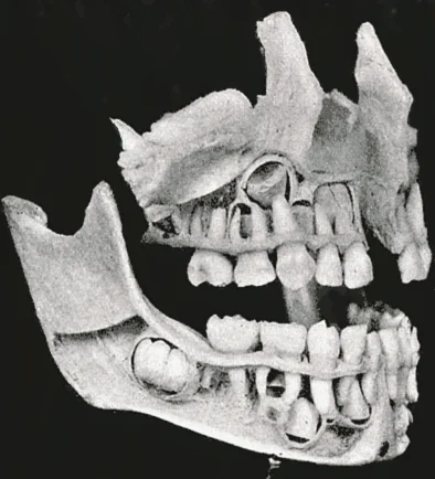
Long description
The complete temporary dentition (about three years), showing the relation of the developing permanent teeth. (Noyes.)

Figure 1: The complete temporary dentition (about three years), showing the relation of the developing permanent teeth (Noyes.).
The oral cavity is lined with stratified squamous epithelium, which can be keratinized or non-keratinized depending on its location. The teeth consist of three main hard tissues: enamel, dentin, and cementum. Enamel is the hardest substance in the human body, composed primarily of hydroxyapatite crystals, and covers the crown of the tooth. Dentin surrounds the pulp cavity and is less mineralized than enamel, containing dentinal tubules that extend from the pulp to the cementum-enamel junction. Cementum covers the root of the tooth and provides attachment for the periodontal ligament. [1]
The periodontium consists of four specialized tissues: gingiva, periodontal ligament, alveolar bone, and cementum. These tissues work together to provide physical and mechanical support for the teeth. The gingiva is keratinized stratified squamous epithelium that protects the underlying bone. The periodontal ligament is a fibrous connective tissue that anchors the tooth to the alveolar bone and contains collagen fibers arranged in specific groups. Alveolar bone is the specialized bone that forms the tooth sockets, and cementum is a bonelike substance that covers the root surface. [3]
Figure 2Gray1004
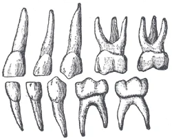
Long description
Deciduous teeth. Left side. Labelled illustration from Wikimedia Commons, used under its open licence.

Figure 2: Deciduous teeth (left side).
Figure 3Gray1002
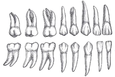
Long description
Permanent teeth. Right side. (Burchard.)

Figure 3: Permanent teeth (right side).
Figure 4Gray1001
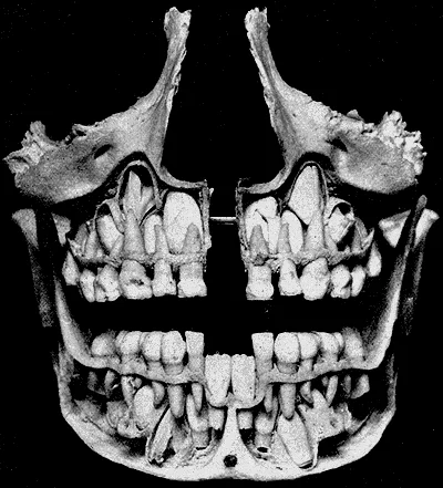
Long description
Front view of the skull shown in Fig. 1000. Note the relation of the permanent incisors and cuspids to each other and the roots of the temporary teeth. (Noyes.)

Figure 4: Front view of the skull shown in Fig (1000).
Figure 5Gray995
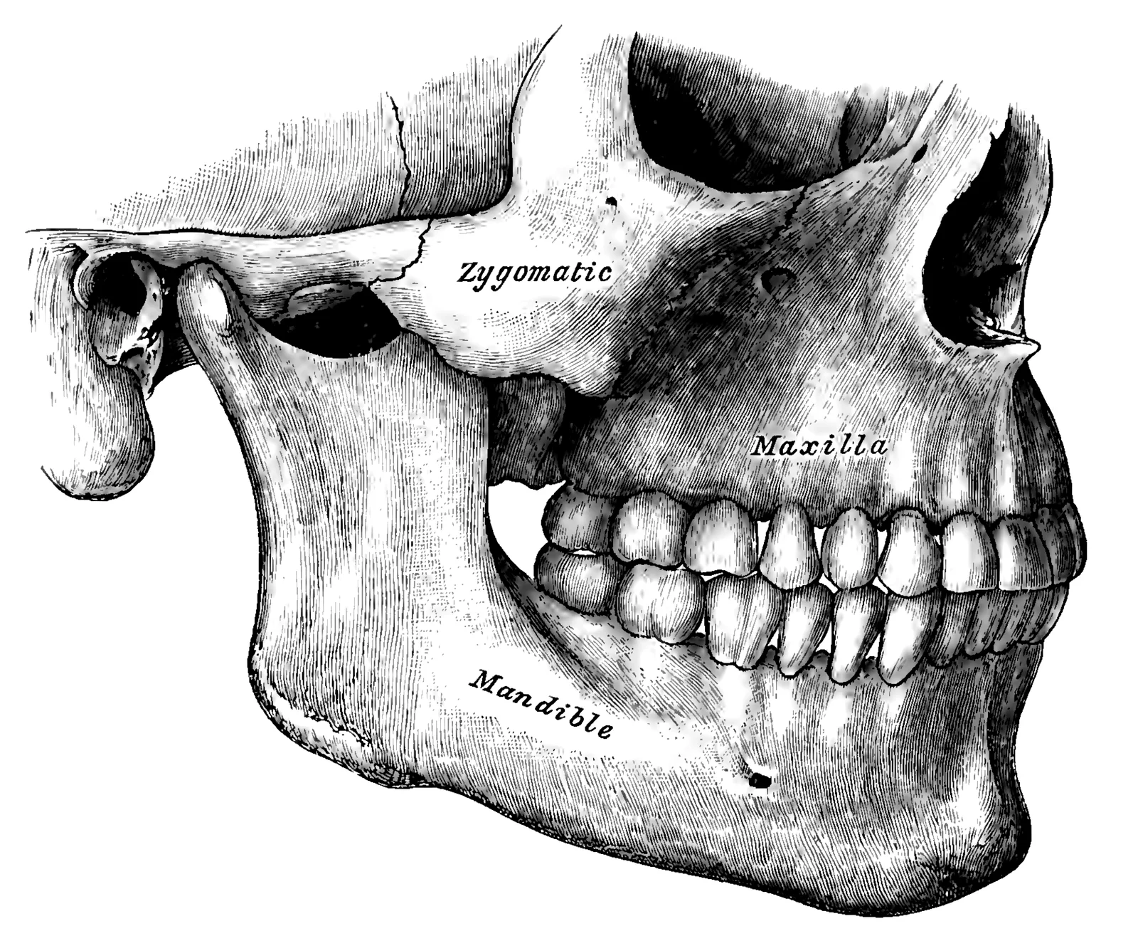
Long description
Side view of the teeth and jaws. Labelled illustration from Wikimedia Commons, used under its open licence.

Figure 5: Side view of the teeth and jaws.
The pulp cavity contains nerves, blood vessels, and connective tissue, and is responsible for dentin formation and tooth nutrition. The odontoblasts lining the pulp cavity are responsible for dentin production, forming dentinal tubules as they lay down dentin matrix. The ameloblasts, located in the inner enamel epithelium, are responsible for enamel formation during tooth development. [1]
The oral cavity also contains numerous minor salivary glands that secrete mucus and serous fluids, aiding in lubrication and digestion. The major salivary glands (parotid, submandibular, and sublingual) produce saliva, which contains enzymes, antibodies, and other protective substances. These antibodies help protect the oral cavity from pathogens and maintain oral health. [1]
Special stains are histological techniques used to highlight specific components of oral cavity and teeth tissues that are not visible with standard hematoxylin and eosin (H&E) staining. These stains are essential for identifying structures such as collagen fibers, basement membranes, microorganisms, and specific extracellular matrix components. [1] [4]
DefinitionSpecial stains
Histological techniques that highlight specific tissue components not visible with standard H&E staining.
In oral histology, special stains help visualize the periodontal ligament, which secures teeth in the alveolar processes of the maxilla and mandible. This connective tissue structure can be specifically stained using Masson's trichrome, which highlights collagen fibers in blue, allowing clear visualization of the ligament fibers that connect the tooth cementum to the alveolar bone.
For examining the gingivae (gums), which line the alveolar processes and surround the necks of the teeth, special stains like Verhoeff's stain can be used to demonstrate elastic fibers. These fibers provide flexibility to the gingival tissue, allowing it to withstand the mechanical forces of mastication.
When studying biofilm formation on oral surfaces, bacterial adhesins can be highlighted using immunohistochemical stains. Many pathogenic oral bacteria possess adhesins that bind to major components of the oral pellicle such as agglutinin, amylase, fibrinogen, fibronectin, or mucins. Specific antibodies against these bacterial proteins can be applied to tissue sections to identify the distribution and density of bacterial colonization. [4]
For examining the interface between dental implants and surrounding tissues, special stains like toluidine blue can highlight the organization of collagen fibers and the presence of inflammatory cells.
The teeth (dentes) function as organs for tearing and grinding food. [1]
The tooth is divided into two primary anatomical regions: the crown and the root. The crown is the portion that projects above the gum line, while the root is embedded within the mandible or maxilla. [1]
DefinitionPulp Cavity
The central space of the tooth containing loose connective tissue, nerves, and blood vessels.
The internal architecture consists of the following layers:
- Pulp Cavity: The innermost core. The specific segment of this cavity extending through the root is termed the root canal. [1]
- Dentin: A bone-like tissue that surrounds the pulp cavity. [1]
- Enamel: The outermost layer of the crown, which covers the dentin and is the hardest substance in the human body. [1]
- Cementum: A layer of modified bone that covers the dentin specifically within the root. [1]
The outermost layer of the crown is the enamel, which meets the gum line. Beneath the enamel lies the dentin, forming the bulk of the tooth. In the centre of the tooth is the pulp cavity, which contains the neurovascular bundle. The pulp cavity narrows as it descends into the root, forming the root canal. Surrounding the dentin in the root area is the cementum, which serves as the interface between the tooth and the supporting alveolar bone. [1]
The tongue is positioned over the floor of the oral cavity. It is anchored to the hyoid bone, the styloid processes of the temporal bones, and the mandible. A medial septum runs the entire length of the tongue, dividing the organ into two symmetrical halves. [1]
NoteHyoid Bone
The hyoid is unique because it only articulates indirectly or distantly with other bones. [1]
The oropharynx wall shares a similar histological structure with the oral cavity, featuring a mucosa composed of a stratified squamous epithelium that contains mucus-producing glands. This epithelial structure provides protection against mechanical stress and pathogens while the mucus glands contribute to lubrication. The remarkable stability of secretory IgA (SIgA) makes it well suited to function in protease-containing secretions such as whole saliva. [1]
The tonsils are part of the Waldeyer's ring and represent a specialized component of the mucosa-associated lymphoid tissue (MALT). They are covered by stratified squamous epithelium that extends into crypts, creating invaginations that increase the surface area for immune surveillance. These crypts are lined with reticular epithelium, which contains specialized antigen-presenting cells that sample antigens from the oral environment. The structural organization of the tonsils supports their function in immune defense by providing a large surface area for antigen sampling while maintaining the barrier function of the mucosal lining.
Beneath the epithelium lies the tonsillar tissue, which consists of lymphoid follicles with germinal centers similar to other lymphoid organs. These follicles are densely packed with lymphocytes, B cells, and T cells, organized to facilitate immune responses. The tonsils also contain numerous plasma cells that produce secretory IgA (SIgA), which plays a crucial role in mucosal immunity.
During swallowing, the elevator skeletal muscles of the pharynx contract, raising and expanding the pharynx to receive the bolus of food. Once received, these muscles relax and the constrictor muscles of the pharynx contract, forcing the bolus into the esophagus and initiating peristalsis. This mechanical action facilitates the sampling of pathogens by the tonsillar tissue while also preventing excessive exposure to potential threats. The tonsils are strategically located at the entrance of the respiratory and dig [1]
DefinitionTonsil structure
The tonsils consist of stratified squamous epithelium extending into crypts, underlying lymphoid follicles with germinal centers, and plasma cells producing SIgA, all positioned to sample antigens from the oral environment.
The stratified squamous epithelium covers the surface and extends into crypts, which increase surface area for antigen sampling. The reticular epithelium lining the crypts contains specialized antigen-presenting cells. Beneath this lies the lymphoid tissue with germinal centers containing B and T lymphocytes. The deep tonsillar tissue contains plasma cells that produce SIgA. The tonsils are positioned at the oropharyngeal junction, allowing them to sample antigens from both respiratory and digestive pathways during swallowing.
When examining histological slides of the oral cavity and teeth, several key structures should be identified. The soft palate is visible as a muscular structure covered by mucosa, with the uvula hanging down from its posterior midline. The uvula appears as a fleshy projection composed of connective tissue and glands, covered by stratified squamous epithelium. Adjacent to the uvula, two muscular folds extend downward: the palatoglossal arch (anterior) and palatopharyngeal arch (posterior). Between these arches, the palatine tonsils appear as clusters of lymphoid tissue embedded in the mucosa.
Figure 6Gray997
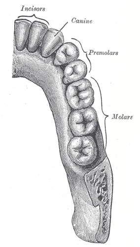
Long description
Permanent teeth of right half of lower dental arch, seen from above.

Figure 6: Permanent teeth of right half of lower dental arch, seen from above.
Figure 7Gray996
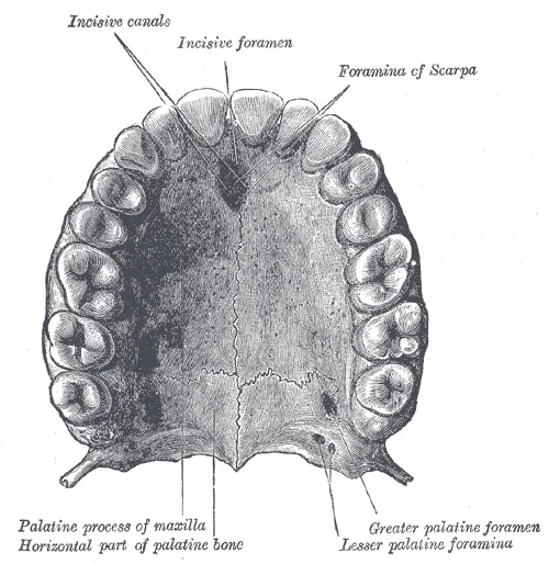
Long description
Permanent teeth of upper dental arch, seen from below.

Figure 7: Permanent teeth of upper dental arch, seen from below.
The tongue is a muscular organ with both extrinsic and intrinsic muscles. Extrinsic muscles include the palatoglossus, hyoglossus, styloglossus, and genioglossus, which originate outside the tongue and insert into its connective tissues. The lingual tonsils are located at the base of the tongue and appear as aggregates of lymphoid tissue.
The teeth consist of several distinct layers. The enamel is the outermost layer, appearing as the most radiopaque structure on histological slides. Dentin surrounds the pulp and appears less radiopaque than enamel. The dental pulp contains blood vessels, nerves, and connective tissue. Cementum covers the root surface and is similar in appearance to bone but lacks Haversian canals.
Figure 8Gray1003
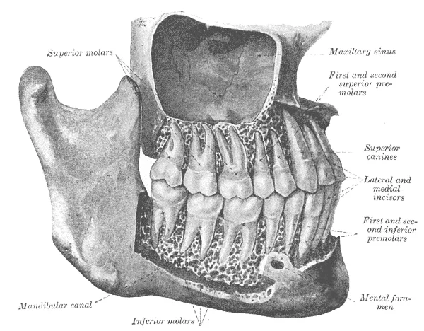
Long description
The permanent teeth, viewed from the right. The external layer of bone has been partly removed and the maxillary sinus has been opened. (Spalteholz.)

Figure 8: The permanent teeth, viewed from the right.
DefinitionSecretory Component
A carbohydrate-rich glycoprotein of approximately 100 kDa constitutively expressed basolaterally on secretory epithelial cells, exhibiting strong non-covalent affinity for J chain-containing pIgA and pentameric IgM. [2]
| Structure | Location | Histological appearance |
|---|---|---|
| Soft palate | Posterior hard palate | Mucosa covering muscle with seromucous glands |
| Uvula | Midline posterior soft palate | Fleshy projection with connective tissue core |
| Palatine tonsils | Between palatoglossal and palatopharyngeal arches | Lymphoid tissue crypts |
| Lingual tonsils | Base of tongue | Lymphoid tissue aggregates |
| Extrinsic tongue muscles | Originating outside tongue | Skeletal muscle fibers with specific orientations |
When examining salivary gland histology, note the presence of secretory epithelial cells that express the polymeric Ig receptor (pIgR), also known as secretory component. The J chain is crucial for binding pIgA and pentameric IgM to this receptor. [2]
The dental structures demonstrate the enamel covering the crown, dentin beneath, and the dental pulp containing blood vessels and nerves.
The oral cavity is lined by a mucous membrane whose epithelial type varies with its functional role. Deep to this epithelium lie connective tissue and the buccinator muscles.
Moving right, the vermilion border marks the transition to the red lip, where the epithelium is still keratinised but thinner, and the underlying dermis is densely packed with capillaries. The orbicularis oris muscle fibres appear in cross-section beneath the mucosa.
The tongue is a muscular organ covered by specialised mucosa. Its dorsal surface bears papillae: filiform papillae are numerous, conical, and keratinised, giving a rough texture; fungiform papillae are scattered among them, mushroom-shaped, with taste buds on their upper surface; circumvallate papillae form a V-shaped row anterior to the sulcus terminalis, each surrounded by a deep trench lined with taste buds. The lingual tonsils lie at the root of the tongue as lymphoid nodules beneath the epithelium.
The teeth are composed of enamel, dentine, cementum, and pulp. Enamel is the hardest tissue in the body, formed by ameloblasts, and is acellular, avascular, and non-innervated. Dentine, produced by odontoblasts, is a mineralised connective tissue traversed by dentinal tubules containing odontoblast processes. Cementum covers the root and anchors the periodontal ligament fibres. The pulp contains blood vessels, nerves, and connective tissue.
Salivary glands are compound tubuloacinar glands. Ducts are lined by cuboidal epithelium, with striated ducts showing basal infoldings and mitochondria, and interlobular ducts lined by stratified cuboidal or columnar epithelium. Inflammatory changes, such as in sialadenitis, show acinar atrophy, fibrosis, and lymphocytic infiltration.
Histopathological correlates of oral disease include leukoplakia, a white patch of hyperkeratosis or epithelial dysplasia that cannot be scraped off; erythroplakia, a red, atrophic area with a high risk of malignant transformation; and squamous cell carcinoma, showing nests of atypical keratinocytes invading the underlying connective tissue. In dental caries, demineralisation of enamel is followed by bacterial invasion of dentine, with inflammation of the pulp (pulpitis) and later periapical abs
- Enamel is susceptible to mechanical and chemical erosion, leading to tooth decay when bacteria release acids that degrade calcium crystals. This process begins when bacterial colonies feeding on sugars in the mouth release acids, causing soft tissue inflammation and degradation of the enamel's calcium crystals. [1]
- The pharynx is a short tube of skeletal muscle lined with mucous membrane, running from the posterior oral and nasal cavities to the esophagus and larynx. It serves as a pathway for both air and food, connecting the upper respiratory and digestive systems. [1]
- The pharynx has three subdivisions: nasopharynx (breathing and speech), oropharynx, and laryngopharynx (both breathing and digestion). The nasopharynx is the most superior portion, while the oropharynx begins inferior to it and continues with the laryngopharynx. [1]
- NALT supplies secretory effector sites of the upper aerodigestive tract with activated pIgA+ precursor cells. This process involves specific homing molecules that differ from those used in the small intestine, creating a dichotomy in immune cell trafficking. [2]
- The leukocyte integrin α4β7 is important for B-cell extravasation into the gut lamina propria but not for homing to airways and salivary glands. This selectivity is due to differences in chemokine receptor-chemokine interactions between these regions. [2]
- At least one fourth of IgA+ PCs in human ileal lamina propria produce polyreactive antibodies that are somatically mutated with signs of antigen-driven selection. This represents a significant portion of the mucosal immune response in the gut. [2]
- Two major antibody classes in saliva are SIgA, synthesized by plasma cells in salivary glands, and IgG, derived from serum by passive diffusion. SIgA provides mucosal immunity, while IgG represents systemic immunity that enters saliva through gingival crevices. [2]
- The periodontium consists of four specialized tissues: gingiva, periodontal ligament, alveolar bone, and cementum. These structures work together to provide physical and mechanical support for the teeth. [3]
- Severe periodontitis leads to loss of periodontal attachment, a major cause of tooth loss in adults. This condition results in the destruction of the supporting structures around the teeth. [3]
- Polished gold alloys accumulate less bacteria compared to natural teeth in vivo, though biofilm composition is comparable. This suggests that material choice may influence bacterial adhesion in dental restorations. [4]
- Biofilm formation on gold, titanium, or zirconium abutment materials does not show major differences in a 14-day in situ study. [4]
Exam trapConfusing types of epithelium
A common mistake is assuming the oral cavity has keratinized epithelium throughout. In fact, the oral mucosa contains non-keratinized stratified squamous epithelium in most areas, which provides protection without the rigidity of keratinization.
Exam trapMisunderstanding salivary immunoglobulin ratios
Students often incorrectly assume IgA is the dominant immunoglobulin in saliva by concentration. While amylase is the most abundant protein in saliva, IgA represents only a minor fraction of total salivary protein. [2]
Exam trapOverestimating IgM's role in saliva
Many students mistakenly believe IgM plays a significant role in salivary defense. In reality, the IgA-to-IgM concentration ratio in parotid secretion is favored by a factor of approximately five (or 12-fold on a molar basis) due to diffusion restrictions for the large IgM pentamer. [2]
Exam trapAssuming salivary IgA directly correlates with systemic disease
A frequent error is believing salivary IgA levels directly indicate susceptibility to caries or periodontal disease. While early studies suggested an inverse relationship between salivary IgA activity and caries susceptibility, the relationship is complex due to compensatory mechanisms like increased SIgM when SIgA is deficient. [2]
Exam trapMisinterpreting bacterial adhesion mechanisms
Students often incorrectly assume bacterial adhesion depends primarily on contact area. In fact, adhesive strength is mainly determined by the amount and nature of contacts between surfaces and macromolecules on the bacterial surface, not the contact area. [4]
Exam trapIgnoring the maturation of the secretory immune system
The secretory immune system undergoes significant maturation, making it particularly difficult to study in young children. [2]
NoteClinical applications of salivary IgA
Knowledge of salivary IgA is clinically relevant in diagnosing conditions like coeliac disease, where IgA antibodies to gluten induced in the gut are reflected as diagnostic IgA titers in whole saliva. [2]
NoteVaccine development
Understanding salivary IgA responses is crucial for developing caries vaccines. Research shows that baseline levels of salivary IgA antibodies to glucan-binding protein in children at 3-4 years of age may predict caries risk, though this relationship becomes clearer in older children. [2]
NoteImmunodeficiency assessment
In patients with primary immunodeficiencies, particularly selective IgA deficiency, assessment of salivary immune components helps understand compensatory mechanisms like increased SIgM that reacts with commensal oral bacteria.
NoteUnderstanding bacterial adhesion
Knowledge of bacterial adhesion mechanisms informs strategies to prevent dental caries and periodontal disease. The Smoluchowski-Levich approximation (j0′=0.538D∞CrhPex1/3) helps understand initial bacterial transport to surfaces, while understanding that surface appendages can pierce energy barriers explains how bacteria establish firm anchorage.
Salivary IgA Assessment in Clinical Practice
- Patient presents with oral symptomsConsider salivary IgA assessment
- Question: Is patient at risk for immunodeficiency?
Yes
- Test for selective IgA deficiency
No
- Consider other diagnostic approaches
- Question: Is patient being evaluated for coeliac disease?
Yes
- Test for IgA antibodies to gluten in saliva
No
- Consider caries risk assessment
- Interpret results in context of age and maturationRemember secretory immune system matures over time
Clinical pearlAge-specific interpretation
When assessing salivary IgA levels, it's essential to consider the patient's age. The secretory immune system undergoes significant maturation, making interpretation particularly complex in young children.
- The oral mucosa contains non-keratinized stratified squamous epithelium, which provides protection without the rigidity of keratinization
- IgA represents only a minor fraction of total salivary protein, with amylase being the most abundant protein
- The IgA-to-IgM concentration ratio in parotid secretion is favored by a factor of approximately five (or 12-fold on a molar basis)
- Salivary IgA antibodies to gluten in patients with coeliac disease provide a diagnostic alternative to blood testing
- Compensatory mechanisms like increased SIgM can mask the effects of SIgA deficiency
- Baseline levels of salivary IgA antibodies to glucan-binding protein may predict caries risk in children
- Bacterial adhesion strength depends on the amount and nature of surface contacts, not contact area
Answer each one in your head first, then open the answer.
What type of epithelium lines most of the oral cavity?
Show answerHide answer
Non-keratinized stratified squamous epitheliumWhich protein is the most abundant in saliva?
Show answerHide answer
AmylaseHow much more concentrated is IgA compared to IgM in parotid secretion on a molar basis?
Show answerHide answer
12-foldWhat determines bacterial adhesive strength to surfaces?
Show answerHide answer
The amount and nature of contacts between surfaces and macromolecules
- Betts JG, Young KA, Wise JA, et al. The Mouth, Pharynx, and Esophagus. OpenStax, Anatomy and Physiology 2e (CC BY-NC-SA 4.0). Accessed .
- Brandtzaeg P. Secretory immunity with special reference to the oral cavity. Journal of oral microbiology 2013;5. doi:10.3402/jom.v5i0.20401. Accessed .
- Li J, Wang Y, Tang M, et al. New insights into nanotherapeutics for periodontitis: a triple concerto of antimicrobial activity, immunomodulation and periodontium regeneration. Journal of nanobiotechnology 2024;22(1):19. doi:10.1186/s12951-023-02261-y. Accessed .
- Sterzenbach T, Helbig R, Hannig C, Hannig M. Bioadhesion in the oral cavity and approaches for biofilm management by surface modifications. Clinical oral investigations 2020;24(12):4237-4260. doi:10.1007/s00784-020-03646-1. Accessed .
Figure credits
- Figure 1: Henry Vandyke Carter, via Wikimedia Commons, Public domain, Public domain.
- Figure 2: Henry Vandyke Carter, via Wikimedia Commons, Public domain, Public domain.
- Figure 3: Henry Vandyke Carter, via Wikimedia Commons, Public domain, Public domain.
- Figure 4: Henry Vandyke Carter, via Wikimedia Commons, Public domain, Public domain.
- Figure 5: Henry Vandyke Carter, via Wikimedia Commons, Public domain, Public domain.
- Figure 6: Henry Vandyke Carter, via Wikimedia Commons, Public domain, Public domain.
- Figure 7: Henry Vandyke Carter, via Wikimedia Commons, Public domain, Public domain.
- Figure 8: Henry Vandyke Carter, via Wikimedia Commons, Public domain, Public domain.
Educational content for medical students and doctors. It is not a substitute for clinical judgement or local guidelines.