Connective tissue
Connective tissue connects, supports and protects body structures.
Connective tissue serves to connect tissues and organs throughout the body, functioning differently from epithelial tissue by having cells dispersed within a matrix rather than being closely packed. The matrix, which is a major component of connective tissue, typically includes a large amount of extracellular material produced by the connective tissue cells embedded within it. This matrix usually consists of a ground substance that is often crisscrossed by protein fibers, and while the ground substance is typically fluid, it can also be mineralized and solid as in bones. [1]
The three broad categories of connective tissue are classified according to the characteristics of their ground substance and the types of fibers found within the matrix: connective tissue proper (including loose and dense connective tissue), supportive connective tissue (bone and cartilage), and fluid connective tissue. [1]
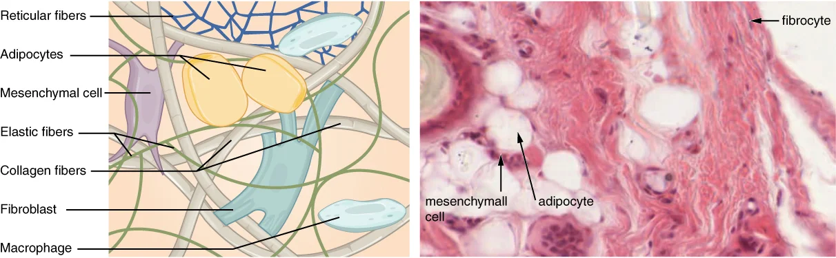
Connective tissue proper includes various cell types such as fibroblasts, fibrocytes, adipocytes, and mesenchymal cells, which are fixed cells that remain within the connective tissue. Other cells like macrophages, mast cells, lymphocytes, plasma cells, and phagocytic cells move in and out of connective tissue in response to chemical signals and are part of the immune system. Mast cells, found in connective tissue proper, contain cytoplasmic granules with histamine and heparin, and when irritated or damaged, they release histamine causing vasodilation, increased blood flow, and inflammatory responses.
Figure 1408 Connective Tissue
Long description
Fibroblasts produce this fibrous tissue. Connective tissue proper includes the fixed cells fibrocytes, adipocytes, and mesenchymal cells. LM × 400. (Micrograph provided by the Regents of University of Michigan Medical School © 2012)

Figure 1: Fibroblasts produce this fibrous tissue.
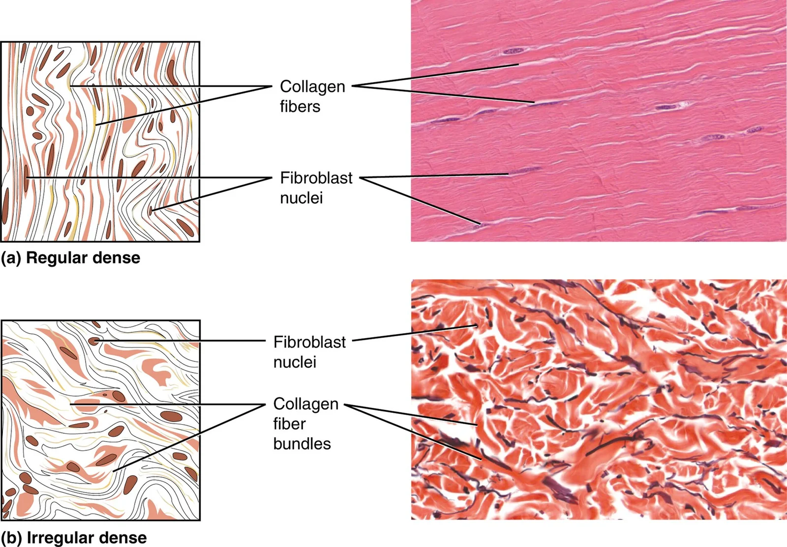
Loose connective tissue has loosely organized fibers with large spaces in between, while dense connective tissue contains more collagen fibers and displays greater resistance to stretching. Dense connective tissue is categorized as regular or irregular, with dense regular having parallel fibers enhancing tensile strength in the direction of fiber orientation, as found in ligaments and tendons. Dense regular elastic tissue contains elastin fibers in addition to collagen, allowing structures like ligaments in the vocal folds and between vertebrae to return to their original length after stretching. [1]
Figure 2411 Reg Dense-Irregular Dense
Long description
(a) Dense regular connective tissue consists of collagenous fibers packed into parallel bundles. (b) Dense irregular connective tissue consists of collagenous fibers interwoven into a mesh-like network. From top, LM × 1000, LM × 200. (Micrographs provided by the Regents of University of Michigan Med

Figure 2: (a) Dense regular connective tissue consists of collagenous fibers packed into parallel bundles.
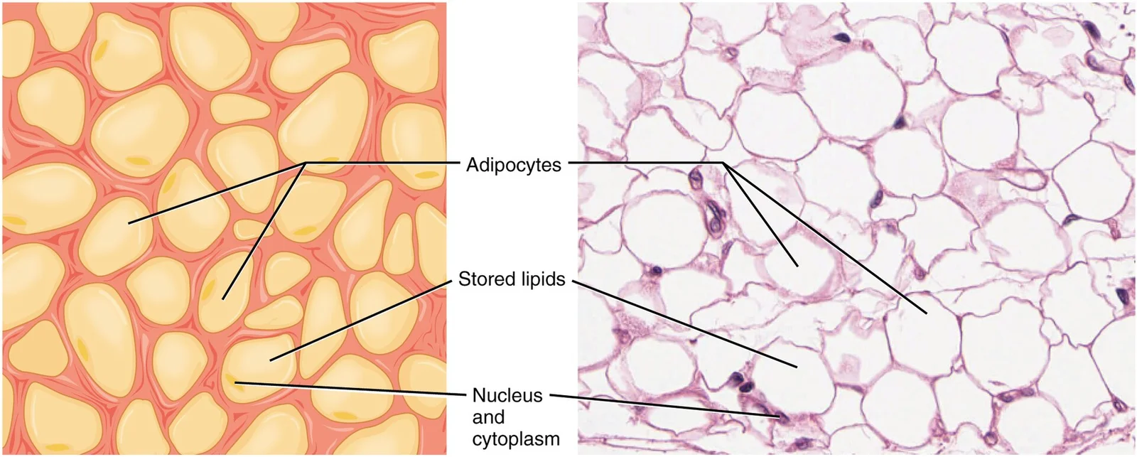
Adipose tissue, a type of loose connective tissue, serves to isolate and store energy reserves and responds dynamically to alterations in calorie excess through adipocyte hypertrophy and hyperplasia[1]. During obesity, macrophages constitute up to 40% of all adipose tissue cells and contribute to obesity-induced inflammation[1]. These macrophages differentiate into proinflammatory M1 macrophages forming "crown-like structures" around necrotic adipocytes and secrete inflammatory cytokines like IL-1β, MCP-1, TNF-α, and IL-6[1]. [3]
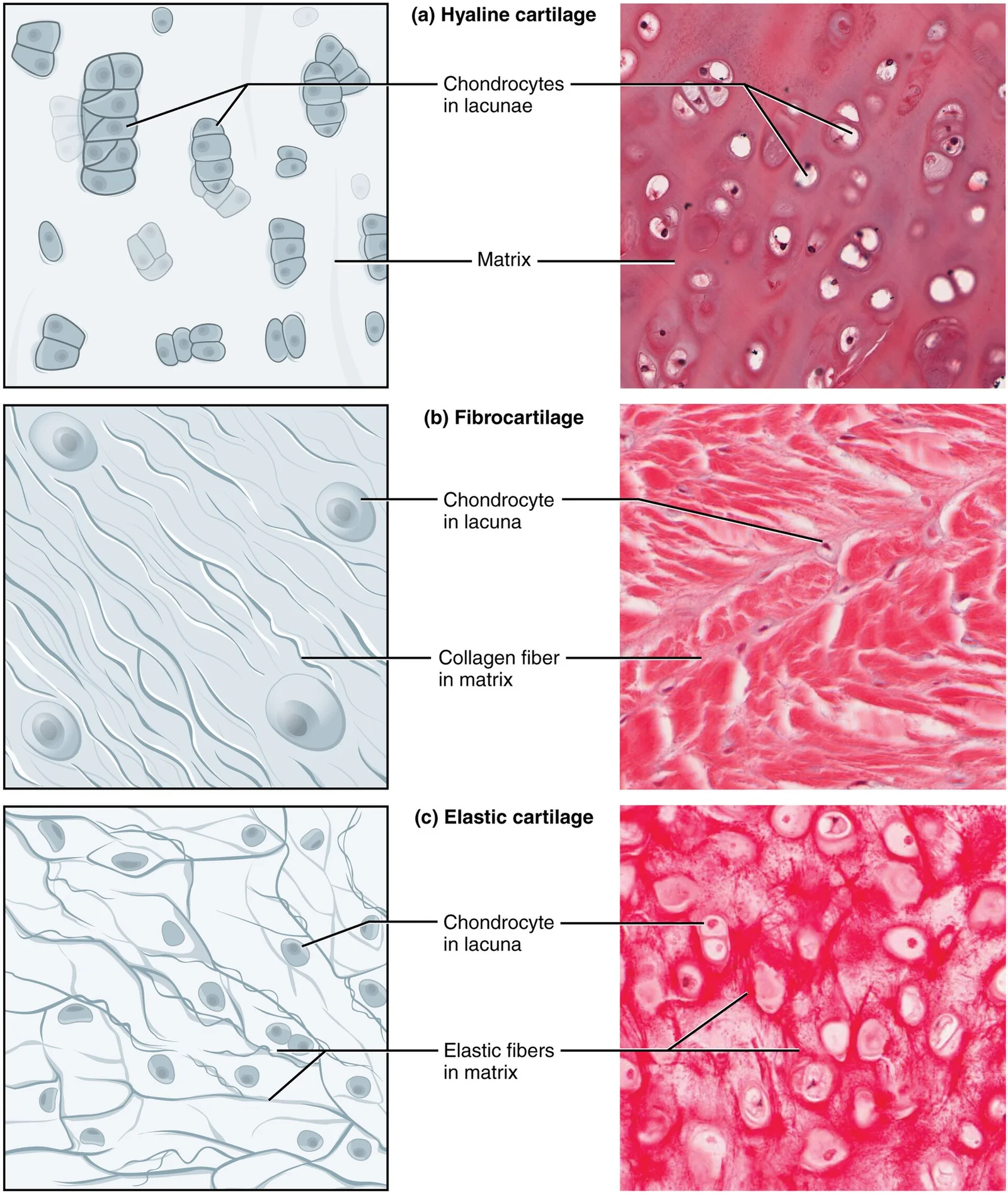
Figure 3412 Types of Cartilage-new
Long description
Cartilage is a connective tissue consisting of collagenous fibers embedded in a firm matrix of chondroitin sulfates. (a) Hyaline cartilage provides support with some flexibility. The example is from dog tissue. (b) Fibrocartilage provides some compressibility and can absorb pressure. (c) Elastic car

Figure 3: Cartilage is a connective tissue consisting of collagenous fibers embedded in a firm matrix of chondroitin sulfates.
Connective tissue is a heterogeneous system where cells are embedded in an extracellular matrix stabilized by proteins. Under light microscopy, the most abundant cell type in connective tissue proper is the fibroblast, which secretes polysaccharides and proteins that combine with extracellular fluids to form a viscous ground substance. A less active form of the fibroblast, the fibrocyte, is the second most common cell type in this tissue. The mesenchymal cell acts as a multipotent adult stem cell capable of differentiating into any type of connective tissue cell needed for repair and healing. The structural variety of connective tissue reflects its diverse functions, including separating and cushioning organs, providing support, and assisting movement. [1]
Figure 4409 Adipose Tissue
Long description
This is a loose connective tissue that consists of fat cells with little extracellular matrix. It stores fat for energy and provides insulation. LM × 800. (Micrograph provided by the Regents of University of Michigan Medical School © 2012)

Figure 4: This is a loose connective tissue that consists of fat cells with little extracellular matrix.
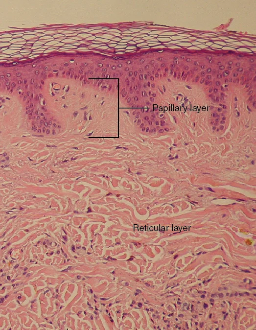
The extracellular matrix consists of a viscous ground substance with embedded fibrous proteins. In dense irregular connective tissue, the direction of fibers is random, which gives the tissue greater strength in all directions. In some tissues, fibers crisscross and form a mesh, while in others, stretching in several directions is achieved by alternating layers where fibers run in the same orientation in each layer, and these layers are stacked at an angle. The dermis of the skin is a prime example of dense irregular connective tissue rich in collagen fibers. Dense irregular elastic tissues provide strength and the ability to regain original shape after stretching, a property evident in arterial walls. [1]
| Cell Type | Function | Structural Feature |
|---|---|---|
| Fibroblast | Most abundant; secretes matrix components | Produces polysaccharides and proteins for ground substance |
| Fibrocyte | Less active form of fibroblast | Second most common cell in connective tissue proper |
| Mesenchymal cell | Multipotent adult stem cell | Differentiates into various connective tissue cells for repair |
Collagen fibers are a major component of the extracellular matrix. In the epimysium, a thick connective tissue layer surrounding the entire muscle, coarse collagen fibers are arranged in a proteoglycan matrix. The arrangement of these fibers varies between muscles of different shapes and functions. For instance, in the relatively thin epimysium of the long strap-like M. sternomandibularis in the cow, two sets of collagen fibers run at approximately 55 degrees to the muscle long axis. In contrast, the thicker epimysium of M. semitendinosus contains close-packed fibers parallel to the muscle long axis, which merge into the tendon. In pennate muscles, the thicker epimysium forms a sheet-like aponeurosis that acts as a wide base of muscle attachment. [4]
Figure 5506 Layers of the Dermis
Long description
This stained slide shows the two components of the dermis—the papillary layer and the reticular layer. Both are made of connective tissue with fibers of collagen extending from one to the other, making the border between the two somewhat indistinct. The dermal papillae extending into the epidermis b

Figure 5: This stained slide shows the two components of the dermis—the papillary layer and the reticular layer.
DefinitionDense Irregular Connective Tissue
A connective tissue where the direction of fibers is random, providing greater strength in all directions and less strength in one particular direction.
NoteEpimysium Structure
The epimysium is a thick connective tissue layer composed of coarse collagen fibers in a proteoglycan matrix that surrounds the entire muscle and defines its volume. [4]
Special stains are essential techniques in histology that allow for the visualization of specific components within connective tissue that are not apparent with standard hematoxylin and eosin staining. These stains highlight particular structures such as collagen fibers, elastic fibers, reticular fibers, and various extracellular matrix components. [3]
DefinitionSpecial stains
Histological techniques that use specific dyes to highlight particular tissue components not visible with standard staining.
One of the most commonly used special stains for connective tissue is Masson's trichrome stain. This stain differentiates between collagen fibers and cytoplasm, making it particularly useful for examining the organization of connective tissue in various organs. The staining process involves several steps, including rinsing the tissue in transport buffer to remove debris and clotted pieces before applying the specific stains. [2]
Another important technique is the Verhoeff-Van Gieson stain, which is particularly valuable for demonstrating elastic fibers. This stain uses a combination of hematoxylin, elastic tissue stain, and picric acid counterstain, resulting in elastic fibers appearing black while collagen stains yellow and nuclei remain black. This distinction is crucial when examining tissues like arteries, lung parenchyma, and ligaments where both fiber types are present.
For connective tissue rich in reticular fibers, such as in lymph nodes, spleen, and bone marrow, the silver impregnation technique (Gomori's method) is employed. This method stains reticular fibers black against a yellow background, highlighting the delicate network that supports these organs.
The tissue sample includes areas of dense irregular connective tissue surrounding blood vessels, with the collagen fibers arranged in multiple directions to provide strength and support.
When examining connective tissue using special stains, it's important to understand the specific properties of each stain and the structures they highlight. This knowledge is essential for accurate interpretation of histological sections and for identifying normal versus pathological changes in connective tissue.
Under electron microscopy, connective tissue reveals a complex arrangement of fibers and ground substance. The distinctive appearance of cartilage is due to polysaccharides called chondroitin sulfates, which bind with ground substance proteins to form proteoglycans[1]. Embedded within the cartilage matrix are chondrocytes, or cartilage cells, and the space they occupy are called lacunae (singular = lacuna)[1]. A layer of dense irregular connective tissue, the perichondrium, encapsulates the cartilage[1].
Cartilaginous tissue is avascular, thus all nutrients need to diffuse through the matrix to reach the chondrocytes[1]. This is a factor contributing to the very slow healing of cartilaginous tissues[1]. In loose connective tissue, which is found between many organs where it acts both to absorb shock and bind tissues together, the structure allows water, salts, and various nutrients to diffuse through to adjacent or embedded cells and tissues[1].
The extracellular matrix (ECM) structures within skeletal muscle play an important role in muscle development, function and adaptation. Each individual muscle is surrounded by epimysial connective tissue and within the muscle there are two distinct extracellular matrix (ECM) structures, the perimysium and endomysium. Together, these three ECM structures make up the intramuscular connective tissue (IMCT). There are large variations in the amount and composition of IMCT between functionally different muscles. [4]
Periodic junctions are formed by branching collagen fiber bundles from the perimysium inserting into the surface of the endomysium. PJPs are staggered at the surface of each muscle fiber and separated by a distance of approximately 300 μm. Transmission electron microscopy and immunohistochemistry studies of PJPs have revealed a concentration of muscle fiber nuclei and mitochondria in the muscle fiber underneath the PJP. This suggests that PJP's may be a point of transmission of mechanical information and stimuli into the muscle fibers which affects expression in the cell (i.e., points of mechanotransduction). [4]
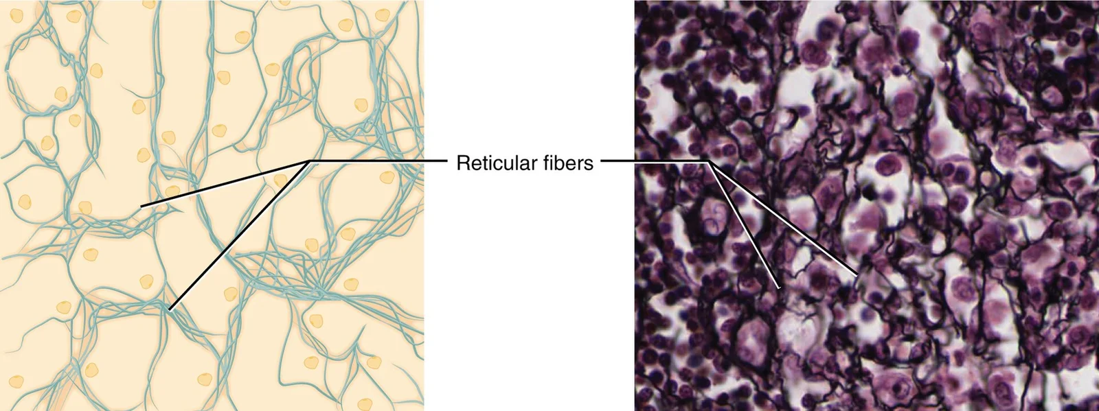
Connective tissue is characterized by its diverse structural components that enable specific functions throughout the body. The fundamental organization includes cells, fibers, and ground substance, each contributing to tissue properties. Reticular tissue forms a mesh-like supportive framework for soft organs such as lymphatic tissue, the spleen, and the liver. Reticular cells produce reticular fibers that create a network onto which other cells attach, deriving its name from the Latin reticulum meaning "little net". [1]
Figure 6410 Reticular Tissue
Long description
This is a loose connective tissue made up of a network of reticular fibers that provides a supportive framework for soft organs. LM × 1600. (Micrograph provided by the Regents of University of Michigan Medical School © 2012)

Figure 6: This is a loose connective tissue made up of a network of reticular fibers that provides a supportive framework for soft organs (lm × 1600).
Intramuscular connective tissue (IMCT) demonstrates a hierarchical organization essential for muscle function. Each muscle is surrounded by an outer extracellular matrix layer called the epimysium. Internally, muscles are divided into fascicles by a continuous network of connective tissue termed perimysium, which connects to the epimysium at the muscle's surface. Within each fascicle, the endomysium forms a continuous network between individual muscle fibers. This organization allows for coordinated force transmission and accommodation of shape changes during muscle contraction. [4]
The mechanical properties of connective tissue depend on its composition and structure. Most muscles undergo shape changes during contraction, with fusiform muscles bulging in mid-section as they contract, similar to fan-shaped and pennate muscles. This shape change requires that certain elements within the tissue can shear past neighboring elements, with shear displacements likely accommodated between fascicles rather than within them. [4]
Connective tissue growth factor (CTGF, CCN2) represents an important regulatory protein in connective tissue biology. As a member of the CCN family, CTGF acts through integrin- and heparan sulfate proteoglycan-mediated adhesive signaling to directly modulate adhesion and indirectly influence the functional activities of other extracellular ligands such as cytokines, growth factors, morphogens, and matrix components. [5]
Connective tissue is characterized by an extracellular matrix and cells that are not in direct contact with each other. The key features include the presence of various cell types embedded in a matrix that can be fluid, gel-like, or solid. Connective tissue proper includes loose (areolar) connective tissue, dense irregular connective tissue, dense regular connective tissue, and adipose tissue. [4]
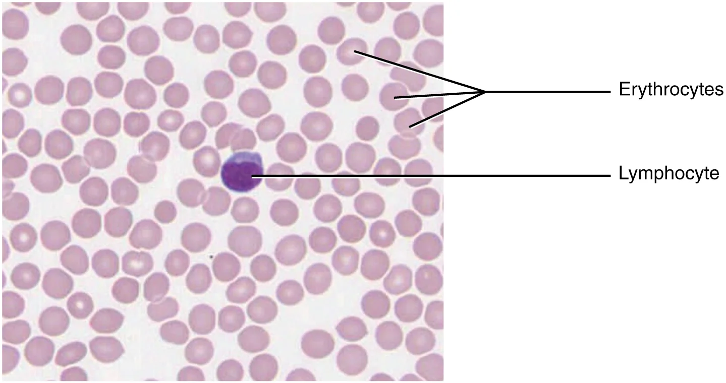
Figure 7424 Blood A Fluid Connective Tissue-new
Long description
Blood is a fluid connective tissue containing erythrocytes and various types of leukocytes that circulate in a liquid extracellular matrix. LM × 1600. (Micrograph provided by the Regents of University of Michigan Medical School © 2012)

Figure 7: Blood is a fluid connective tissue containing erythrocytes and various types of leukocytes that circulate in a liquid extracellular matrix (lm × 1600).
When examining connective tissue under a microscope, look for the following characteristics:
- Cells: Fibroblasts (most common), adipocytes, mast cells, macrophages, and plasma cells
- Fibers: Collagen fibers (white, thick), elastic fibers (yellow, thin), and reticular fibers (fine, branching)
- Ground substance: Amorphous material between cells and fibers
The fibroblasts appear as spindle-shaped cells with elongated nuclei. Elastic fibers, if present, appear as thin, dark-staining fibers that can be stretched. The ground substance appears as a pale, amorphous material surrounding the cells and fibers. [4]
When identifying connective tissue, it's important to distinguish it from other tissue types. Muscle tissue contains elongated cells with striations (skeletal) or spindle-shaped cells with a single nucleus (smooth). Nervous tissue consists of neurons with processes and glial cells. Epithelial tissue consists of cells arranged in sheets with little to no extracellular matrix.
Adipose tissue is a specialized form of connective tissue where adipocytes are the predominant cell type. These cells appear as large, empty circles (due to fat being dissolved during processing) with a thin rim of cytoplasm and a flattened nucleus pushed to the side. [4]
Cartilage, another specialized connective tissue, has a firm matrix with chondrocytes in lacunae. Hyaline cartilage appears as a homogeneous, glassy matrix with chondrocytes often arranged in groups. [1]
Bone tissue, the most rigid connective tissue, shows osteocytes in lacunae within a calcified matrix. Compact bone appears as dense, organized structure with Haversian systems, while cancellous bone has a spongy appearance with trabeculae. [1]
Connective tissue pathologies often present with characteristic histological features that aid in diagnosis and understanding disease mechanisms. In conditions involving excessive scarring and fibrosis, such as systemic sclerosis or keloid formation, the histopathology reveals increased deposition of collagen fibers and alterations in the extracellular matrix. The figure displays a high-power view of the dermis, where thick, eosinophilic collagen bundles are arranged in haphazard patterns rather than the organized parallel bundles seen in healthy tissue. This disorganization contributes to the reduced elasticity characteristic of fibrotic conditions.
The CCN2 protein, also known as connective tissue growth factor, is over-expressed in various connective tissue pathologies, including excessive scarring and fibrosis, as well as in the stroma surrounding tumors. This over-expression appears to selectively mediate or sustain specific actions of TGFβ in mesenchymal cells. The mechanism involves CCN2 decreasing Smad7 availability and increasing Smad2, thereby enhancing TGFβ signaling. However, it's important to note that CCN2-deficient fibroblasts show only a partial impairment of TGFβ responses, with no defect in Smad3-dependent responses. This suggests that while CCN2 plays a significant role in TGFβ-mediated fibrosis, other pathways also contribute to this process. [5]
In the context of adipose tissue, which is a specialized form of connective tissue, histopathological changes are associated with metabolic dysfunction. The accumulation of visceral adipose tissue (VAT) is the strongest predictor of insulin resistance, while subcutaneous adipose tissue (SAT) accumulation correlates with the onset of type 2 diabetes and cardiovascular disease. Histologically, adipose tissue expansion in obesity can occur through hyperplasia (increased number of adipocytes) or hypertrophy (increased size of adipocytes). In hypertrophic obesity, adipocytes become enlarged and release pro-inflammatory cytokines, creating a microenvironment that further impairs adipogenesis and contributes to insulin resistance. [3]
The epigenetic regulation of adipogenesis is another important histopathological consideration. In hypertrophic obese individuals, hypermethylation occurs at CpG dinucleotides within the promoter region of the ZNF423 gene, which correlates with reduced ZNF423 expression in adipocyte precursor cells. This epigenetic modification impairs the differentiation of precursor cells into mature adipocytes, contributing to adipose tissue dysfunction. Conversely, BMP4 causes demethylation of the Zfp423 promoter, which can commit otherwise non-adipogenic cells to the adipogenic lineage. [3]
- Adipocyte Morphology: White adipocytes store lipids as a single large drop, whereas brown adipocytes store lipids as many droplets. [1]
- Metabolic Activity: Brown adipocytes possess high metabolic activity, contrasting with the lower metabolic activity of white fat adipocytes. [1]
- Macrophage Origin: Macrophages are large cells derived from monocytes, a type of blood cell, that enter the connective tissue matrix from blood vessels. [1]
- Cytokine Function: When stimulated, macrophages release cytokines, which are small proteins acting as chemical messengers to recruit other immune cells. [1]
- Macrophage Types: Roaming macrophages move rapidly by amoeboid movement to engulf debris, while fixed macrophages are permanent residents of their tissues. [1]
- Fluid Connective Tissue: Blood and lymph are classified as fluid connective tissues where cells circulate in a liquid extracellular matrix. [1]
- Hematopoietic Origin: The formed elements circulating in blood are all derived from hematopoietic stem cells located in the bone marrow. [1]
- Cell Functions: Erythrocytes transport oxygen, leukocytes defend against microorganisms, and platelets are cell fragments involved in blood clotting. [1]
- Endomysium Structure: The endomysium contains a fibrous network layer estimated to be between 0.2 and 1.0 micrometers in thickness. [4]
- Basement Membrane: Each muscle fiber is bounded by a sarcolemma and an external basement membrane layer that is 50 nanometers thick. [4]
- Perimysium Geometry: Collagen bundles in the perimysium lie at plus 55 degrees and minus 55 degrees to the muscle fiber direction at rest length. [4]
- Force Transfer: Forces are transferred laterally between adjacent muscle fibers by shear through the endomysium, explaining length changes in non-activated fibers. [4]
- Adipocyte Droplets: Do not confuse white and brown adipocytes; white cells have one large drop, while brown cells have many small droplets.
- Macrophage Movement: Distinguish between roaming macrophages that move amoeboidly and fixed macrophages that remain stationary in tissue.
- Blood Components: Remember that platelets are cell fragments, not whole cells, and are specifically involved in clotting.
- Muscle Connective Tissue: Do not confuse the endomysium (around single fibers) with the perimysium (around fascicles); the perimysium has a criss-cross lattice structure.
- Insulin Signaling: In skeletal muscle, diacylglycerols and ceramides can activate PKC epsilon, which phosphorylates IRS1 on serine residues, impairing insulin signaling. [3]
Exam trapMuscle fiber length misconception
A common mistake is assuming that muscle fibers typically run the entire length of a muscle fascicle, inserting onto tendons at both ends. In reality, many muscles have fibers that do not span the entire fascicle. [4]
Exam trapIMCT function misunderstanding
Students often incorrectly believe that intramuscular connective tissue (IMCT) structures primarily work in tension to transmit contractile force. Current understanding shows that their through-plane shear properties are more important than tensile properties. [4]
Exam trapElastin distribution
It's commonly thought that elastin is either absent or present in minimal amounts in muscle connective tissue. In fact, while present in small amounts in most muscles, elastin can be dramatically increased in specific muscles like bovine latissimus dorsi and semitendinosus, where it acts as an elastic energy store. [4]
Exam trapCollagen changes with age
Many students incorrectly assume that increased intramuscular collagen content with aging is simply due to muscle fiber volume loss. While this may contribute, there is evidence of actual increase in collagen concentration and changes in IMCT properties due to age-related covalent crosslinking.
Exam trapConsent form specificity
A frequent error is believing that consent forms for biological samples must be highly specific. Broad consent forms are acceptable and advantageous, as they allow collected samples to be used for multiple research projects not specified at the time of consent. [2]
Exam trapWithdrawal implications
Students often misunderstand what happens when a donor withdraws from a study. The consent form should clearly explain that upon withdrawal, identifiable samples and associated data will be destroyed unless they have already been used for research. [2]
Exam trapBMAd contamination
When working with bone marrow-derived adipocytes (BMAd), it's important to recognize that the enriched fraction contains a significant amount of contaminating hematopoietic and immune cells. Results from this fraction must be interpreted within this context. [2]
NoteClinical examination
Knowledge of connective tissue is essential when examining patients for muscle injuries. The connections within muscle tissue afforded by IMCT and myotendinous joint connections are predominant sites of lesions in muscle strain injuries, as highlighted in recent meta-analyses. [4]
NoteSurgical planning
In the operating theatre, understanding the continuity of connections between IMCT and fascia helps surgeons coordinate the action of agonistic muscles during procedures. This knowledge is particularly important when reconstructing damaged muscle tissue or planning tendon transfers.
NoteImaging interpretation
When interpreting imaging studies, radiologists must understand the mechanical properties of skeletal muscle, which can be described by a contractile element (CE), an elastic element in series with this (SE), and another elastic component in parallel to the CE and SE. This "Hill-type three-element model" helps explain the stress-strain behavior of muscle.
NoteResearch consent
In clinical research, proper consent forms for biological samples must clearly state that donors can withdraw at any point and specify acceptable withdrawal methods. The forms should also explain that identifiable samples and data will be destroyed upon withdrawal unless already used for research.
Muscle Injury Assessment
- Patient presents with muscle injuryHistory and physical examination
- Question: Is there tenderness at myotendinous junction or IMCT connections?
Yes
- Suspect strain injuryIMCT/myotendinous junction involvement likely
- Consider imagingUltrasound or MRI to assess extent
No
- Consider other diagnosesDirect trauma, compartment syndrome, etc.
- Treatment planningBased on location and extent of injury
Clinical pearlAging considerations
In geriatric practice, understanding age-related changes in connective tissue is crucial. After maturity, there is an increase in intramuscular collagen content and changes in IMCT properties due to covalent crosslinking, which affects muscle elasticity and function.
Clinical pearlEnergy storage mechanisms
In sports medicine, recognizing that elastin in certain muscles acts as an elastic energy store helps explain the biomechanics of activities requiring rapid lengthening and shortening of muscles, such as running and jumping.
- Many muscle fibers do not span the entire fascicle length, terminating either on tendinous insertions or within the fascicle itself
- Intramuscular connective tissue (IMCT) structures primarily function through shear properties rather than tension for force transmission
- Elastin can be dramatically increased in specific muscles where it acts as an elastic energy store
- The Hill-type three-element model (CE, SE, PE) helps describe the mechanical properties of skeletal muscle
- IMCT and myotendinous joint connections are predominant sites of lesions in muscle strain injuries
- Broad consent forms allow biological samples to be used for multiple research projects
- Donors can withdraw from studies at any point, with identifiable samples and data destroyed unless already used
- BMAd-enriched fractions contain significant contaminating hematopoietic and immune cells
Answer each one in your head first, then open the answer.
What is a common misconception about muscle fiber length in muscle fascicles?
Show answerHide answer
Many muscle fibers do not span the entire fascicle lengthWhat property of IMCT structures is now considered more important than tensile properties?
Show answerHide answer
Through-plane shear propertiesWhere is elastin dramatically increased in certain muscles and what is its function?
Show answerHide answer
In muscles like bovine latissimus dorsi and semitendinosus, where it acts as an elastic energy storeWhat are the predominant sites of lesions in muscle strain injuries?
Show answerHide answer
The connections within muscle tissue afforded by IMCT and myotendinous joint connections
- Betts JG, Young KA, Wise JA, et al. Connective Tissue Supports and Protects. OpenStax, Anatomy and Physiology 2e (CC BY-NC-SA 4.0). Accessed .
- Lucas S, Tencerova M, von der Weid B, et al. Guidelines for Biobanking of Bone Marrow Adipose Tissue and Related Cell Types: Report of the Biobanking Working Group of the International Bone Marrow Adiposity Society. Frontiers in endocrinology 2021;12:744527. doi:10.3389/fendo.2021.744527. Accessed .
- Longo M, Zatterale F, Naderi J, et al. Adipose Tissue Dysfunction as Determinant of Obesity-Associated Metabolic Complications. International journal of molecular sciences 2019;20(9):E2358. doi:10.3390/ijms20092358. Accessed .
- Purslow PP. The Structure and Role of Intramuscular Connective Tissue in Muscle Function. Frontiers in physiology 2020;11:495. doi:10.3389/fphys.2020.00495. Accessed .
- Leask A, Parapuram SK, Shi-Wen X, Abraham DJ. Connective tissue growth factor (CTGF, CCN2) gene regulation: a potent clinical bio-marker of fibroproliferative disease?. Journal of cell communication and signaling 2009;3(2):89-94. doi:10.1007/s12079-009-0037-7. Accessed .
Figure credits
- Figure 1: OpenStax College, via Wikimedia Commons, CC BY 3.0, CC BY 3.0.
- Figure 2: OpenStax College, via Wikimedia Commons, CC BY 3.0, CC BY 3.0. resized.
- Figure 3: OpenStax College, via Wikimedia Commons, CC BY 3.0, CC BY 3.0. resized.
- Figure 4: OpenStax College, via Wikimedia Commons, CC BY 3.0, CC BY 3.0. resized.
- Figure 5: OpenStax College, via Wikimedia Commons, CC BY 3.0, CC BY 3.0.
- Figure 6: OpenStax College, via Wikimedia Commons, CC BY 3.0, CC BY 3.0. resized.
- Figure 7: OpenStax College, via Wikimedia Commons, CC BY 3.0, CC BY 3.0.
Educational content for medical students and doctors. It is not a substitute for clinical judgement or local guidelines.