Epithelial tissue
Epithelial tissue forms protective linings and glands throughout the body.
Epithelial tissue is highly cellular with little or no extracellular material present between cells. Adjoining cells form specialized intercellular connections called cell junctions. Epithelial cells exhibit polarity with differences between the apical surface and basal surface. The basal lamina, a mixture of glycoproteins and collagen, provides an attachment site for the epithelium, separating it from underlying connective tissue.
Nutrients must come by diffusion or absorption from underlying tissues or the surface. Many epithelial tissues are capable of rapidly replacing damaged and dead cells.
Cells of epithelia are closely connected and are not separated by extracellular material. Three basic types of connections allow varying degrees of interaction between cells: tight junctions, anchoring junctions, and gap junctions. [1]
DefinitionEpithelial polarity
The structural and functional differences between the apical and basal surfaces of epithelial cells.
Epithelial tissues are classified by cell shape and arrangement. The three basic cell shapes are squamous (thin and scale-like), cuboidal (cube-shaped), and columnar (tall and rectangular). Single cell layers form simple epithelia, whereas stacked cells form stratified epithelia. [1]
| Type | Description | Location |
|---|---|---|
| Simple squamous | Single layer of thin, flat cells | Alveoli of lungs, kidney tubules, lining of capillaries |
| Simple cuboidal | Single layer of cube-shaped cells | Kidney tubules, glandular ducts |
| Simple columnar | Single layer of tall, rectangular cells | Digestive system, female reproductive tract |
| Stratified squamous | Multiple layers with apical squamous cells | Skin (keratinized), mouth cavity (un-keratinized) |
| Stratified cuboidal | Multiple layers with apical cuboidal cells | Glands, ducts |
| Stratified columnar | Multiple layers with apical columnar cells | Glands, ducts |
| Pseudostratified | Single layer appearing stratified | Respiratory tract [1] |
| Transitional | Specialized stratified that stretches | Urinary bladder |
NoteSurface specializations
Epithelial cells may have microvilli (finger-like projections for absorption), cilia (hair-like structures for movement), or stereocilia (long microvilli found in the male reproductive system).
The main functions of epithelia are protection from the environment, coverage, secretion and excretion, absorption, and filtration. Cells are bound together by tight junctions that form an impermeable barrier. Gap junctions allow free exchange of soluble molecules between cells, while anchoring junctions attach cell to cell or cell to matrix. [1]
Epithelial renewal is a critical process where damaged or dead cells are replaced. In the intestinal epithelium, this process can be affected by energy deficiency diseases like ulcerative colitis, where diminished butyrate oxidation leads to alterations in mucus synthesis and crypt cell maturation[1]. Mitochondrial dysfunction in intestinal epithelial cells is associated with reduced stemness and Paneth cell dysfunction[1]. [2]
Metaplasia is the conversion of one type of epithelial tissue to another, often in response to chronic irritation. Dysplasia refers to abnormal changes in epithelial cell size, shape, and organization that may precede malignant transformation. Carcinoma is malignant cancer that originates in epithelial tissue.
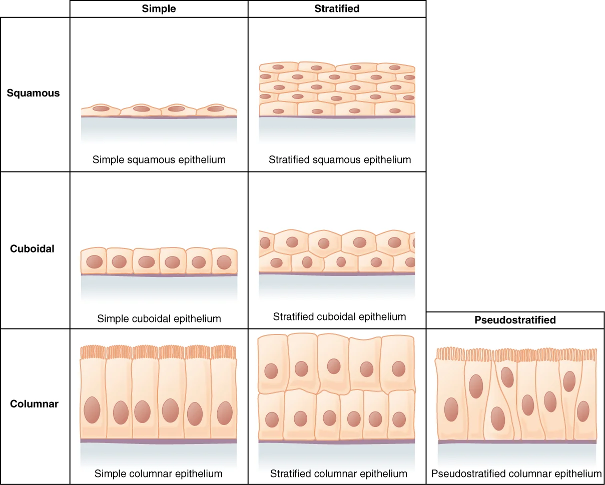
Figure 1403 Epithelial Tissue
Long description
Simple epithelial tissue is organized as a single layer of cells and stratified epithelial tissue is formed by several layers of cells.

Figure 1: Simple epithelial tissue is organized as a single layer of cells and stratified epithelial tissue is formed by several layers of cells.
Epithelial tissues are classified according to the shape of the cells and number of cell layers formed. Cell shapes can be squamous (flattened and thin), cuboidal (boxy, as wide as it is tall), or columnar (rectangular, taller than it is wide). Similarly, the number of cell layers in the tissue can be one—where every cell rests on the basal lamina—which is a simple epithelium, or more than one, which is a stratified epithelium and only the basal layer of cells rests on the basal lamina. Pseudostratified (pseudo- = "false") describes tissue with a single layer of irregularly shaped cells that give the appearance of more than one layer. Transitional describes a form of specialized stratified epithelium in which the shape of the cells can vary. [1]
| Cell Shape | Description | Arrangement |
|---|---|---|
| Squamous | Flattened and thin | Simple or stratified |
| Cuboidal | Boxy, as wide as it is tall | Simple or stratified |
| Columnar | Rectangular, taller than it is wide | Simple, pseudostratified, or stratified |
| Transitional | Variable shape | Stratified |
Both simple and pseudostratified columnar epithelia are heterogeneous epithelia because they include additional types of cells interspersed among the epithelial cells. For example, a goblet cell is a mucus-secreting unicellular "gland" interspersed between the columnar epithelial cells of mucous membranes. [1]
Epithelial cell junctions are specialized structures that connect adjacent cells and provide stability and selective barrier functions. At one end of the spectrum is the tight junction, which separates the cells into apical and basal compartments. When two adjacent epithelial cells form a tight junction, there is no extracellular space between them and the movement of substances through the extracellular space between the cells is blocked. This enables the epithelia to act as selective barriers. [1]
Anchoring junctions include several types of cell junctions that help stabilize epithelial tissues. Anchoring junctions are common on the lateral and basal surfaces of cells where they provide strong and flexible connections. There are three types of anchoring junctions: desmosomes, hemidesmosomes, and adherens. Desmosomes occur in patches on the membranes of cells. The patches are structural proteins on the inner surface of the cell's membrane. The adhesion molecule, cadherin, is embedded in these patches and projects through the cell membrane to link with the cadherin molecules of adjacent cells. These connections are especially important in holding cells together. [1]
Hemidesmosomes, which look like half a desmosome, link cells to the extracellular matrix, for example, the basal lamina. While similar in appearance to desmosomes, they include the adhesion proteins called integrins rather than cadherins. Adherens junctions use either cadherins or integrins depending on whether they are linking to other cells or matrix. The junctions are characterized by the presence of the contractile protein actin located on the cytoplasmic surface of the cell membrane. The actin can connect isolated patches or form a belt-like structure inside the cell. These junctions influence the shape and folding of the epithelial tissue. [1]
The tight junctions appear as points of close contact between adjacent cells near the apical surface, forming a seal that prevents substances from passing between cells. Adherens junctions appear as belt-like structures or patches along the lateral surfaces, containing actin filaments that help maintain cell shape and tissue organization.
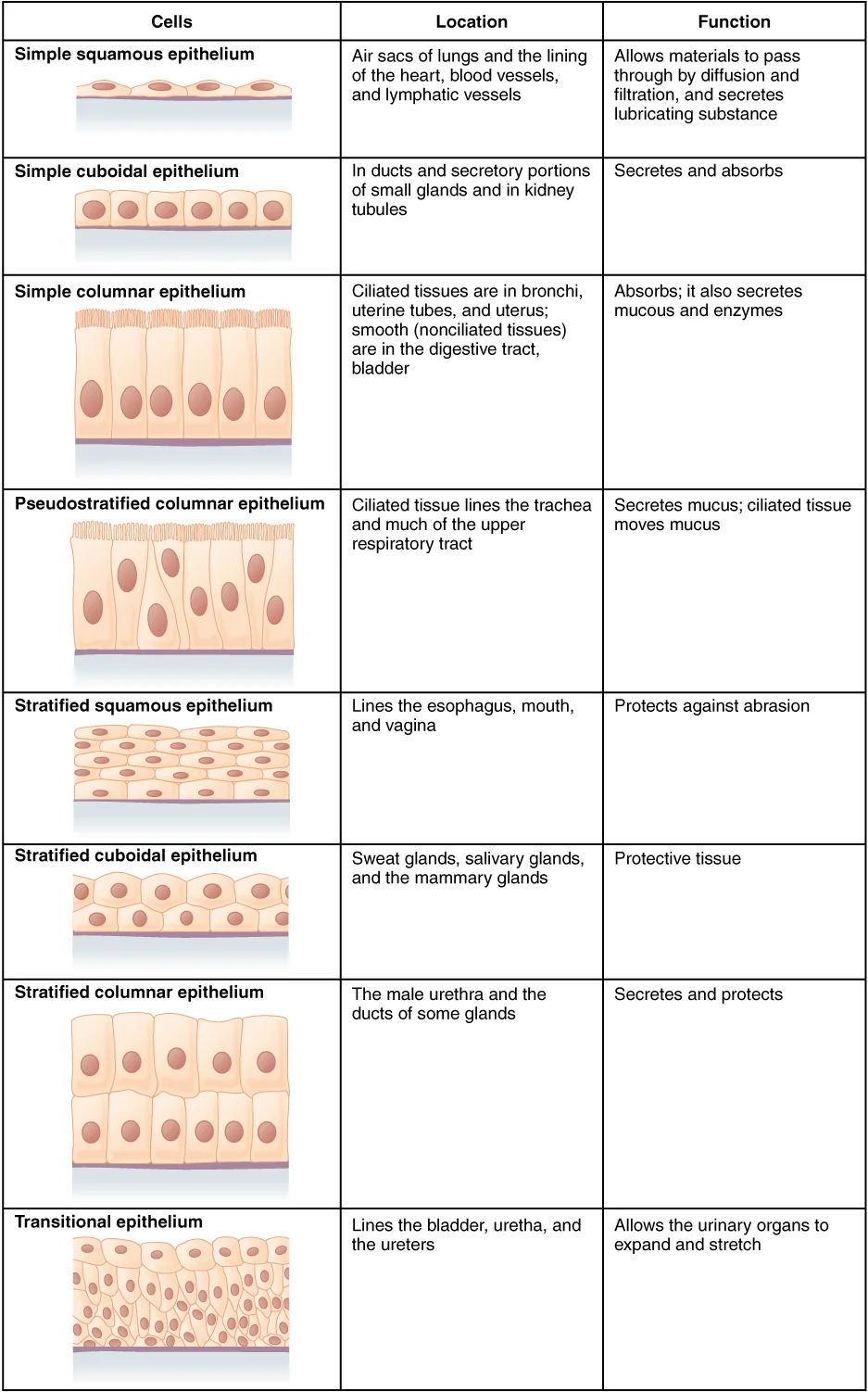
Figure 2423 Table 04 02 Summary of Epithelial Tissue CellsN
Long description
423_Table_04_02_Summary_of_Epithelial_Tissue_CellsN.jpg

Figure 2: 423_Table_04_02_Summary_of_Epithelial_Tissue_CellsN.jpg.
Special stains are histological techniques that highlight specific components of epithelial tissue that are not visible with standard hematoxylin and eosin (H&E) staining. These stains are essential for identifying different types of epithelial cells and their specialized structures.
DefinitionSpecial Stain
A histological technique that uses specific dyes to highlight particular cellular components or tissue elements not visible with standard staining.
One important application of special stains is the visualization of cilia on the apical surface of certain epithelial cells. Cilia are hair-like projections that extend from the apical surface of epithelial cells and are involved in movement or sensory functions. Special stains such as silver impregnation or immunohistochemistry can highlight these structures, allowing for their identification and characterization.
Another important use of special stains is in identifying different types of epithelial cells based on their metabolic characteristics. For example, in the intestinal epithelium, special stains can help identify cells with different metabolic profiles along the crypt-villus axis. Crypt cells, including stem cells and transit amplifying cells, primarily rely on glycolysis, while differentiated cells show increased dependence on mitochondrial oxidative phosphorylation. Special stains for mitochondrial enzymes or metabolic markers can help visualize these differences. [2] [1]
| Stain | Target | Application |
|---|---|---|
| PAS (Periodic Acid-Schiff) | Glycogen, basement membrane | Highlights glycogen in epithelial cells and basement membrane |
| Masson's Trichrome | Collagen, cytoplasm | Differentiates collagen (blue/green) from epithelial cytoplasm (red) |
| Alcian Blue | Acidic mucopolysaccharides | Identifies goblet cells in epithelium |
| Silver Impregnation | Cilia, nerve fibers | Highlights ciliated epithelial surfaces |
| Immunohistochemistry | Specific proteins | Identifies epithelial cell types based on protein expression |
The cilia appear as dark, hair-like projections extending from the apical surface of the epithelial cells. These cilia are located on the free surface of the epithelium and are responsible for moving mucus and trapped particles away from the underlying tissue. The basal surface of the epithelium, which rests on the basement membrane, appears lighter in comparison.
Epithelial tissue forms the continuous lining of body surfaces and cavities, serving as a critical interface between the internal environment and the external world. Its primary function is to provide a smooth and protective surface. This protective role is evident in mesothelial cells, which are squamous epithelial cells that secrete a fluid that lubricates the mesothelium. Such lubrication reduces friction in moving organs, allowing for efficient physiological movement without tissue damage. [1]
Beneath the microvilli lies the terminal web, a network of actin filaments that anchors these projections. The lateral membrane features tight junctions near the apex, sealing the intercellular space to maintain barrier integrity. Desmosomes are located slightly below the tight junctions, providing mechanical adhesion between adjacent cells. Basal infoldings increase the surface area for transport processes. The nucleus is centrally located, with mitochondria distributed throughout the cytoplasm to support high metabolic demands.
The intestinal epithelium is a multicellular interface located in close proximity to a complex and dense microbial milieu. Simultaneously intestinal epithelial cells absorb nutrients, and form a physical and immune-mediated barrier against adverse components of the luminal environment. This barrier function is paramount, as the intestinal epithelium represents the frontline of the complex pathogenesis, lying at the interface of luminal inflammatory triggers such as the microbiome and host immune cells. A breach of this well-structured barrier is suggested as cornerstone of chronic inflammation. Consequently, episodes of tissue injury and incomplete healing of the intestinal epithelium are a prerequisite for immune reactivation and account for recurrent, chronically progressing phenotypes of IBD. [2]
DefinitionEpithelial Polarity
Epithelial cells exhibit distinct apical, lateral, and basal domains, each specialized for specific functions such as absorption, adhesion, and anchorage.
Clinical pearlRegenerative Capacity
The intestinal epithelium renews itself every 3–5 days, a rate unmatched by most other tissues in the body.
| Cell Type | Location | Primary Function |
|---|---|---|
| Enterocytes | Small and Large Intestine | Nutrient absorption and barrier formation |
| Goblet Cells | Entire Digestive Tract | Mucus secretion for lubrication and protection |
| Paneth Cells | Small Intestine Crypts | Antimicrobial defence and stem cell support |
| Mesothelial Cells | Body Cavities | Secretion of lubricating fluid |
This redundancy ensures that the epithelial barrier can be restored even after significant damage. The structural integrity of the epithelium is thus maintained not only by its cellular composition but also by its remarkable ability to regenerate from multiple stem cell pools. [2]
Epithelial tissue forms the fundamental lining of body surfaces and cavities, serving as a selective barrier between the internal environment and the external world. Its primary function is protection, absorption, secretion, and sensation. The structural integrity of this tissue is paramount; a breach in barrier integrity is associated with inflammatory pathologies, and requires immediate action to restore barrier function and to induce mucosal healing. [2]
A significant functional specialization of epithelial tissue is the formation of glands. A gland is a structure made up of one or more cells modified to synthesize and secrete chemical substances. Most glands consist of groups of epithelial cells. These glands are classified based on their mode of secretion and their structural arrangement. [1]
In contrast, an exocrine gland is one whose secretions leave through a duct that opens directly, or indirectly, to the external environment (exo- = “outside”). [1]
The mechanism of secretion further distinguishes exocrine glands. Merocrine glands secrete products as they are synthesized. Apocrine glands release secretions by pinching off the apical portion of the cell. Holocrine gland cells store their secretions until they rupture and release their contents. In this case, the cell becomes part of the secretion. [1]
The functional capacity of epithelial cells, particularly in the intestine, is tightly regulated by mitochondrial metabolism. Not only mitochondrial metabolism, but also mitochondrial proteostasis and the associated mitochondrial unfolded protein response (UPRmt) contribute to these processes and are involved in intestinal pathologies by controlling self-renewal and the proliferative capacity of the epithelium. This is not only critical to maintain epithelial homeostasis, but also crucial during inflammation and for dedifferentiation processes associated with wound healing. [2]
For example, mitochondria contribute to inflammatory processes by production of ROS.
Clinical pearlMetabolic Interface
Epithelial metabolism acts as an interface for microbiome and immune cell-derived signals, highlighting its potential as a therapeutic target.
Immune cells and their cytokines reciprocally regulate IECs metabolism and function.
The apical surface faces the lumen. The basal surface rests on the basement membrane. Mitochondria are abundant in the basal cytoplasm. This supports high energy demands. The structure enables selective transport.
| Gland Type | Secretion Mechanism | Example |
|---|---|---|
| Merocrine | Secrete products as synthesized | Salivary glands |
| Apocrine | Pinch off apical portion | Mammary glands |
| Holocrine | Cell ruptures to release content | Sebaceous glands |
To identify epithelial tissue on a histological slide, you must first recognise its fundamental architecture. This includes the airways, the digestive tract, and the urinary and reproductive systems. A key identifying feature is the presence of a basement membrane, which anchors the epithelium to the underlying connective tissue. The cells are tightly packed with minimal extracellular matrix, creating a continuous barrier. [1]
DefinitionEpithelium
Epithelial tissues provide the body’s first line of protection from physical, chemical, and biological wear and tear. [1]
When examining a slide, look for the polarity of the cells. The apical surface faces the lumen or external environment, while the basal surface rests on the basement membrane. You may observe specialized structures on the apical surface. For instance, cilia are microscopic extensions of the apical cell membrane that are supported by microtubules. These cilia beat in unison and move fluids as well as trapped particles. In the airway, this forms a mucociliary escalator that sweeps particles of dust and pathogens trapped in the secreted mucus toward the throat. [1]
Identifying Epithelial Types
- Identify Cell ShapeLook at the apical surface
- Question: Is it stratified?
Yes
- Check Apical Cell ShapeIf flat, it is stratified squamous; if dome-shaped, consider transitional
No
- Check for Cilia or MicrovilliCilia indicate respiratory or ventricular epithelium
The cells are arranged in a single layer, confirming it is simple epithelium. The nuclei are oval and aligned in a row, further supporting this identification.
To distinguish it, note that endothelium lines vascular spaces, whereas epithelium lines external or internal body surfaces. Another look-alike is mesothelium, which lines body cavities. [1]
Clinical pearlKey Distinction
Remember that epithelium covers external surfaces and lines internal tracts, while endothelium lines blood vessels.
In glandular epithelium, look for secretory units. Holocrine secretion involves the rupture and destruction of the entire gland cell. The cell accumulates its secretory products and releases them only when it bursts. New gland cells differentiate from cells in the surrounding tissue to replace those lost by secretion. The sebaceous glands that produce the oils on the skin and hair are holocrine glands/cells. [1]
Epithelial tissue is defined by its cellular origin and functional specialisation, which directly informs its histological appearance. Epithelial cells derive from all three major embryonic layers[1]. The epithelia lining the skin, parts of the mouth and nose, and the anus develop from the ectoderm[1]. Cells lining the airways and most of the digestive system originate in the endoderm[1]. The epithelium that lines vessels in the lymphatic and cardiovascular system derives from the mesoderm and is called an endothelium[1]. This distinction is critical when identifying tissue type in cross-sections.
DefinitionEndothelium
The specific name for the simple squamous epithelium lining the interior surface of blood vessels and lymphatic vessels, derived from the mesoderm.
Paneth cells contribute to stem cell homeostasis and anti-microbial defence exclusively in the small intestine, while goblet cells are spread throughout the entire epithelial lining of the digestive tract. [2]
Clinical pearlSecretory Polarity
The apical surface faces the lumen or external environment, while the basal surface attaches to the basement membrane. Secretory products are released exclusively from the apical domain.
Exocrine glands release their contents through a duct that leads to the epithelial surface[1]. Mucous, sweat, saliva, and breast milk are all examples of secretions from exocrine glands[1]. They are all discharged through tubular ducts[1]. Secretions into the lumen of the gastrointestinal tract, technically outside of the body, are of the exocrine category[1]. Histologically, exocrine glands are identified by their ductal systems connecting the secretory acini or tubules to the epithelial surface.
| Type | Example | Duct Structure |
|---|---|---|
| Exocrine | Mucous, Sweat, Saliva | Tubular ducts leading to surface |
| Endocrine | Hormones | No ducts; released into blood |
The central lumen is lined by columnar cells with basal nuclei. Apical cytoplasm contains secretory granules. Surrounding the epithelium is the lamina propria, containing connective tissue and immune cells. The basement membrane separates the epithelium from the underlying stroma.
Exam trapLumen vs Stroma
Do not confuse the glandular lumen (secretory space) with the interstitial stroma. The lumen is continuous with the external environment in exocrine glands.
Understanding these structural correlates allows for accurate diagnosis. The embryonic origin predicts the tissue's response to injury and its potential for regeneration. Secretory function determines the histological staining characteristics, such as mucicarmine for mucin or PAS for glycogen. The presence of ducts confirms exocrine function. These features are consistent across species and are fundamental to medical histology.
DefinitionSerous Glands
Glands named after their product; serous glands produce watery, plasma-like secretions rich in enzymes such as alpha amylase. [1]
Serous glands are characterised by watery, blood-plasma-like secretions that are rich in enzymes, specifically alpha amylase. These are frequently found in the salivary glands of the mouth.
DefinitionMucous Glands
Mucous glands release watery to viscous products rich in the glycoprotein mucin. [1]
In contrast, mucous glands release products ranging from watery to viscous, which are rich in the glycoprotein mucin. Like serous glands, they are common in the oral salivary glands. [1]
DefinitionMixed Glands
Mixed exocrine glands contain both serous and mucous components, releasing both types of secretions.
Mixed exocrine glands contain both serous and mucous elements and consequently release both types of secretions. This combination is also observed in the salivary glands. [1]
These progenitor cells eventually differentiate into mature secretory and absorptive cells.
Exam trapOlfm4 Specificity
Olfm4 is an alternative marker for highly proliferative intestinal stem cells, co-expressed with but not restricted to Lgr5+ stem cells. [2]
Its numbers expand during mucosal healing and it is implicated in colorectal cancer.
It encodes a putative secreted glycoprotein involved in these pathological changes.
Intestinal epithelial cells are highly adapted to their anatomical location.
This highlights the metabolic distinction between small and large intestine epithelia.
During inflammation, the need for nucleotide substrates increases substantially. Supplementation of hypoxanthine or selective colonization with purine-producing bacteria improved cellular energetics, promoted mucus generation and epithelial barrier function. [2]
Activated type I receptors phosphorylate intracellular transcriptional regulators.
NoteSmad Complex
Activated R-Smads form complexes with the common Smad, Smad4. [3]
Activated R-Smads form complexes with the common Smad, Smad4, and translocate to the nucleus to modulate gene transcription.
This is associated with endothelial-to-mesenchymal transition and myofibroblast infiltration.
The nuclei of neighboring cells appear at different levels rather than clustered in the basal end, creating the illusion of stratification. [1]
Another common mistake involves the classification of exocrine glands, specifically confusing merocrine secretion with other modes. Students often overlook that merocrine secretion is the most common type of exocrine secretion. In this process, secretions are enclosed in vesicles that move to the apical surface of the cell where the contents are released by exocytosis. It is crucial to distinguish this from apocrine or holocrine methods. For instance, watery mucous containing the glycoprotein mucin, a lubricant that offers some pathogen protection is a merocrine secretion. The eccrine glands that produce and secrete sweat are another example. Confusing the mechanism of release can lead to errors in identifying gland types in histological slides. [1]
A third pitfall relates to the functional role of the intestinal epithelium in maintaining homeostasis. Students may focus solely on the barrier function, ignoring the active engagement with the microbiome. The mucosal interface not only responds to infections, but also to non-pathogenic bacteria. This finding seeded the idea that intestinal tissue homeostasis requires active engagement of multifaceted microbial (such as pattern recognition receptors) and chemical sensors (including olfactory re [2]
Binding of LAP to mature TGF-β confers latency, preventing binding of the growth factor to its receptors. [3]
Knowledge of epithelial histology is fundamental at the bedside and in the operating theatre. When examining respiratory specimens, identifying pseudostratified columnar epithelium helps confirm the tissue origin. Pseudostratified columnar epithelium is found in the respiratory tract, where some of these cells have cilia. This distinction is vital when differentiating between upper and lower respiratory tract pathology. For example, the presence of cilia indicates a functional mucociliary escalator, which is compromised in conditions like primary ciliary dyskinesia.
In surgical pathology, recognising merocrine glands aids in the diagnosis of sweat gland disorders. The eccrine glands that produce and secrete sweat are another example. Understanding that these glands use exocytosis to release watery mucous containing the glycoprotein mucin, a lubricant that offers some pathogen protection, helps explain the composition of sweat and its role in thermoregulation and skin integrity. This knowledge is applied when evaluating hidradenitis suppurativa or other adnexal disorders.
On imaging and in clinical practice, understanding the intestinal epithelial interface is crucial for managing inflammatory bowel disease (IBD). The mucosal interface not only responds to infections, but also to non-pathogenic bacteria. This finding seeded the idea that intestinal tissue homeostasis requires active engagement of multifaceted microbial (such as pattern recognition receptors) and chemical sensors (including olfactory receptors and purinergic receptors (ATP receptors)). Clinicians use this concept to target therapies that modulate immune responses to the microbiome, rather than just suppressing inflammation broadly.
In the context of fibrosis, understanding TGF-β activation is key to emerging therapeutic strategies. Because TGF-β is secreted in a latent form, generation of bioactive TGF-β is the major mechanism regulating TGF-β actions in fibrotic tissues. Spatially restricted release of active TGF-β that binds to fibroblast TGF-β receptors (TβRs) to activate profibrotic signaling may involve formation of a profibrotic cellular niche comprising immune cells and fibroblasts. This knowledge guides the development of drugs that target the latent complex or its activation mechanisms, rather than the mature cytokine itself. [3]
Epithelial Tissue Identification
- Identify Epithelial LayerCheck cell shape and nuclear arrangement
- Question: Single layer with offset nuclei?
Yes
- Pseudostratified ColumnarCheck for cilia/mucin; found in respiratory tract
No
- Question: Glandular structure?
Yes
- Exocrine GlandCheck secretion mode: Merocrine (exocytosis)
No
- Other EpitheliumRe-evaluate histological features
Pseudostratified epithelium is a single layer of irregularly shaped and differently sized columnar cells. Nuclei appear at different levels rather than clustered in the basal end. All cells contact the basal lamina, though some do not reach the apical surface. [1] Merocrine secretion is the most common type of exocrine secretion. Secretions are enclosed in vesicles that move to the apical surface for exocytosis. Watery mucous containing the glycoprotein mucin is a merocrine secretion. Intestinal homeostasis requires active engagement of microbial and chemical sensors. TGF-β is secreted in a latent form, requiring activation for bioactivity.
Answer each one in your head first, then open the answer.
What structural feature distinguishes pseudostratified epithelium from true stratified epithelium?
Show answerHide answer
All cells are in contact with the basal laminaWhich secretion mode is described as the most common type of exocrine secretion?
Show answerHide answer
Merocrine secretionWhat component of watery mucous offers some pathogen protection?
Show answerHide answer
The glycoprotein mucinWhat happens to TGF-β binding when LAP is bound to mature TGF-β?
Show answerHide answer
It confers latency, preventing binding of the growth factor to its receptorsWhere is pseudostratified columnar epithelium primarily found?
Show answerHide answer
In the respiratory tract
- Betts JG, Young KA, Wise JA, et al. Epithelial Tissue. OpenStax, Anatomy and Physiology 2e (CC BY-NC-SA 4.0). Accessed .
- Rath E, Haller D. Intestinal epithelial cell metabolism at the interface of microbial dysbiosis and tissue injury. Mucosal immunology 2022;15(4):595-604. doi:10.1038/s41385-022-00514-x. Accessed .
- Frangogiannis N. Transforming growth factor-β in tissue fibrosis. The Journal of experimental medicine 2020;217(3):e20190103. doi:10.1084/jem.20190103. Accessed .
Figure credits
Educational content for medical students and doctors. It is not a substitute for clinical judgement or local guidelines.