Connective tissues
Overview of connective tissue classification, structure, and key examples including ligaments, tendons, and supportive tissues.
ALIASE: CT, Connective tissue proper, Supporting tissue
Connective tissue is one of the four basic types of human tissue, primarily composed of fibrous extracellular matrix (ECM) components[1]. All connective tissues derive from the mesodermal layer of the embryo. Clusters of mesenchymal cells are scattered throughout adult tissue and supply the cells needed for replacement and repair after a connective tissue injury. [3]
The three broad categories of connective tissue are classified according to the characteristics of their ground substance and the types of fibers found within the matrix. These categories are connective tissue proper, supportive tissue, and fluid tissue. Connective tissue proper includes loose connective tissue and dense connective tissue. Both tissues have a variety of cell types and protein fibers suspended in a viscous ground substance. [1] [2]
DefinitionConnective Tissue Proper
Includes loose and dense connective tissues, characterized by cells and fibers suspended in a viscous ground substance.
Supportive connective tissue, comprising bone and cartilage, provides structure and strength to the body and protects soft tissues. A few distinct cell types and densely packed fibers in a matrix characterize these tissues. In bone, the matrix is rigid and described as calcified because of the deposited calcium salts. [1] [2]
DefinitionSupportive Connective Tissue
Comprises bone and cartilage, providing structural integrity and protection through densely packed fibers and rigid or flexible matrices.
Fibroblasts are present in all connective tissue proper. Fibrocytes, adipocytes, and mesenchymal cells are fixed cells, which means they remain within the connective tissue. Other cells move in and out of the connective tissue in response to chemical signals. Macrophages, mast cells, lymphocytes, plasma cells, and phagocytic cells are found in connective tissue proper but are actually part of the immune system protecting the body. [1]
NoteCellular Dynamics
Fixed cells like fibrocytes and adipocytes remain in place, while immune cells such as macrophages and lymphocytes migrate in response to signals.
Connective tissues support and link organs, and are composed of specialized fibroblasts derived from mesenchymal stem cells. A feature of connective tissues is the presence of extracellular matrix conferring specific biomechanical properties and functions. Tendons, ligaments, adipose tissue, bone, cartilage, and intervertebral disc are connective tissues involved in diverse physiological roles including nutrient storage, endocrine function, and providing structural integrity[1]. [2][3]
Dense connective tissue contains more collagen fibers than does loose connective tissue. As a consequence, it displays greater resistance to stretching. There are two major categories of dense connective tissue: regular and irregular. Dense regular connective tissue fibers are parallel to each other, enhancing tensile strength and resistance to stretching in the direction of the fiber orientations. [1]
Loose connective tissue proper includes adipose tissue, areolar tissue, and reticular tissue. These serve to hold organs and other tissues in place and, in the case of adipose tissue, isolate and store energy reserves. The matrix is the most abundant feature for loose tissue although adipose tissue does not have much extracellular matrix. In loose connective tissue, the fibers are loosely organized, leaving large spaces in between. [1]
DefinitionDense Connective Tissue
Characterized by abundant collagen fibers providing tensile strength, elasticity, and protection, with greater resistance to stretching than loose tissue.
Dense connective tissue proper is richer in fibers and may be regular, with fibers oriented in parallel as in ligaments and tendons, or irregular, with fibers oriented in several directions. Organ capsules (collagenous type) and walls of arteries (elastic type) contain dense irregular connective tissue. The dense connective tissues can also be divided into two subtypes: (1) the regular connective tissue, which refers to tendons/ligaments built with regular collagen fibers and (2) the irregular connective tissues embedding organs, composed of irregular collagen fibers, such as skeletal muscle connective tissue, pericardium, or peritoneum. [1] [2]
Exam trapDense Irregular vs Regular
Regular fibers are parallel for unidirectional strength; irregular fibers are oriented in several directions to resist multidirectional stress.
Ligaments and tendons are made of dense regular connective tissue, but in ligaments not all fibers are parallel. Dense regular elastic tissue contains elastin fibers in addition to collagen fibers, which allows the ligament to return to its original length after stretching. The ligaments in the vocal folds and between the vertebrae in the vertebral column are elastic. [1]
DefinitionLigaments and Tendons
Both are dense regular connective tissue; tendons have parallel fibers, while ligaments may have non-parallel fibers and sometimes elastin for elasticity.
| Feature | Tendon | Ligament |
|---|---|---|
| Primary Function | Connects muscle to bone | Connects bone to bone |
| Fiber Orientation | Parallel | Mostly parallel, but not all |
| Elasticity | Low | Variable; some contain elastin |
Adipose tissue is a loose tissue populated by large, lipid-containing adipocytes[1]. It serves to isolate and store energy reserves. The physical properties of connective tissues are similarly varied, perhaps best exemplified by contrasting adipose tissue... with bone, a hard tissue consisting primarily of mineralized collagen fibrils[1]. [3]
Cartilage and bone are supportive tissue. Cartilage contains chondrocytes and is somewhat flexible. Bone provides structure and strength to the body and protects soft tissues. The matrix in bone is rigid and described as calcified because of the deposited calcium salts. [1]
DefinitionSupportive Tissue
Includes cartilage (flexible, chondrocyte-containing) and bone (rigid, calcified matrix), providing structural integrity and protection.
NoteAdipose Tissue
A loose connective tissue characterized by large lipid-containing adipocytes, serving to isolate and store energy reserves.
Connective tissue is defined by its structural triad: cells, amorphous ground substance, and protein fibers. Unlike epithelial tissue, where cells are closely packed with minimal extracellular space, connective tissue cells are dispersed within a matrix that often contains a large amount of extracellular material. This matrix is typically a fluid ground substance crisscrossed by protein fibers, though it can be mineralized and solid, as seen in bone. The classification of connective tissues is primarily based on the physical properties and arrangement of their extracellular matrix (ECM), which dictates their specific functions.
Loose connective tissue is found between many organs, where it acts to absorb shock and bind tissues together. It allows water, salts, and various nutrients to diffuse through to adjacent or embedded cells and tissues. Adipose tissue is a proper loose connective tissue mainly composed of adipocytes, included in a disorganized network of collagen fibers[1]. Adipose cells store surplus energy in the form of fat and contribute to the thermal insulation of the body. [2]
Dense irregular connective tissue features a random direction of fibers, which gives the tissue greater strength in all directions. In some tissues, fibers crisscross and form a mesh, while in others, stretching in several directions is achieved by alternating layers where fibers run in the same orientation in each layer. The dermis of the skin is an example of dense irregular connective tissue rich in collagen fibers. Dense irregular elastic tissues give arterial walls the strength and the ability to regain original shape after stretching. [1]
Specialized cells in connective tissue defend the body from microorganisms that enter the body. Transport of fluid, nutrients, waste, and chemical messengers is ensured by specialized fluid connective tissues, such as blood and lymph. Connective tissues separate and cushion organs, protecting them from shifting or traumatic injury. They also provide support, assist movement, store and transport energy molecules, protect against infections, and contribute to temperature homeostasis. [1]
| Type | Key Features | Examples |
|---|---|---|
| Loose | Dispersed cells, shock absorption, nutrient diffusion | Adipose tissue, tissue between organs[1] |
| Dense Irregular | Random fiber direction, strength in all directions | Dermis, arterial walls |
| Fluid | Transport of fluids, nutrients, and messengers | Blood, lymph |
| Mineralized | Solid matrix, structural support | Bone |
DefinitionExtracellular Matrix
The extracellular matrix is the non-cellular component of connective tissue, consisting of ground substance and protein fibers that stabilize the tissue structure.
NoteFibroblast Heterogeneity
Fibroblasts are interstitial cells that produce extracellular matrix, but they display remarkable heterogeneity of molecular signatures and phenotypes across different connective tissues[1].
Connective tissues provide structural support, transport, and defence. They are defined by cells embedded in an extracellular matrix composed of fibres and ground substance. The most abundant cell in connective tissue proper is the fibroblast. These cells secrete polysaccharides and proteins that combine with extra-cellular fluids to produce a viscous ground substance. This ground substance, along with embedded fibrous proteins, forms the extra-cellular matrix. A less active form, the fibrocyte, is the second most common cell type in connective tissue proper. [1]
DefinitionConnective Tissue Proper
Tissue where fibroblasts and fibrocytes are the primary cells, embedded in a matrix of collagen, elastin, and ground substance.
Fluid connective tissues include blood and lymph. In these tissues, cells circulate in a liquid extracellular matrix. The formed elements in blood are derived from hematopoietic stem cells located in bone marrow. Erythrocytes, or red blood cells, transport oxygen and some carbon dioxide. Leukocytes, or white blood cells, defend against potentially harmful microorganisms or molecules. Platelets are cell fragments involved in blood clotting. Some white blood cells can cross the endothelial layer lining blood vessels to enter adjacent tissues. Nutrients, salts, and wastes are dissolved in the liquid matrix and transported through the body. [1]
DefinitionBlood
A fluid connective tissue where cells circulate in a liquid extracellular matrix, derived from hematopoietic stem cells.
Tendon is a proper dense regular connective tissue that links muscle to bone. It is involved in the transmission of forces generated by muscle contraction to bone. Tendon is a key component of the musculo-skeletal system, which allows body movement. The main structural and functional component of the tendon is type I collagen. However, the presence of type I collagen is not specific to tendon since it is expressed in many other connective tissues such as bone and adipose tissue. Tendon specificity is given by the spatial organization of type I collagen fibrils parallel to the tendon axis. [2]
DefinitionTendon
Dense regular connective tissue linking muscle to bone, characterized by parallel type I collagen fibrils.
Figure 1412 Types of Cartilage-new
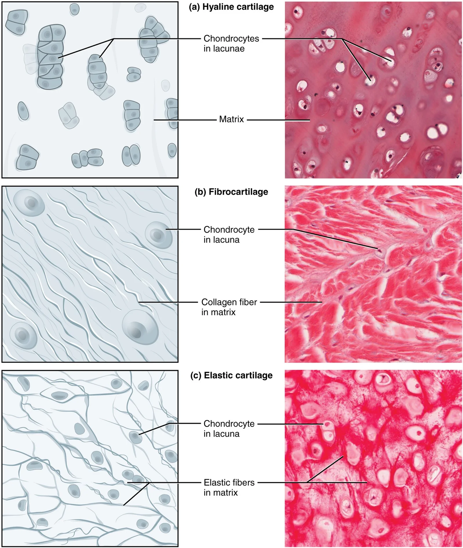
Long description
Cartilage is a connective tissue consisting of collagenous fibers embedded in a firm matrix of chondroitin sulfates. (a) Hyaline cartilage provides support with some flexibility. The example is from dog tissue. (b) Fibrocartilage provides some compressibility and can absorb pressure. (c) Elastic car

Figure 1: Cartilage is a connective tissue consisting of collagenous fibers embedded in a firm matrix of chondroitin sulfates.
Surrounding the fascicle is the endotenon, containing blood vessels. The outer layer is the epitenon. The parallel arrangement ensures efficient force transmission.
The endotenon surrounds each fascicle. The epitenon covers the entire tendon.
NoteCollagen Variation
Collagen content varies widely, from 80 μg/mg in muscle to 708 μg/mg in articular cartilage.
| Tissue | Collagen (μg/mg dry tissue) |
|---|---|
| Intervertebral disc (IVD) | 385 |
| Articular cartilage | 708 |
| Skeletal muscle | 80 |
| Tendon | 149 |
| Adipose tissue | 294 |
Connective tissues provide the essential structural framework for the human body, maintaining posture and protecting internal organs through two major forms of supportive tissue: cartilage and bone. The mechanical integrity of these tissues relies heavily on the extracellular matrix (ECM), which is a composite of cell-secreted molecules offering both biochemical and structural support. The ECM’s composition in humans consists broadly of water, protein, and polysaccharides, with the precise balance reflecting the functional requirements of the specific tissue. [1] [3]
The structural properties of the ECM are determined by its protein composition, particularly the abundance of collagen and elastin. Fibroblasts secrete three main types of fibers: collagen, elastic, and reticular fibers. Collagen fibers are formed from fibrous protein subunits linked to create long, straight structures. Although flexible, these fibers possess great tensile strength and resist stretching, providing ligaments and tendons with their characteristic resilience and strength. This strength allows connective tissues to hold together even during body movement. [3] [1]
Cartilage exhibits a distinctive appearance due to polysaccharides known as chondroitin sulfates, which bind with ground substance proteins to form proteoglycans. Embedded within the cartilage matrix are chondrocytes, or cartilage cells, which occupy spaces called lacunae. A layer of dense irregular connective tissue, the perichondrium, encapsulates the cartilage. Crucially, cartilaginous tissue is avascular, meaning all nutrients must diffuse through the matrix to reach the chondrocytes. This lack of direct blood supply is a primary factor contributing to the very slow healing of cartilaginous tissues. [1]
The mechanical properties of the matrix extend beyond simple structural integrity, as surrounding cells can sense matrix rigidity through integrin-mediated interactions. These mechanical signals are interpreted by cells, affecting their motility, proliferation, differentiation, and apoptosis. Pathological changes in the ECM composition are evident in various conditions. For instance, osteoarthritis, disk degeneration, and scoliosis lead to distinct changes in ECM composition, particularly regarding the abundance of collagen and elastin. In scoliosis, there is a reduction in collagen abundance, particularly in the annulus and endplate, supporting the theory that abnormalities in the intervertebral disc ECM are involved in its pathophysiology. [3]
Clinical pearlCartilage Healing
Cartilage is avascular, so nutrients must diffuse through the matrix to reach chondrocytes, resulting in very slow healing.
| Component | Source/Location | Function |
|---|---|---|
| Collagen Fibers | Secreted by fibroblasts | Provide tensile strength and resilience to ligaments and tendons. |
| Chondroitin Sulfates | Cartilage matrix | Bind with proteins to form proteoglycans, giving cartilage its distinctive appearance. |
| Perichondrium | Dense irregular connective tissue | Encapsulates cartilage. |
| Lacunae | Cartilage matrix | Spaces occupied by chondrocytes. |
Connective tissues are broadly categorised into fluid and solid types, each serving distinct physiological roles. Fluid connective tissues, specifically lymph and blood, consist of specialised cells circulating within a watery medium. This medium contains essential salts, nutrients, and dissolved proteins that support cellular metabolism and homeostasis. [1]
The immune defence within connective tissue relies heavily on macrophages. These large cells originate from monocytes, a type of blood cell, which migrate from blood vessels into the connective tissue matrix. Macrophages are critical for defending against pathogens and clearing degraded host cells. They function in two primary states: roaming and fixed. Roaming macrophages move rapidly via amoeboid movement to engulf infectious agents and cellular debris. In contrast, fixed macrophages are permanent residents within specific tissues, providing constant surveillance. When stimulated, macrophages release cytokines, which are small proteins acting as chemical messengers. These cytokines recruit other immune cells to infected sites and stimulate their activities. [1]
Pathologies of connective tissue often stem from alterations in the extracellular matrix (ECM). Osteoarthritis is a common disorder affecting cartilage, occurring in 60% of adults over the age of 65 in Europe and North America. This condition leads to pathological changes in the cartilage matrix structure and composition, coinciding with excessive ECM degradation. Similarly, intervertebral disc (IVD) degeneration is linked to excessive matrix catabolism. While incidence rates vary, some reports suggest disc degeneration affects up to 70% of certain populations. This disorder is frequently associated with back pain. [3]
Quantitative analysis of ECM components reveals significant variations in healthy and diseased states. In articular cartilage, tendon, and skeletal muscle, sulfated glycosaminoglycans are key constituents. Fibronectin is identified in tendon, ligament, and articular cartilage, while elastin is present in tendon and IVD cartilage. In degenerating IVD and osteoarthritic articular cartilage, collagen content in the annulus fibrosus increases significantly. Conversely, collagen content decreases in scoliotic IVD. Elastin content also increases in degenerating discs. These quantitative estimates provide crucial insights into tissue function in both physiology and pathology. [3]
Exam trapMacrophage Origin
Macrophages are derived from monocytes, not directly from stem cells within the tissue itself.
DefinitionCytokines
Small proteins released by macrophages that act as chemical messengers to recruit and stimulate other immune cells. [1]
| Component | Tissues Present |
|---|---|
| Sulfated GAGs | Articular cartilage, tendon, skeletal muscle |
| Fibronectin | Tendon, ligament, articular cartilage |
| Elastin | Tendon, IVD cartilage |
Clinical pearlOsteoarthritis Prevalence
OA affects 60% of adults over 65 in Europe and North America.
NoteDisc Degeneration
Rates can reach 70% in some populations and are linked to back pain.
DefinitionAmoeboid Movement
The rapid movement method used by roaming macrophages to engulf debris.
Exam trapCollagen in Degeneration
Collagen increases in degenerating IVD and osteoarthritic cartilage, but decreases in scoliotic IVD.
Clinical pearlFixed vs Roaming
Fixed macrophages are permanent residents; roaming macrophages move rapidly to sites of infection.
NoteECM Quantification
Quantitative breakdowns help understand composition changes in physiology and pathology.
DefinitionFluid Connective Tissue
Lymph and blood, containing cells in a watery fluid with salts, nutrients, and proteins.
Exam trapElastin Location
Elastin is found in tendon and IVD cartilage, not typically in articular cartilage.
Clinical pearlCytokine Function
Cytokines recruit immune cells and stimulate their activities at infected sites.
NoteMonocyte Migration
Monocytes enter the connective tissue matrix from blood vessels to become macrophages.
Students frequently confuse the functional roles of different connective tissues, particularly regarding energy metabolism and structural support. A primary error is assuming all adipose tissue functions identically. Brown adipose tissue is thermogenic, meaning that as it breaks down fats, it releases metabolic heat, rather than producing adenosine triphosphate (ATP), a key molecule used in metabolism. This distinction is critical when discussing metabolic rate and temperature regulation, as white adipose tissue primarily stores energy in the form of ATP and lipids, whereas brown tissue dissipates energy as heat. [1]
Another frequent mistake involves the classification and microscopic appearance of cartilage types. Students often overlook that hyaline cartilage is the most common type of cartilage in the body. It consists of short and dispersed collagen fibers and contains large amounts of proteoglycans. Under the microscope, tissue samples appear clear. The surface of hyaline cartilage is smooth. Both strong and flexible, it is found in the rib cage and nose and covers bones where they meet to form moveable joints. It makes up a template of the embryonic skeleton before bone formation. A plate of hyaline cartilage at the ends of bone allows continued growth until adulthood. Confusing this with fibrocartilage, which is tough because it has thick bundles of collagen fibers dispersed through its matrix, leads to errors in identifying joint structures. Menisci in the knee joint and the intervertebral discs are examples of fibrocartilage. [1]
A third pitfall relates to the molecular regulation of connective tissue homeostasis. Egr1 is involved in the development, homeostasis, and healing processes of these tissues, mainly via the regulation of extracellular matrix. In addition, Egr1 is often involved in the abnormal production of extracellular matrix in fibrotic conditions, and Egr1 deletion is seen as a target for therapeutic strategies to fight fibrotic conditions. This generic EGR1 function in matrix regulation has little-explored implications but is potentially important for tendon repair. Students may incorrectly assume that Egr1 solely promotes fibrosis, ignoring its essential role in normal development and repair. [2]
Bone biology presents further misconceptions regarding cell regulation. Bone is a supportive connective tissue composed of cells, fibers, and a mineralized solid ground substance. Egr1 expression is detected in several areas undergoing endochondral bone formation, such as hypertrophic cartilage and periostal regions of the developping long bones. Egr1 is a negative regulator of cartilage markers. Egr1−/− mice display bone loss. The bone loss upon Egr1 deletion is a consequence of an increased bone resorption, via the increased production of the colony stimulating factor-1 CSF-1/M-CSF known to positively regulate osteoclast differentiation. Bone development and homeostasis is controlled by the interplay between bone-forming osteoblasts and bone-resorbing osteoclasts , and this equilibrium is lost in Egr1−/− mice. EGR1 is a negative regulator of the osteoclastogenic cytokine CSF-1 production by stromal cells. Failing to recognise Egr1 as a negative regulator of osteoclastogenesis leads to incorrect predictions about bone density in genetic models. [2]
Exam trapCartilage Composition
Hyaline cartilage appears clear under the microscope due to short, dispersed collagen fibers and high proteoglycan content, unlike the thick collagen bundles of fibrocartilage.
This knowledge is applied at the bedside, in the operating theatre, on imaging and in examinations through the understanding of tissue mechanics and healing. Mechanical signals are the driving force for tendon cell differentiation in different contexts. Although the scheme is not complete, one attempt to hierarchize mechanical and molecular signals would be that mechanical signals activate molecular signals such as EGR1 transcription factor, which then in turn activate the expression of tendon genes such as Scx and Tnmd and Col1a genes. [2]
Egr1 expression is increased within 15 min in response to loading during the healing process in injured rat tendons , indicating that Egr1 expression reflects a rapid transcriptional response following loading changes in healing tendons. The increase of Egr1 expression in injured Achilles tendons after mechanical loading is followed by an increase of tendon strength, indicating a beneficial role for Egr1 in tendon healing. These observations confirm the mechanosensor role for EGR1 during tendon healing. Surgeons use this principle to prescribe early mobilisation protocols rather than prolonged immobilisation for tendon repairs. [2]
Clinical pearlTendon Healing
Mechanical loading upregulates EGR1 within 15 minutes, promoting tendon strength and matrix gene expression.
On imaging, recognising the structural differences between cartilage types aids in diagnosing joint pathology. Hyaline cartilage covers bones where they meet to form moveable joints. Its smooth surface and flexibility are essential for low-friction movement. Fibrocartilage, found in menisci and intervertebral discs, provides toughness due to thick collagen bundles. Degeneration of IVD was associated with a significant increase in collagen abundance in the annulus fibrosus. This is consistent with the current literature, which asserts that the dry-weight of the nucleus is shifted towards proteoglycans. Pathological conditions were associated with significant and disease-specific changes in cartilage collagen content. Radiologists look for these changes to assess degenerative joint disease. [3]
Tendon Loading Response
- Mechanical LoadingApplied to healing tendon
- Question: EGR1 Expression?
Increased
- Activate Scx, Tnmd, Col1aMatrix gene expression
- Increase Tendon StrengthBeneficial healing outcome
Decreased/No Change
- Poor HealingLack of mechanosignalling
In examinations, students must distinguish between the roles of different connective tissues. Bone is a supportive connective tissue composed of cells, fibers, and a mineralized solid ground substance. Egr1 is a negative regulator of cartilage markers. Egr1−/− mice display bone loss. The bone loss upon Egr1 deletion is a consequence of an increased bone resorption, via the increased production of the colony stimulating factor-1 CSF-1/M-CSF known to positively regulate osteoclast differentiation. Bone development and homeostasis is controlled by the interplay between bone-forming osteoblasts and bone-resorbing osteoclasts , and this equilibrium is lost in Egr1−/− mice. EGR1 is a negative regulator of the osteoclastogenic cytokine CSF-1 production by stromal cells. Understanding this regulatory loop is essential for questions on bone metabolism and pathology.
| Type | Key Feature | Primary Location |
|---|---|---|
| Hyaline | Smooth, clear, flexible | Rib cage, nose, joint surfaces |
| Fibrocartilage | Tough, thick collagen bundles | Menisci, intervertebral discs |
| Elastic | Elastic fibers | External ear, epiglottis |
NoteBone Homeostasis
EGR1 negatively regulates osteoclast differentiation by suppressing CSF-1 production, maintaining bone density.
Brown adipose tissue releases metabolic heat rather than producing ATP. Hyaline cartilage is the most common type, appearing clear under the microscope. Fibrocartilage contains thick collagen bundles and is found in menisci and discs. Egr1 regulates extracellular matrix development and healing in connective tissues. EGR1 is a negative regulator of osteoclastogenic cytokine CSF-1 production. Mechanical loading increases EGR1 expression within 15 minutes in healing tendons. Lack of mechanical stimulation is deleterious for tendon healing. [2] Bone is a supportive connective tissue with a mineralized solid ground substance.
Answer each one in your head first, then open the answer.
What is the primary metabolic function of brown adipose tissue?
Show answerHide answer
It releases metabolic heat rather than producing ATP.Which cartilage type is described as having thick bundles of collagen fibers?
Show answerHide answer
Fibrocartilage.What is the role of EGR1 in osteoclast differentiation?
Show answerHide answer
It is a negative regulator of CSF-1 production.How soon does EGR1 expression increase in response to loading in healing tendons?
Show answerHide answer
Within 15 minutes.Where is hyaline cartilage found in the body?
Show answerHide answer
In the rib cage, nose, and covering bones at moveable joints.
- Betts JG, Young KA, Wise JA, et al. Connective Tissue Supports and Protects. OpenStax, Anatomy and Physiology 2e (CC BY-NC-SA 4.0). Accessed .
- Havis E, Duprez D. EGR1 Transcription Factor is a Multifaceted Regulator of Matrix Production in Tendons and Other Connective Tissues. International journal of molecular sciences 2020;21(5):E1664. doi:10.3390/ijms21051664. Accessed .
- McKee TJ, Perlman G, Morris M, Komarova SV. Extracellular matrix composition of connective tissues: a systematic review and meta-analysis. Scientific reports 2019;9(1):10542. doi:10.1038/s41598-019-46896-0. Accessed .
Figure credits
Educational content for medical students and doctors. It is not a substitute for clinical judgement or local guidelines.