Types of muscles
Classification of muscle tissues and their structural organisation from cellular to organ level.
Skeletal muscle is defined by its attachment to the skeleton via strong bands of dense, regular connective tissue known as tendons. This tissue is enclosed in a three-level connective tissue scaffolding. The entire muscle is wrapped in the epimysium, a sheath of dense irregular connective tissue. Inside, muscle fibers are bundled into fascicles by the perimysium. Each individual muscle fiber is covered by the endomysium. These layers compartmentalize the muscle and allow independent movement. [1] [2]
Muscle architecture describes the arrangement of fascicles relative to the tendon. Parallel muscles have fascicles running in the same direction as the long axis. This is the most common organisation. Some parallel muscles are flat sheets, while others are rotund with a central belly. The belly is the plump middle section between origin and insertion. [1]
Pennate muscles blend into a central tendon, resembling a feather quill. Fibers pull at an angle, limiting range of motion but allowing more fibers per size. This design produces relatively more tension for its size. Other arrangements include circular, convergent, fusiform, and triangular. [1]
Actions are defined by the bone movement. The origin attaches to a fixed bone. The insertion attaches to the bone being pulled. The prime mover causes the movement. Synergists assist this action. The antagonist has the opposite action. [1]
Skeletal muscles maintain posture and stabilise joints. They prevent excess movement and dislocation. They also control internal tracts for swallowing and defecation. Each muscle is an organ with blood vessels and nerves. Every fiber is supplied by a somatic motor neuron. Contraction requires this neural signal. [2]
Deep fascia divides muscles into compartments. In the leg, these are anterior, lateral, and posterior. Tendons connect muscle to bone. Aponeuroses are broad flat tendons. Bursae and synovial sheaths reduce friction. [4]
DefinitionFascicle
A bundle of muscle fibers surrounded by perimysium.
| Type | Arrangement | Force | Range of Motion |
|---|---|---|---|
| Parallel | Same as axis | Lower | Greater |
| Pennate | Angled to tendon | Higher | Lesser |
Muscle Action Classification
- Identify Movement...
- Question: Primary mover?
Yes
- Prime Mover
No
- Question: Opposite action?
Yes
- Antagonist
No
- Question: Assists?
Yes
- Synergist
No
- Fixator
Muscles are broadly categorised by their tissue type and functional role. Skeletal muscle is striated and voluntary, yet its actions extend beyond simple locomotion. While many skeletal muscles pull against the skeleton to produce movement, others serve distinct physiological purposes. For instance, facial muscles have their insertions and origins in the skin, allowing them to produce expressions such as a smile or frown. Similarly, skeletal muscles in the tongue and the external urinary and anal sphincters allow for voluntary regulation of urination and defecation. The diaphragm also contracts to change the volume of the pleural cavities without moving the skeleton. [1] [2]
Skeletal muscles do not work in isolation; they are arranged in pairs based on their functions. For muscles attached to the bones of the skeleton, the connection determines the force, speed, and range of movement. These characteristics depend on each other and explain the general organization of the muscular and skeletal systems. [1]
Muscles of the shoulder and upper limb are divided into four groups: muscles that stabilize and position the pectoral girdle, muscles that move the arm, muscles that move the forearm, and muscles that move the wrists, hands, and fingers. The pectoral girdle, or shoulder girdle, consists of the lateral ends of the clavicle and scapula, along with the proximal end of the humerus. The girdle creates a base from which the head of the humerus, in its ball-and-socket joint with the glenoid fossa of the scapula, can move the arm in multiple directions. [3]
Muscles that position the pectoral girdle are located either on the anterior thorax or on the posterior thorax. The anterior muscles include the subclavius, pectoralis minor, and serratus anterior. The posterior muscles include the trapezius, rhomboid major, and rhomboid minor. When the rhomboids are contracted, your scapula moves medially, which can pull the shoulder and upper limb posteriorly. [3]
Similar to the muscles that position the pectoral girdle, muscles that cross the shoulder joint and move the humerus bone of the arm include both axial and scapular muscles. The two axial muscles are the pectoralis major and the latissimus dorsi. The pectoralis major is thick and fan-shaped, covering much of the superior portion of the anterior thorax. [3][4]
Wrist, hand, and finger movements are facilitated by two groups of muscles. The forearm is the origin of the extrinsic muscles of the hand. The palm is the origin of the intrinsic muscles of the hand. The intrinsic muscles of the hand both originate and insert within it. These muscles allow your fingers to also make precise movements for actions, such as typing or writing. These muscles are divided into three groups. The thenar muscles are on the radial aspect of the palm. The hypothenar muscles are on the medial aspect of the palm, and the intermediate muscles are midpalmar. [3] [4]
| Group | Muscles |
|---|---|
| Anterior Thoracic | Subclavius, Pectoralis minor, Serratus anterior |
| Posterior Thoracic | Trapezius, Rhomboid major, Rhomboid minor |
DefinitionPectoral Girdle
The clavicle and scapula make up the pectoral girdle, which provides a stable origin for the muscles that move the humerus. [3]
Upper Limb Muscle Groups
- Upper Limb MusclesDivide into four groups
- Question: Function?
Stabilize Girdle
- Anterior: Subclavius, Pec minor, Serratus anterior
- Posterior: Trapezius, Rhomboids
Move Arm
- Axial: Pec major, Latissimus dorsi
- Scapular: Deltoid, Rotator cuff muscles
Move Forearm
- Cross elbow joint
Move Hand/Fingers
- Extrinsic (Forearm origin)
- Intrinsic (Hand origin)
NoteIntrinsic Hand Muscles
The intrinsic muscles of the hands allow our fingers to make precise movements, such as typing and writing. They both originate and insert within the hand. [3]
| Group | Location | Muscles |
|---|---|---|
| Thenar | Lateral palm | Abductor pollicis brevis, Opponens pollicis, Flexor pollicis brevis, Adductor pollicis |
| Hypothenar | Medial palm | Abductor digiti minimi, Flexor digiti minimi brevis, Opponens digiti minimi |
| Intermediate | Midpalmar | Lumbricals, Palmar interossei, Dorsal interossei |
DefinitionThenar Muscles
The thenar muscles, which are located on the lateral part of the palm, are the abductor pollicis brevis, opponens pollicis, flexor pollicis brevis, and adductor pollicis. [3]
DefinitionHypothenar Muscles
The hypothenar muscles, which are located on the medial part of the palm, are the abductor digiti minimi, flexor digiti minimi brevis, and opponens digiti minimi. [3]
DefinitionIntermediate Muscles
The intermediate muscles, located in the middle of the palm, are the lumbricals, palmar interossei, and dorsal interossei. [3]
NoteFoot Intrinsic Muscles
The foot also has intrinsic muscles, which originate and insert within it (similar to the intrinsic muscles of the hand). These muscles primarily provide support for the foot and its arch, and contribute to movements of the toes. [4]
DefinitionPlantar Aponeurosis
The principal support for the longitudinal arch of the foot is a deep fascia called plantar aponeurosis, which runs from the calcaneus bone to the toes. [4]
NotePlantar Fasciitis
Inflammation of this tissue is the cause of “plantar fasciitis,” which can affect runners. [4]
DefinitionDorsal Foot Muscles
The dorsal group includes only one muscle, the extensor digitorum brevis. [4]
DefinitionPlantar Foot Muscles
The second group is the plantar group, which consists of four layers, starting with the most superficial. [4]
DefinitionIliopsoas Group
The psoas major and iliacus make up the iliopsoas group. [4]
NoteGluteal Muscles
Some of the largest and most powerful muscles in the body are the gluteal muscles or gluteal group. The gluteus maximus is the largest; deep to the gluteus maximus is the gluteus medius, and deep to the gluteus medius is the gluteus minimus, the smallest of the trio. [4]
DefinitionHamstring Group
The three long muscles on the back of the knee are the hamstring group, which flexes the knee. These are the biceps femoris, semitendinosus, and semimembranosus. [4]
NotePopliteal Fossa
The tendons of these muscles form the popliteal fossa, the diamond-shaped space at the back of the knee. [4]
Muscles are classified by their structure, control mechanism, and location. Skeletal muscles are voluntary and striated, attaching to bones via tendons or aponeuroses to facilitate movement. They also protect internal organs, particularly in the abdominal and pelvic regions, by acting as an external shield against trauma and supporting organ weight. [2]
Figure 1Multinuclear muscle fiber cells and some associated mononuclear cells
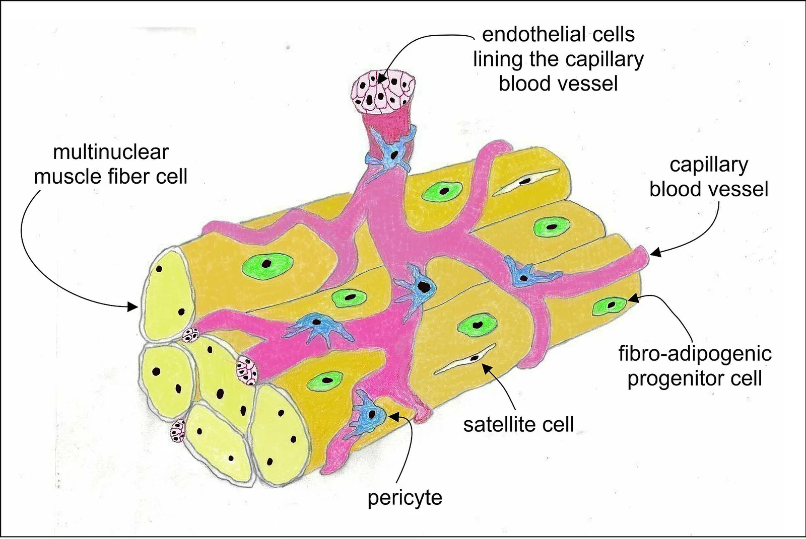
Long description
Skeletal muscle cell types include: very large multinuclear muscle fiber cells; small endothelial cells that line the inside of capillary blood vessels; small fibro-adipogenic progenitor cells (FAPs) which are muscle-fib

Figure 1: Skeletal muscle cell types include: very large multinuclear muscle fiber cells; small endothelial cells that line the inside of capillary blood vessels; small fibro-adipogenic progenitor cells (FAPs) which are muscle-fib.
The quadriceps femoris is the primary muscle group here, comprising four distinct muscles: the rectus femoris, vastus lateralis, vastus medius, and vastus intermedius[1]. These muscles converge to form the quadriceps tendon, which inserts into the patella and continues as the patellar ligament to attach to the tibia[1]. The sartorius, the longest muscle in the body, runs obliquely across the anterior thigh within this compartment[1]. The anterior compartment is responsible for extending the knee joint[1]. To extend the knee, this group of four muscles is activated as agonists. Conversely, flexion of the knee joint is achieved by the antagonistic set known as the hamstrings, located in the posterior compartment. The hamstrings include the biceps femoris, semitendinosus, and semimembranosus[1]. [4]
Circular muscles, also termed sphincters, consist of concentrically arranged bundles of muscle fibers. When these fibers contract, the size of the opening shrinks to the point of closure; when they relax, the opening increases in size. The orbicularis oculi surrounds each eye, serving a similar protective and expressive function. The term 'orbicularis' derives from 'orb', meaning circular, referencing the round structure these muscles enclose. [1]
In the shoulder, muscles originate from the scapula to allow diverse arm movements. The deltoid is a thick muscle that creates the rounded contour of the shoulder and acts as the major abductor of the arm. It also facilitates flexing, medial rotation, extension, and lateral rotation. The subscapularis originates on the anterior scapula and medially rotates the arm. The supraspinatus, located superior to the spine of the scapula, and the infraspinatus, located inferior to it, abduct and laterally rotate the arm, respectively. The teres major, thick and flat, lies inferior to the teres minor and extends the arm while assisting in adduction and medial rotation. [3]
The hand features intrinsic thenar muscles, including the abductor pollicis brevis, opponens pollicis, flexor pollicis brevis, and adductor pollicis. These form the thenar eminence, the rounded contour at the base of the thumb, and enable precise thumb movements. Extrinsic muscles originate in the forearm and insert into the hand to facilitate crude movements. The superficial anterior compartment of the forearm produces flexion and includes the flexor carpi radialis, palmaris longus, flexor carpi ulnaris, and flexor digitorum superficialis. The deep anterior compartment also produces flexion via the flexor pollicis longus and flexor digitorum profundus. Extension is performed by muscles in the posterior compartments, such as the extensor carpi radialis longus, extensor carpi radialis brevis, extensor digitorum, extensor digiti minimi, and extensor carpi ulnaris in the superficial layer. The deep posterior compartment includes the abductor pollicis longus, extensor pollicis brevis, extensor pollicis longus, and extensor indicis. [3]
In the leg, the anterior compartment contains the tibialis anterior, extensor hallucis longus, and extensor digitorum longus, which raise the front of the foot[1]. The fibularis tertius, associated with the extensor digitorum longus, is variable in presence[1]. The lateral compartment includes the fibularis longus and fibularis brevis[1]. The posterior compartment contains large, strong muscles that keep humans upright[1]. The superficial muscles, including the gastrocnemius and soleus, insert onto the calcaneal tendon[1]. The plantaris runs obliquely between them, absent in about seven percent of individuals[1]. Deep posterior muscles include the popliteus, flexor digitorum longus, flexor hallucis longus, and tibialis posterior[1]. [4]
Muscle function is defined by specific roles within a movement. The prime mover, or agonist, is the principal muscle responsible for an action. For example, the biceps brachii acts as the prime mover when lifting a cup. Other muscles may assist this action. The brachialis serves as a synergist in this movement. A synergist can also function as a fixator. This stabilizes the bone that serves as the origin for the prime mover. [1]
Skeletal muscles maintain homeostasis by generating heat. Muscle contraction requires energy. The breakdown of ATP produces heat. This effect is noticeable during exercise. Sustained muscle movement causes body temperature to rise. Shivering produces random skeletal muscle contractions to generate heat in extreme cold. [1]
The excitation of skeletal muscle is triggered by the nervous system[1]. Signals originate from the somatic motor division[1]. This sequence begins with these neural signals[1]. [2]
Forearm actions include flexion, extension, pronation, and supination. Flexion involves the biceps brachii, brachialis, and brachioradialis. Extension involves the triceps brachii and anconeus. Pronation is performed by the pronator teres and pronator quadratus. Supination is performed by the supinator muscle. The forearm is supinated when it faces anteriorly. It is pronated when it faces posteriorly. [3]
The anterior compartment of the forearm contains flexors. These muscles originate on the humerus. They insert onto different parts of the hand. Superficial muscles include the flexor carpi radialis, palmaris longus, flexor carpi ulnaris, and flexor digitorum superficialis. The flexor digitorum superficialis flexes the hand and digits. This allows rapid finger movements. Poor ergonomics can irritate these tendons. This can pinch the median nerve in the carpal tunnel. This causes Carpal Tunnel Syndrome. [3]
The superficial posterior compartment contains extensors. These muscles originate on the humerus. They include the extensor radialis longus and brevis. They also include the extensor digitorum, extensor digiti minimi, and extensor carpi ulnaris. The deep posterior compartment originates on the radius and ulna. Muscles include the abductor pollicis longus. It includes the extensor pollicis brevis and longus. It also includes the extensor indicis. [3]
Retinacula sheath tendons at the wrist. The flexor retinaculum covers the palmar surface. The extensor retinaculum covers the dorsal surface. [3]
The pelvic girdle stabilizes the lower limbs. It connects to the sacrum. There is very little movement of the pelvic girdle. It was designed to stabilize and support the body. [4]
The thigh has medial, anterior, and posterior compartments. The medial compartment adducts the femur. Muscles include the adductor longus, brevis, and magnus. The pectineus and gracilis are also included. [4]
The anterior thigh flexes the thigh and extends the leg. It contains the quadriceps femoris group. This group comprises four muscles. They extend and stabilize the knee. The rectus femoris is anterior. The vastus lateralis is lateral. The vastus medialis is medial. The vastus intermedius is deep. The quadriceps tendon inserts into the patella. The patellar ligament attaches to the tibial tuberosity. The sartorius extends from the anterior superior iliac spine. It inserts on the medial side of the proximal tibia. [4]
The leg muscles move the foot and toes. The anterior compartment includes the tibialis anterior. It includes the extensor hallucis longus. It includes the extensor digitorum longus. It includes the fibularis tertius. The lateral compartment houses the fibularis longus. It houses the fibularis brevis. The superficial posterior compartment has the gastrocnemius. It has the soleus. It has the plantaris. The deep posterior compartment has the popliteus. It has the tibialis posterior. It has the flexor digitorum longus. It has the flexor hallucis longus. [4]
DefinitionPrime Mover
The principal muscle involved in an action is called the prime mover, or agonist. [1]
| Compartment | Key Muscles |
|---|---|
| Anterior Superficial | Flexor carpi radialis, Palmaris longus, Flexor carpi ulnaris, Flexor digitorum superficialis |
| Posterior Superficial | Extensor radialis longus, Extensor carpi radialis brevis, Extensor digitorum, Extensor digiti minimi, Extensor carpi ulnaris |
| Posterior Deep | Abductor pollicis longus, Extensor pollicis brevis, Extensor pollicis longus, Extensor indicis |
Exam trapCarpal Tunnel
Poor ergonomics can irritate tendons and pinch the median nerve, causing Carpal Tunnel Syndrome. [3]
Clinical pearlPelvic Girdle
The pelvic girdle has less range of motion because it was designed to stabilize and support the body. [4]
1. Antagonist Function An antagonist is a muscle with the opposite action of the prime mover. It maintains body or limb position, such as holding the arm out or standing erect. It also controls rapid movement, as in shadow boxing without landing a punch. [1]
2. Convergent Muscle Architecture When fascicles spread over a sizable area but attach to a single point, the muscle is convergent. The attachment point may be a tendon, aponeurosis, or raphe. The temporalis muscle of the cranium is another example. [1]
3. Pennate Muscle Arrangements In unipennate muscles, fascicles are located on one side of the tendon. Bipennate muscles have fascicles on both sides of the tendon. The deltoid muscle is a common multipennate example. [1]
4. Musculoskeletal Levers Bones act as stiff levers in the musculoskeletal system. The load is an object being lifted or resistance to movement. Effort is the applied force from contracting skeletal muscle. [1]
5. Sarcomere Structure The sarcomere is the functional unit of the muscle fiber. It is bordered by Z-discs, to which actin myofilaments are anchored. Each sarcomere is approximately 2 μm in length. Myofibrils are only approximately 1.2 μm in diameter. [2]
6. Myofilament Arrangement Striated appearance is due to sequential arrangement of actin and myosin. The actin and its troponin-tropomyosin complex form the thin filament. [2]
7. Motor Neuron Pathways Motor neurons originate in the spinal cord for most skeletal muscles. A smaller number are located in the brainstem for face, head, and neck muscles. Axons transmit action potentials all the way from the spinal cord to the muscle. [2]
8. Arm Flexors The biceps brachii, brachialis, and brachioradialis flex the forearm[1]. The biceps brachii also supinates the forearm at the radioulnar joints[1]. The brachialis provides additional power in flexing the forearm[1]. These muscles form the anterior compartment of the arm[1]. [3]
9. Forearm Extensors and Rotators The extensors of the forearm are the triceps brachii and anconeus[1]. The pronators are the pronator teres and the pronator quadratus[1]. [3]
10. Pelvic Girdle Stability The pelvic girdle attaches the lower limbs to the torso. The body’s center of gravity is in the area of the pelvis. Leg muscles make up for limited range of motion with size and power. This facilitates stabilization, posture, and movement. [4]
11. Hip Joint Muscles The hip joint is where the pelvic girdle and leg come together. The psoas major and iliacus form the iliopsoas. [4]
12. Thigh Adductors On the medial thigh, adductor longus, brevis, and magnus adduct the thigh. These muscles also medially rotate the thigh. The pectineus muscle adducts and flexes the femur at the hip. [4]
Students frequently confuse the anatomical definitions of muscle attachment points, leading to errors in describing joint mechanics. The origin is the attachment to the fixed or stabilized bone, whereas the insertion is the moveable end attached to the bone being pulled[1]. A common error is assuming the origin never moves; in reality, if the distal attachment is fixed, the roles can reverse during specific movements. Another frequent mistake involves muscle shape classification. Students often mislabel any long muscle as fusiform, but fusiform specifically describes a parallel muscle with a central, large belly that tapers as it extends to its origin and insertion[1].
A significant conceptual trap lies in the mechanism of contraction. Students often believe that calcium directly causes the myosin heads to pull actin. In fact, the action potential sweeps along the sarcolemma and triggers calcium release from the sarcoplasmic reticulum. The calcium then interacts with shielding proteins, forcing them to move aside so actin-binding sites are available for myosin attachment. The myosin head performs the actual pulling. Furthermore, students may overlook the critical role of the neuromuscular junction. Every skeletal muscle fiber is innervated by a motor neuron at this junction, and excitation signals from the neuron are the only way to functionally activate the fiber to contract. [2]
Another pitfall involves the electrical properties of muscle cells. Students sometimes confuse the resting membrane potential with the action potential. The resting potential is typically around -60 to -90 mV, relative to the outside. The action potential is a wave of depolarization that travels along the membrane, allowing signals to be transmitted quickly over long distances. It is crucial to remember that the sarcolemma is the plasma membrane of the muscle fiber, and the sarcoplasm is its cytoplasm. The specialized smooth endoplasmic reticulum that stores calcium is the sarcoplasmic reticulum. [2]
Students also struggle with the structural organization of the muscle fiber. They may not realize that skeletal muscle fibers are long, multinucleated cells. The striations seen in skeletal muscle are created by the organization of actin and myosin, resulting in the banding pattern of myofibrils. The functional unit is the sarcomere, which contains the contractile myofilaments. A common error is ignoring the T-tubules. These invaginations in the sarcolemma ensure the membrane gets close to the sarcoplasmic reticulum, forming a triad with the SR membranes. This structure is essential because the muscle fiber diameter can be up to 100 μm, requiring T-tubules to carry the action potential into the interior. [2]
Finally, students often forget the termination of the signal. After depolarization, the membrane repolarizes, and acetylcholinesterase degrades acetylcholine in the synaptic cleft to prevent unwanted extended contraction. Without this degradation, the muscle would remain in a state of tetanic contraction. Understanding these distinctions prevents confusion between the electrical excitation phase and the mechanical contraction phase. [2]
At the bedside, knowledge of muscle origins and insertions is vital for assessing joint function and identifying lesions. For example, the brachioradialis assists the brachialis during forearm flexion[1]. If a patient cannot flex the elbow, testing the brachialis and brachioradialis helps localize the nerve injury. In the thigh, the tensor fascia latae acts as a synergist of the gluteus medius and iliopsoas in flexing and abducting the thigh. It also stabilizes the lateral aspect of the knee by pulling on the iliotibial tract. Weakness in hip abduction may indicate gluteus medius or tensor fascia latae dysfunction. [4]
The adductor group is clinically significant for hip movement. The adductor longus flexes the thigh, whereas the adductor magnus extends it. The pectineus adducts and flexes the femur at the hip. These muscles are located in the femoral triangle, which also contains the femoral nerve, femoral artery, femoral vein, and deep inguinal lymph nodes. Understanding this anatomy is crucial during femoral vein cannulation or inguinal hernia repair to avoid vascular or neural injury. [4]
In the operating theatre, precise knowledge of muscle relations is essential. The piriformis, obturator internus, obturator externus, superior gemellus, inferior gemellus, and quadratus femoris laterally rotate the femur at the hip. These muscles lie deep to the gluteus maximus. During hip surgery, retracting the gluteus maximus exposes these lateral rotators. Damage to the nerve supply of these muscles can result in gait abnormalities. [4]
On imaging, recognizing muscle shapes aids in identifying pathology. A fusiform muscle has a central belly that tapers at the ends[1]. Abnormal thickening or atrophy can be assessed by comparing the muscle belly to its expected tapering. The striations of skeletal muscle are visible on high-resolution MRI due to the organization of actin and myosin. Pathologies affecting the sarcoplasmic reticulum or T-tubules can disrupt calcium release, leading to weakness. The triad structure, consisting of a T-tubule and adjacent sarcoplasmic reticulum membranes, is critical for excitation-contraction coupling. Disruption here can cause severe muscle dysfunction. [2]
In examinations, students must accurately describe muscle actions and innervations. The versatile muscle that flexes the leg at the knee and flexes, abducts, and laterally rotates the leg at the hip allows us to sit cross-legged. Identifying this muscle requires understanding its multi-joint actions. The neuromuscular junction is where the motor neuron terminal meets the muscle fiber. Questions often test the role of acetylcholinesterase in terminating the signal. Understanding that calcium release from the sarcoplasmic reticulum initiates contraction is a key concept. The sarcomere is the fun [2][4]
Excitation-Contraction Coupling
- Action Potential arrives at NMJMotor neuron releases ACh
- Question: ACh binds receptors?
Yes
- Depolarization of sarcolemmaNa+ influx
No
- No contractionBlockade or degradation
- AP propagates via T-tubulesReaches SR triads
- Question: Ca++ released from SR?
Yes
- Ca++ binds troponinMoves shielding proteins
No
- No contractionSR dysfunction
- Myosin binds actinCross-bridge cycling
- Muscle contractionFilament sliding
The origin is the fixed attachment, while the insertion is the moveable attachment[1]. Fusiform muscles have a central belly that tapers at the origin and insertion[1]. The sarcolemma is the plasma membrane, and sarcoplasm is the cytoplasm. The neuromuscular junction is where motor neurons activate skeletal muscle fibers. Action potentials travel along the sarcolemma and T-tubules to trigger calcium release. Calcium from the sarcoplasmic reticulum allows myosin to bind actin. The tensor fascia latae stabilizes the knee via the iliotibial tract. The femoral triangle contains the femoral nerve, artery, vein, and lymph nodes.
Answer each one in your head first, then open the answer.
What is the moveable end of a muscle called?
Show answerHide answer
InsertionWhich enzyme degrades acetylcholine to prevent extended contraction?
Show answerHide answer
AcetylcholinesteraseWhat structure carries the action potential into the interior of the muscle fiber?
Show answerHide answer
T-tubulesWhich muscle helps stabilize the lateral aspect of the knee by pulling on the iliotibial tract?
Show answerHide answer
Tensor fascia lataeWhat is the functional unit of a skeletal muscle fiber?
Show answerHide answer
Sarcomere
- Betts JG, Young KA, Wise JA, et al. Interactions of Skeletal Muscles, Their Fascicle Arrangement, and Their Lever Systems. OpenStax, Anatomy and Physiology 2e (CC BY-NC-SA 4.0). Accessed .
- Betts JG, Young KA, Wise JA, et al. Skeletal Muscle. OpenStax, Anatomy and Physiology 2e (CC BY-NC-SA 4.0). Accessed .
- Betts JG, Young KA, Wise JA, et al. Muscles of the Pectoral Girdle and Upper Limbs. OpenStax, Anatomy and Physiology 2e (CC BY-NC-SA 4.0). Accessed .
- Betts JG, Young KA, Wise JA, et al. Appendicular Muscles of the Pelvic Girdle and Lower Limbs. OpenStax, Anatomy and Physiology 2e (CC BY-NC-SA 4.0). Accessed .
Figure credits
Educational content for medical students and doctors. It is not a substitute for clinical judgement or local guidelines.