Classification of joints
Structural and functional classification of human joints, focusing on fibrous, cartilaginous, and synovial types.
Joints, or articulations, are the junctions between bones or between a bone and cartilage. They are classified structurally based on the material connecting the bones and functionally based on the degree of movement permitted.
Fibrous joints are united by fibrous connective tissue, with no joint cavity present. These joints are generally immobile or slightly mobile. Functionally, they are classified as synarthroses (immobile) or amphiarthroses (slightly moveable). [3]
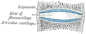
Cartilaginous joints are joined by hyaline cartilage or fibrocartilage. Like fibrous joints, they lack a fluid-filled cavity. They are functionally classified as either synarthroses or amphiarthroses. The pubic symphysis is an amphiarthrosis that strongly unites the right and left hip bones. Intervertebral discs also form amphiarthroses, allowing small movements between adjacent vertebrae. [3] [1]
Synovial joints are the most common type of joint in the body. A key structural characteristic unique to synovial joints is the presence of a joint cavity. This fluid-filled space allows articulating bone surfaces to contact each other without being directly connected by fibrous tissue or cartilage. This structure permits smooth movement and increased mobility. Overall, each type of synovial joint is necessary to provide the body with its great flexibility and mobility. [1] [4] [3]
Figure 1Gray300
Long description
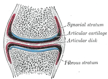
Diagrammatic section of a diarthrodial joint, with an articular disk.

Figure 1: Diagrammatic section of a diarthrodial joint, with an articular disk.
Figure 2Gray299
Long description
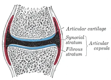
Diagrammatic section of a diarthrodial joint.

Figure 2: Diagrammatic section of a diarthrodial joint.
Functionally, all synovial joints are classified as diarthroses, meaning they are freely moveable. The type of movement allowed is determined by the structural classification of the joint. [2] [3][4]
DefinitionFunctional Classifications
Synarthrosis: essentially immobile joint. Amphiarthrosis: allows limited movement. Diarthrosis: freely moveable joint. [3]
Synovial joints are strengthened by ligaments, which are strong bands of fibrous connective tissue. Ligaments hold bones together, resist excessive movements, and prevent separation. They are classified by their relationship to the fibrous articular capsule: [1]
- Extrinsic ligaments: Located outside the articular capsule.
- Intrinsic ligaments: Fused to or incorporated into the wall of the articular capsule. [1]
- Intracapsular ligaments: Located inside the articular capsule.
Some synovial joints contain an articular disc (meniscus) to provide padding, smooth movements, or strengthen the joint. Muscles and their tendons acting across a joint also provide indirect support by increasing contractile strength when needed. [1]
Each synovial joint is specialized for certain movements, which are determined by its structural type. Movements are generally paired, with one being the opposite of the other. [1] [2][4]
- Uniaxial joints: Allow movement in a single anatomical plane. An example is the elbow.
- Biaxial joints: Allow movement in two planes. An example is the metacarpophalangeal joints of the fingers.
- Multiaxial joints: Allow movement in three planes. Examples include the shoulder and hip joints. [3]
A multiaxial ball-and-socket joint has much more mobility than a uniaxial hinge joint. However, ligaments and muscles restrict the total range of motion. The shoulder has little ligament support, granting a very large range of motion. In contrast, the hip has strong ligaments that reduce range of motion but confer stability during standing and weight bearing. [2]
Stability is often inversely related to mobility. The ball-and-socket joint of the shoulder has little in the way of ligament support, which gives the shoulder a very large range of motion. In contrast, movements at the hip joint are restricted by strong ligaments, which reduce its range of motion but confer stability during standing and weight bearing. [2] [1]
Synovial joints are innervated by nerves that supply the skin over the joint and the muscles that move it. This is known as Hilton’s law. The joint capsule receives sensory innervation from these same nerves, providing proprioception and pain sensation.
Joints are primarily categorised by their degree of mobility. An immobile or nearly immobile joint is termed a synarthrosis. These joints create a strong union between bones, which is essential in areas where bones must protect internal organs. Examples include the fibrous sutures of the skull that protect the brain and the cartilaginous manubriosternal joint that protects the heart. [3]
Conversely, a freely mobile joint is known as a diarthrosis. All synovial joints in the body are classified as diarthroses. These are most prevalent in the appendicular skeleton, allowing the limbs a wide range of motion. Some synovial joints contain a fat pad to act as a cushion between the bones. [4] [3]
Synovial joints are further divided into six types based on the shape of the articulating surfaces and the movements they permit. There are many types of movement that can occur at synovial joints. [4] [1]
| Joint Type | Articulation Characteristic | Examples |
|---|---|---|
| Pivot | Bone held in a ring by a ligament and another bone | C1 (atlas) and C2 (axis) dens; proximal radioulnar joint |
| Hinge | Convex end of one bone fits the concave end of another | Elbow, knee, ankle, and interphalangeal joints [1] |
| Condyloid | Rounded bony area fits into a shallow depression | Metacarpophalangeal joints; radiocarpal joint |
| Saddle | Bones fit together like a rider and a saddle | First carpometacarpal joint (thumb base) |
| Plane | Small, flattened surfaces | Intercarpal, intertarsal, acromioclavicular, and zygapophysial joints |
| Ball-and-socket | Rounded head fits into a large socket | Shoulder and hip joints [1] |
Diarthroses are functionally classified by the number of axes (anatomical planes) in which they allow movement.
Functional Classification of Synovial Joints
- Synovial JointDetermine number of axes of motion
- Question: How many axes?
One (Uniaxial)
- Pivot or Hinge joints
Two (Biaxial)
- Condyloid or Saddle joints
Three (Multiaxial)
- Plane or Ball-and-socket joints
Uniaxial joints, such as pivot and hinge joints, allow movement in only one plane. Hinge joints specifically permit only bending and straightening, while pivot joints allow only rotation around a single axis. [1]
Biaxial joints, including condyloid and saddle joints, allow for abduction and adduction as well as flexion and extension. When these movements are combined sequentially, they produce circumduction. [4]
Multiaxial joints allow movement in all three anatomical planes. These are divided by their range of motion: [3]
- Plane joints: These allow bones to slide or rotate, but the range of motion is usually slight. However, small motions can combine across adjacent joints to produce larger movements, such as the inversion and eversion of the foot or the flexion, extension, and lateral flexion of the vertebral column. [3]
- Ball-and-socket joints: These allow for the greatest range of motion of all synovial joints. They permit flexion-extension, abduction-adduction, circumduction, and both medial (internal) and lateral (external) rotation. [4]
DefinitionJoint
A joint, or articulation, is any location where adjacent bones or bone and cartilage come together to form a connection. [3]
Joints are categorised using two different systems: structural and functional. Structural classification is based on how the bones are held together and how they articulate. Functional classification describes the degree of movement available, which ranges from immobile to slightly mobile, and finally to freely moveable joints. [3]
The functional classification of joints is determined by the amount of mobility found between the adjacent bones. [3]
Structural classifications of the body joints are based on how the bones are held together and articulate with each other. At fibrous joints, the adjacent bones are directly united to each other by fibrous connective tissue. Similarly, at a cartilaginous joint, the adjacent bones are united by cartilage. In contrast, at a synovial joint, the articulating bone surfaces are not directly united to each other, but come together within a fluid-filled joint cavity. [1] [3]
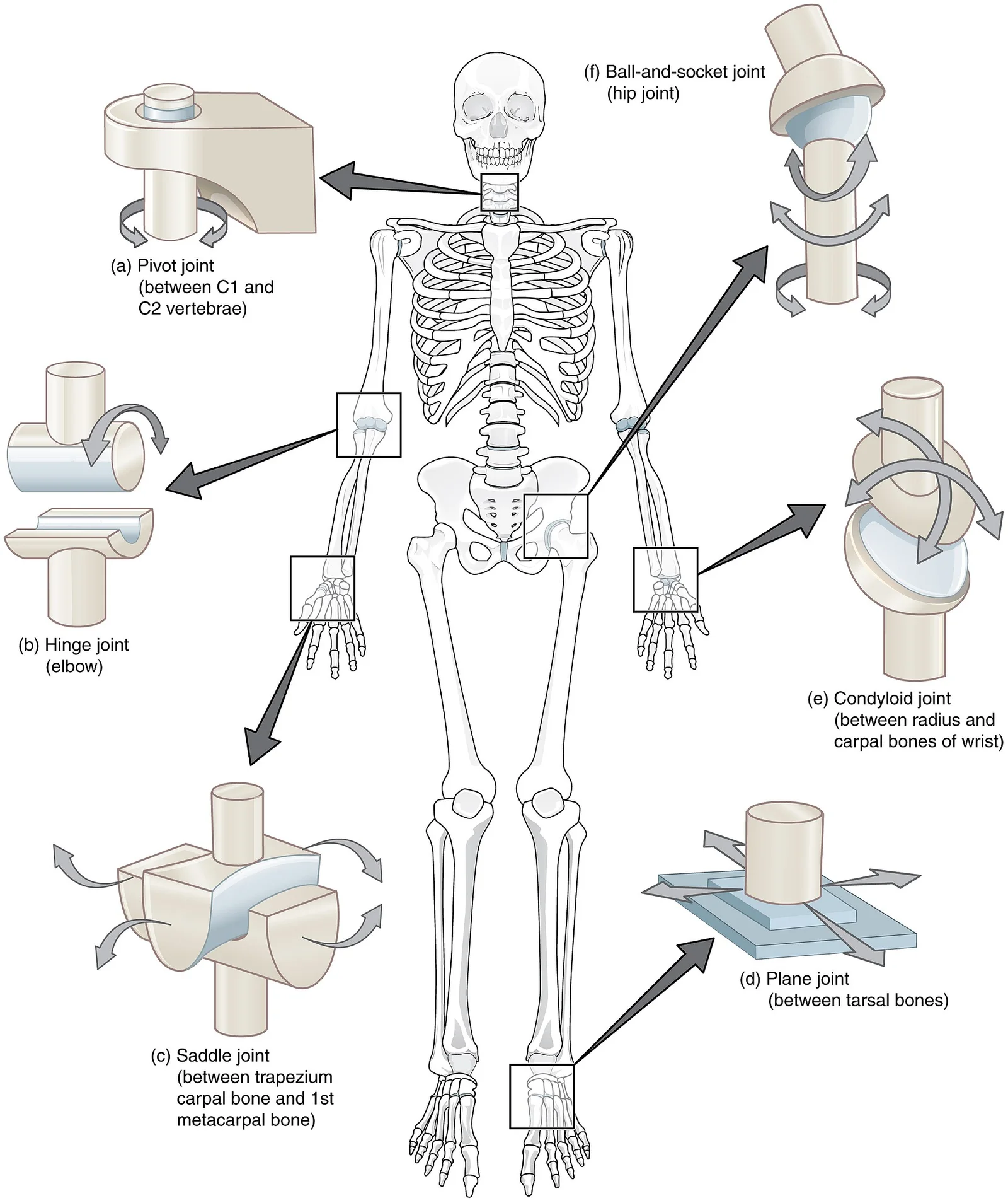
The six types of synovial joints are pivot, hinge, condyloid, saddle, plane, and ball-and socket-joints. [1] [4]
At a condyloid joint (ellipsoid joint), the shallow depression at the end of one bone articulates with a rounded structure from an adjacent bone or bones. The knuckle (metacarpophalangeal) joints of the hand between the distal end of a metacarpal bone and the proximal phalanx bone are condyloid joints. Another example is the radiocarpal joint of the wrist, between the shallow depression at the distal end of the radius bone and the rounded scaphoid, lunate, and triquetrum carpal bones. In this case, the articulation area has a more oval (elliptical) shape. Functionally, condyloid joints are biaxial joints that allow for two planes of movement. One movement involves the bending and straightening of the fingers or the anterior-posterior movements of the hand. [3] [1]
DefinitionArticular Disc
A fibrocartilage structure located between the articulating bones in some synovial joints, which can act to unite bones, provide shock absorption, or smooth movements.
A few synovial joints of the body have a fibrocartilage structure located between the articulating bones. This is called an articular disc, which is generally small and oval-shaped, or a meniscus, which is larger and C-shaped. These structures can serve several functions, depending on the specific joint. In some places, an articular disc may act to strongly unite the bones of the joint to each other. Examples of this include the articular discs found at the sternoclavicular joint or between the distal ends of the radius and ulna bones. At other synovial joints, the disc can provide shock absorption and cushioning between the bones, which is the function of each meniscus within the knee joint. Finally, an articular disc can serve to smooth the movements between the articulating bones, as seen at the temporomandibular joint. [1]
The joints of the upper limb provide for large ranges of motion, which give the upper limb great mobility, thus enabling actions such as the throwing of a ball or typing on a keyboard. The joints of the lower limb are more robust, giving them greater strength and the stability needed to support the body weight during running, jumping, or kicking activities. [2]
The joints of the vertebral column include the symphysis joints formed by each intervertebral disc and the plane synovial joints between the superior and inferior articular processes of adjacent vertebrae. Each of these joints provide for limited motions, but these sum together to produce flexion, extension, lateral flexion, and rotation of the neck and body. The range of motions available in each region of the vertebral column varies, with all of these motions available in the cervical region. While the ball-and-socket joint gives the greatest range of movement at an individual joint, in other regions of the body, several joints may work together to produce a particular movement. [2][3][4]
Figure 3Gray298
Long description
Diagrammatic section of a symphysis. Labelled illustration from Wikimedia Commons, used under its open licence.

Figure 3: Diagrammatic section of a symphysis.
Each of the different structural types of synovial joints also allow for specific motions. The atlantoaxial pivot joint provides side-to-side rotation of the head, while the proximal radioulnar articulation allows for rotation of the radius during pronation and supination of the forearm. Hinge joints, such as at the knee and elbow, allow only for flexion and extension. Similarly, the hinge joint of the ankle only allows for dorsiflexion and plantar flexion of the foot. [4] [2]
Circumduction is the movement of a body region in a circular manner, in which one end of the body region being moved stays relatively stationary while the other end describes a circle. It involves the sequential combination of flexion, abduction, extension, and adduction at a joint. This type of motion is found at biaxial condyloid and saddle joints, and at multiaxial ball-and-sockets joints. [4]
Rotation can also occur at the ball-and-socket joints of the shoulder and hip. Here, the humerus and femur rotate around their long axis, which moves the anterior surface of the arm or thigh either toward or away from the midline of the body. Movement that brings the anterior surface of the limb toward the midline of the body is called medial (internal) rotation. Conversely, rotation of the limb so that the anterior surface moves away from the midline is lateral (external) rotation. [4] [1]
Figure 4909 Types of Synovial Joints
Long description
Illustration from Anatomy & Physiology, Connexions Web site. http://cnx.org/content/col11496/1.6/, Jun 19, 2013.

Figure 4: Illustration from Anatomy & Physiology, Connexions Web site (http://cnx.org/content/col11496/1.6/, jun 19, 2013).
Synovial joints are the most common type of joints in the body[1]. They are characterized by the presence of a joint cavity, inside of which the bones of the joint articulate with each other[1]. The articulating surfaces of the bones at a synovial joint are not directly connected to each other by connective tissue or cartilage, which allows the bones to move freely against each other[1]. The walls of the joint cavity are formed by the articular capsule[1]. [3]
A uniaxial joint only allows for a motion in a single plane (around a single axis). The elbow joint, which only allows for bending or straightening, is an example of a uniaxial joint. The elbow joint is a uniaxial hinge joint formed by the humeroulnar joint, the articulation between the trochlea of the humerus and the trochlear notch of the ulna. Also associated with the elbow are the humeroradial joint and the proximal radioulnar joint. All three of these joints are enclosed within a single articular capsule. [2][3]
At a saddle joint, both of the articulating surfaces for the bones have a saddle shape, which is concave in one direction and convex in the other (see c). This allows the two bones to fit together like a rider sitting on a saddle[1]. Saddle joints are functionally classified as biaxial joints[1]. The primary example is the first carpometacarpal joint, between the trapezium (a carpal bone) and the first metacarpal bone at the base of the thumb[1]. This joint pro
At a plane joint (gliding joint), the articulating surfaces of the bones are flat or slightly curved and of approximately the same size, which allows the bones to slide against each other (see d). The motion at this type of joint is usually small and tightly constrained by surrounding ligaments[1]. Based only on their shape, plane joints can allow multiple movements, including rotation[1]. However, not all of these movements are available to every plane joint due to limi
The joint with the greatest range of motion is the ball-and-socket joint[1]. At these joints, the rounded head of one bone (the ball) fits into the concave articulation (the socket) of the adjacent bone (see f). At the hip joint, the head of the femur articulates with the acetabulum of the hip bone, and at the shoulder joint, the head of the humerus articulates with the glenoid cavity of the scapula[1]. Ball-and-socket joints are classified functionally as multiaxial joi
The knee includes three articulations. The femoropatellar joint is between the patella and distal femur. The patella, a sesamoid bone incorporated into the tendon of the quadriceps femoris muscle of the anterior thigh, serves to protect this tendon from rubbing against the distal femur during knee movements. The medial and lateral tibiofemoral joints, between the condyles of the femur and condyles of the tibia, are modified hinge joints that allow for knee extension and flexion. During these movements, the condyles of the femur both roll and glide over the surface of the tibia. As the knee comes into full extension, a slight medial rotation of the femur serves to “lock” the knee into its most stable, weight-bearing position. The reverse motion, a small lateral rotation of the femur, is required to initiate knee flexion. When the knee is flexed, some rotation of the leg is available. [2]
Inversion and eversion are complex movements that involve the multiple plane joints among the tarsal bones of the posterior foot (intertarsal joints) and thus are not motions that take place at the ankle joint. Inversion is the turning of the foot to angle the bottom of the foot toward the midline, while eversion turns the bottom of the foot away from the midline. These are important motions that help to stabilize the foot when walk [3]
Joint stability is not solely dependent on static structures. In some areas, such as the shoulder, ligaments are relatively weak. In these instances, muscles and their tendons act as dynamic ligaments, where the body increases the strength of muscle contraction as forces on the joint increase to provide indirect support. [1]
DefinitionDynamic Ligament
A muscle and its tendon that provide indirect support to a joint by increasing contraction strength to resist forces.
Synovial joints are classified by their movement capabilities. Pivot joints are uniaxial, meaning rotation is the only motion allowed. Two key examples include the atlantoaxial joint, where the first cervical (C1) vertebra rotates around the dens of the second cervical (C2) vertebra, and the proximal radioulnar joint, which facilitates pronation and supination of the forearm. In contrast, rotation in the vertebral column is produced by the summation of small rotational movements between adjacent vertebrae. [1] [4]
Pivot Joint Mechanics
- Pivot JointUniaxial structure
- Question: Allowed Motion?
Rotation
- Atlantoaxial JointC1 rotates around C2 dens
- Proximal Radioulnar JointRadius rotates for pronation/supination
Other
- Not PermittedRotation is the only motion allowed
Hinge joints, such as the elbow or knee, are particularly susceptible to hyperextension, which is the abnormal extension of a joint beyond its normal range of motion. The knee is supported by a complex arrangement of extrinsic and intracapsular ligaments. [4]
| Ligament | Location and Attachment | Primary Function |
|---|---|---|
| Tibial collateral (MCL) | Medial epicondyle of femur to medial tibia | Resists hyperextension or rotation |
| Fibular collateral (LCL) | Lateral epicondyle of femur to head of fibula | Resists hyperextension or rotation |
| Anterior cruciate (ACL) | Between tibia and inner femoral condyles | Resists hyperextension of the knee [2] |
| Posterior cruciate (PCL) | Between tibia and inner femoral condyles | Prevents anterior sliding of the femur [2] |
Clinical pearlMCL Attachment
The tibial collateral ligament is firmly attached on its deep side to the articular capsule and the medial meniscus. [2]
Bursae are fluid-filled sacs that prevent muscle rubbing during movement. They are classified by location: submuscular bursae, such as the trochanteric bursa between the gluteus maximus and the greater trochanter, and subtendinous bursae, such as the subacromial bursa (protecting shoulder tendons) or the suprapatellar bursa (separating the anterior thigh muscle tendon from the distal femur). [1]
Exam trapThe Broken Hip
A "broken hip" in elderly patients is typically not a hip joint dislocation but a fracture of the femoral neck. [2]
Clinical correlations include inversion ankle sprains, which damage the lateral ankle ligaments, and femoral neck fractures, which can lead to necrosis of the bone and cartilage if the blood supply is disrupted. Additionally, extension of the thigh beyond the anatomical standing position is strictly limited by the hip joint ligaments. [2]
Students frequently confuse the structural classification of joints with their functional classification, leading to errors in describing movement capabilities. A pivot joint is structurally defined by a rounded bone portion enclosed within a ring formed by bone and ligament. Functionally, this structure permits rotation around a single axis, classifying it as a uniaxial diarthrosis. The atlantoaxial joint between C1 and C2 is a primary example, where the dens of the axis rotates within the atla [1]
Another frequent mistake involves the stability of the shoulder joint. The glenohumeral joint is a ball-and-socket joint formed by the humeral head and glenoid cavity. Students often assume its large range of motion implies high stability, but this mobility is achieved at the cost of structural support. The lack of intrinsic bony constraint makes it prone to dislocation, unlike the hip joint which has a deep acetabulum. This trade-off between mobility and stability is a key concept for understanding joint design. [2] [1]
Misidentifying the ligaments of the elbow is also prevalent. The ulnar collateral ligament is located on the medial side, arising from the medial epicondyle and attaching to the proximal ulna. Its strongest part is the anterior portion, which specifically resists hyperextension. Injury to this ligament is associated with repetitive forceful extension, such as in baseball pitchers. Conversely, the radial collateral ligament is on the lateral side, arising from the lateral epicondyle and blending with the annular ligament. The annular ligament encircles the radius head, stabilizing the proximal radioulnar pivot joint. Confusing medial with lateral or the specific attachments of these ligands leads to incorrect clinical assessments. [2]
The ankle joint is often misclassified regarding its movement planes. The talocrural joint is a uniaxial hinge joint allowing only dorsiflexion and plantar flexion. Students may incorrectly attribute inversion and eversion to this joint. These motions actually occur at the subtalar and intertarsal joints through gliding movements. The talocrural joint is stabilized medially by the strong deltoid ligament and laterally by the anterior talofibular, posterior talofibular, and calcaneofibular ligame [P72] [2]
Finally, thumb movements are frequently confused. Opposition is the movement bringing the thumb tip to the finger tip, produced at the saddle joint between the trapezium and first metacarpal. This involves combined flexion and abduction. Abduction of the thumb is an anterior movement bringing it to a 90° position perpendicular to the palm. This is distinct from abduction of other limbs, which occurs in the coronal plane away from the midline. Understanding these specific planes and joint types is essential for accurate anatomical description. [4] [1]
Knowledge of joint classification is fundamental at the bedside for assessing range of motion and identifying pathology. When examining the shoulder, recognizing it as a ball-and-socket joint with limited structural support explains its susceptibility to dislocation. Clinicians must appreciate that the large range of motion comes at the expense of stability. In the elbow, understanding the hinge mechanism helps in evaluating flexion and extension deficits. Assessment of the ulnar collateral ligament is critical in patients with medial elbow pain, particularly those with a history of throwing sports. The anterior portion of this ligament resists hyperextension, and its injury is a known cause of instability. Surgical repair, known as Tommy John surgery, is indicated for such injuries. [2]
In the operating theatre, precise knowledge of joint structures guides surgical approaches and repairs. For the ankle, the talocrural joint's hinge nature dictates the approach for fracture fixation or arthrodesis. Surgeons must protect the deltoid ligament medially and the lateral ligaments to maintain stability. The lateral ligaments resist inversion, so their integrity is crucial for preventing recurrent ankle sprains. In the wrist and hand, the saddle joint of the thumb allows for opposition, a movement vital for fine motor skills. Surgical reconstruction of this joint, such as in thumb base arthritis, aims to restore this complex movement. Understanding the annular ligament's role in the proximal radioulnar joint is essential during forearm surgeries to prevent radial head subluxation. [2]
On imaging, classification aids in interpreting normal anatomy and detecting abnormalities. On X-rays or MRI, the pivot joint of the atlantoaxial region is assessed for alignment of the dens within the atlas. Misalignment can indicate ligamentous injury or fracture. The elbow's hinge joint structure is evaluated for congruency of the humeroulnar and humeroradial articulations. The presence of the annular ligament around the radius head is visible on cross-sectional imaging, confirming the stability of the proximal radioulnar joint. In the ankle, the talocrural joint space is measured to assess for arthritis or ligamentous laxity. The subtalar joint's gliding motions are assessed for inversion and eversion capabilities, distinct from the talocrural joint. [1]
In examinations, students are tested on their ability to classify joints and describe their movements accurately. Questions may ask to identify the type of joint at specific locations, such as the pivot joint at the proximal radioulnar joint. Students must distinguish between uniaxial hinge joints like the elbow and uniaxial pivot joints like the atlantoaxial joint. Descriptions of movements like abduction and adduction must specify the plane and axis. For the thumb, abduction is anterior, while for limbs, it is lateral in the coronal plane. Opposition is a unique movement of the thumb involving the saddle joint. Understanding these distinctions ensures correct answers in both written and practical exams.
Clinical pearlStability vs Mobility
The shoulder joint has the largest range of motion but the least stability due to lack of structural support.
| Joint Type | Structural Feature | Functional Movement | Example |
|---|---|---|---|
| Pivot | Bone in ligamentous ring | Uniaxial rotation | Atlantoaxial joint |
| Hinge | Uniaxial articulation | Flexion/Extension | Elbow joint |
| Ball-and-Socket | Head in cavity | Multiaxial | Glenohumeral joint |
| Saddle | Concave/Convex surfaces | Opposition | Thumb CMC joint |
Elbow Ligament Assessment
- Patient presents with elbow pain...
- Question: Medial or Lateral pain?
Medial
- Assess Ulnar Collateral LigamentCheck anterior portion for hyperextension injury
- Question: History of throwing?
Yes
- Suspect Tommy John InjuryReconstructive repair may be needed
No
- Consider other medial pathologies...
Lateral
- Assess Radial Collateral LigamentCheck attachment to annular ligament
- Question: Instability in rotation?
Yes
- Check Annular LigamentEncircles radius head at pivot joint
No
- Consider lateral epicondylitis...
Pivot joints allow uniaxial rotation within a ligamentous ring. The atlantoaxial joint is a pivot joint allowing head rotation. The proximal radioulnar joint is a pivot joint for forearm rotation. The glenohumeral joint is a ball-and-socket with high mobility but low stability. The elbow is a uniaxial hinge joint for flexion and extension. [2] The ulnar collateral ligament resists hyperextension and is on the medial elbow. The talocrural joint is a uniaxial hinge for dorsiflexion and plantar flexion. Thumb opposition occurs at the saddle joint between trapezium and first metacarpal.
Answer each one in your head first, then open the answer.
What type of joint is the atlantoaxial joint?
Show answerHide answer
Pivot jointWhich ligament resists hyperextension of the elbow?
Show answerHide answer
Ulnar collateral ligamentWhat movement is allowed by the talocrural joint?
Show answerHide answer
Dorsiflexion and plantar flexionWhich joint allows for thumb opposition?
Show answerHide answer
First carpometacarpal jointWhat is the functional classification of a pivot joint?
Show answerHide answer
Uniaxial diarthrosis
- Betts JG, Young KA, Wise JA, et al. Synovial Joints. OpenStax, Anatomy and Physiology 2e (CC BY-NC-SA 4.0). Accessed .
- Betts JG, Young KA, Wise JA, et al. Anatomy of Selected Synovial Joints. OpenStax, Anatomy and Physiology 2e (CC BY-NC-SA 4.0). Accessed .
- Betts JG, Young KA, Wise JA, et al. Classification of Joints. OpenStax, Anatomy and Physiology 2e (CC BY-NC-SA 4.0). Accessed .
- Betts JG, Young KA, Wise JA, et al. Types of Body Movements. OpenStax, Anatomy and Physiology 2e (CC BY-NC-SA 4.0). Accessed .
Figure credits
- Figure 1: Henry Vandyke Carter, via Wikimedia Commons, Public domain, Public domain.
- Figure 2: Henry Vandyke Carter, via Wikimedia Commons, Public domain, Public domain.
- Figure 3: Henry Vandyke Carter, via Wikimedia Commons, Public domain, Public domain.
- Figure 4: OpenStax College, via Wikimedia Commons, CC BY 3.0, CC BY 3.0. resized.
Educational content for medical students and doctors. It is not a substitute for clinical judgement or local guidelines.