Bone development and ossification
Overview of bone formation pathways, growth mechanisms, and developmental timelines.
Bone formation, or osteogenesis, proceeds via two distinct pathways: intramembranous and endochondral ossification. Both pathways result in identical bone tissue, but the embryonic precursors differ fundamentally. In intramembranous ossification, mesenchymal cells differentiate directly into bone-producing cells without a cartilage intermediate. This process is responsible for the formation of the flat bones of the skull vault. The clavicle is unique among appendicular bones as [1][2][3]
In contrast, endochondral ossification involves the replacement of a hyaline cartilage model with bone. The cartilage serves strictly as a template and does not transform directly into bone tissue. This pathway is responsible for the formation of long bones, the bones at the base of the skull, and most of the axial skeleton. The process begins in most appendicular bones by the twelfth fetal week. Initially, mesenchyme within limb buds differentiates into hyaline cartilage to form these model [1][2]
Growth in bone length is driven by the epiphyseal plate, a layer of hyaline cartilage situated between the diaphysis and the epiphysis. This plate continues to grow, enabling longitudinal expansion of the bone. A primary ossification center appears in the diaphysis, followed later by secondary centers in the epiphyses. The number of secondary centers varies by bone size. Small bones, such as the phalanges, typically possess only one secondary center and thus a single epiphyseal plate. Large bones, such as the femur, develop several secondary centers, each associated with its own epiphyseal plate. [1]
Bone diameter increases through appositional growth, a process distinct from lengthening. This involves osteoclasts resorbing old bone lining the medullary cavity while osteoblasts deposit new bone beneath the periosteum. This simultaneous erosion and deposition is termed modeling. Modeling primarily occurs during growth, whereas remodeling in adults involves resorption and replacement on the same surface. Even without injury, approximately 5 to 10 percent of the adult skeleton is remodeled annually. [2]
Once ossification is complete, epiphyseal plates fuse in a specific sequence. The lesser trochanter fuses first at the onset of puberty, around 11 years of age[1]. The greater trochanter follows approximately one year later[1]. The femoral head fuses between 14 and 17 years, while the distal condyles of the femur fuse last, between 16 and 19 years[1]. Radiologically, the cartilage of an epiphyseal plate is less dense than bone, appearing dark on X-rays[1]. This radiolucency can be mistaken for a fracture line in children, making knowledge of fusion ages critical for accurate interpretation.
Bone development, or ossification, is fundamentally classified into two distinct mechanisms based on the precursor tissue: intramembranous ossification and endochondral ossification. The embryonic tissue that gives rise to all bones, cartilages, and connective tissues of the body is called mesenchyme. This undifferentiated connective tissue serves as the common origin for both pathways, though the subsequent differentiation steps diverge significantly. [4]
Intramembranous ossification involves the direct conversion of mesenchyme into bone tissue without an intermediate cartilage stage. This process is responsible for forming the flat bones of the face, most of the cranial bones, and the clavicles. The clavicle is a notable example, as it develops via this method, with ossification beginning during the fifth week of development and continuing until 25 years of age. The process begins when mesenchymal cells in the embryonic skeleton gather together [1][2]
In contrast, endochondral ossification involves the formation of a hyaline cartilage model that is gradually converted into bone. This mechanism gives rise to the bones and joints of the limbs. The limbs initially develop as small limb buds that appear on the sides of the embryo around the end of the fourth week of development. Starting during the sixth week, areas of mesenchyme within the bud begin to differentiate into the hyaline cartilage that will form models for each of the future bones. The synovial joints will form between the adjacent cartilage models, in an area called the joint interzone. Cells at the center of this interzone region undergo cell death to form the joint cavity, while surrounding mesenchyme cells will form the articular capsule and supporting ligaments. The process of endochondral ossification, which converts the cartilage models into bone, begins by the twelfth week of embryonic development. [4] [1] [2]
Throughout fetal development and into childhood growth and development, bone forms on the cartilaginous matrix. As the cartilage grows, capillaries penetrate it. This penetration initiates the transformation of the perichondrium into the bone-producing periosteum. Here, the osteoblasts form a periosteal collar of compact bone around the cartilage of the diaphysis. By the second or third month of fetal life, bone cell development and ossification ramps up and creates the primary ossification center, a region deep in the periosteal collar where ossification begins. [2] [1]
| Feature | Intramembranous Ossification | Endochondral Ossification |
|---|---|---|
| Precursor | Mesenchyme | Hyaline cartilage model |
| Primary Bones | Flat bones of face, cranial bones, clavicles | Limb bones, axial skeleton |
| Key Process | Direct differentiation into osteoblasts | Cartilage model replaced by bone |
| Timeline | Starts week 5; clavicle continues to age 25 | Starts week 12 in limbs |
The axial skeleton begins to form during early embryonic development. However, growth, remodeling, and ossification continue for several decades after birth before the adult skeleton is fully formed. At birth, ossification of much of the bone has occurred, but the hyaline cartilage of the epiphyseal plate will remain throughout childhood and adolescence to allow for bone lengthening. Hyaline cartilage is also retained as the articular cartilage that covers the surfaces of the bones at synovial joints. The epiphyseal plate is retained for many years, until the bone reaches its final, adult size, at which time the epiphyseal plate disappears and the epiphysis fuses to the diaphysis. By the time a fetus is born, most of the cartilage has been replaced with bone. Some additional cartilage will be replaced throughout childhood, and some cartilage remains in the adult skeleton. [3] [1][2][4]
Bone development, or ossification, proceeds via two distinct embryological pathways: intramembranous and endochondral ossification. The choice of mechanism depends on the skeletal region and the nature of the precursor tissue.
Intramembranous ossification involves the direct differentiation of mesenchymal tissue into bone, without a cartilage intermediate. This process is characteristic of the flat bones of the skull and the clavicles.
DefinitionIntramembranous Ossification
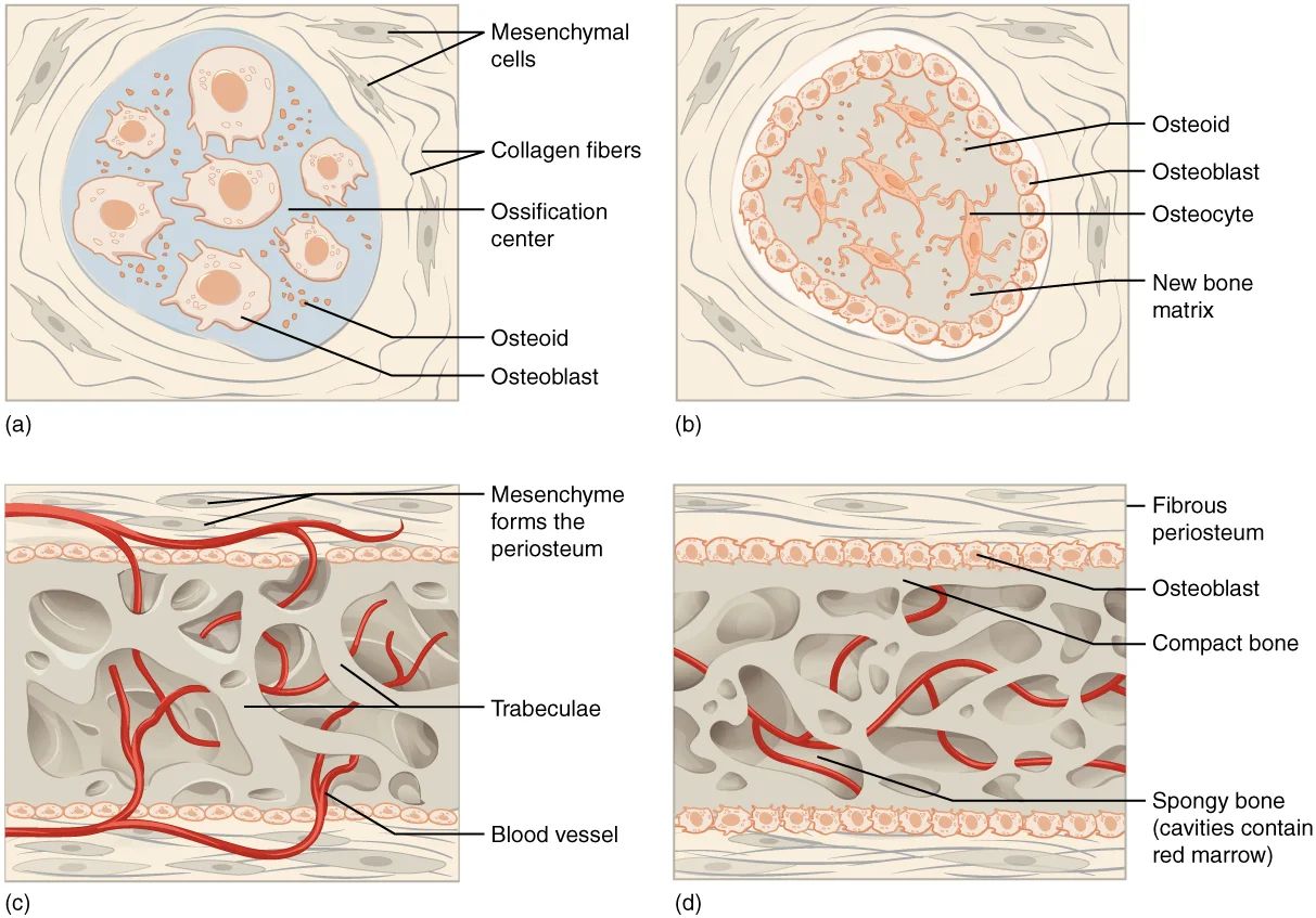
Mesenchymal cells accumulate at specific sites and differentiate directly into osteoblasts. These osteoblasts secrete osteoid that subsequently mineralises to form trabeculae, which interconnect to create spongy bone surrounded by compact bone. The developing skull bones remain separated by dense connective tissue membranes known as fontanelles, allowing deformation of the skull. [1][2]
Figure 1611 Intramembraneous Ossification
Long description
Intramembranous ossification follows four steps. (a) Mesenchymal cells group into clusters, and ossification centers form. (b) Secreted osteoid traps osteoblasts, which then become osteocytes. (c) Trabecular matrix and periosteum form. (d) Compact bone develops superficial to the trabecular bone, an

Figure 1: Intramembranous ossification follows four steps.
At birth, the skull and clavicles are not fully ossified nor are the sutures of the skull closed.
The clavicles are unique among long bones as they ossify via this intramembranous route. The last bones to complete intramembranous ossification are the flat bones of the face, which attain adult dimensions only after the adolescent growth spurt. [2]
Endochondral ossification is the process by which most of the skeleton, including the appendicular skeleton and the base of the skull, is formed. It begins with the formation of a hyaline cartilage model.
By the time the fetal skeleton is fully formed, cartilage only remains at the joint surface as articular cartilage and between the diaphysis and epiphysis as the epiphyseal plate.
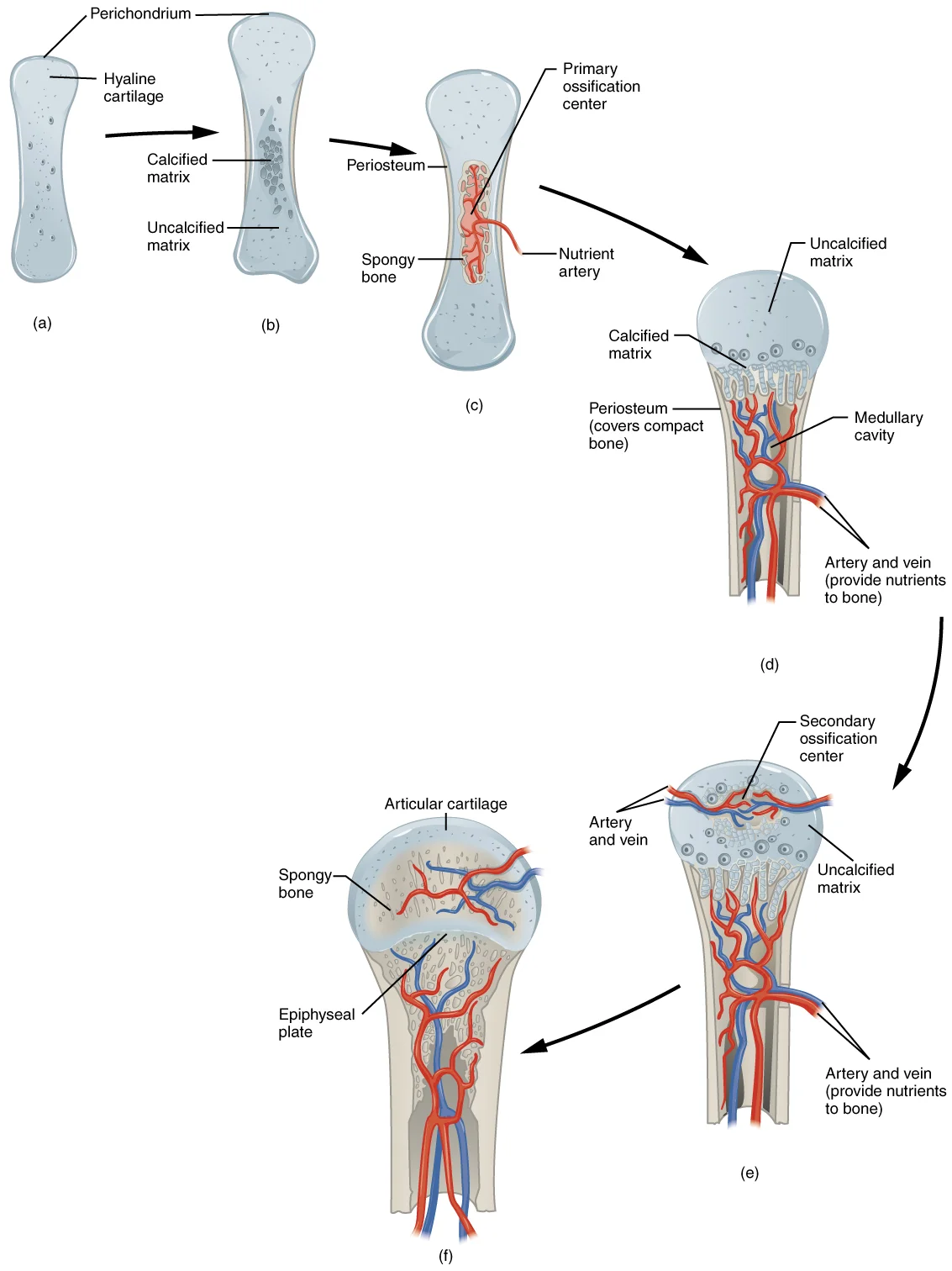
In the limbs, mesenchyme differentiates into a cartilage model. Ossification begins in the diaphysis (shaft) with the invasion of blood vessels and osteogenic cells. Primary ossification centres form, replacing cartilage with bone. Secondary ossification centres appear later in the epiphyses (ends) of the bone. Between the diaphysis and epiphysis lies the epiphyseal plate, a layer of hyaline cartilage responsible for longitudinal bone growth. [2]
Figure 2608 Endochrondal Ossification
Long description
Endochondral ossification follows five steps. (a) Mesenchymal cells differentiate into chondrocytes. (b) The cartilage model of the future bony skeleton and the perichondrium form. (c) Capillaries penetrate cartilage. Perichondrium transforms into periosteum. Periosteal collar develops. Primary ossi

Figure 2: Endochondral ossification follows five steps.
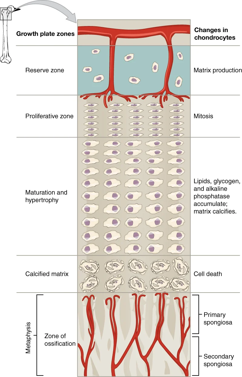
The epiphyseal plate is organised into four distinct zones. The reserve zone, closest to the epiphysis, contains small chondrocytes that anchor the plate to the epiphyseal bone. Moving towards the diaphysis, chondrocytes proliferate, then hypertrophy. In the zone of calcified matrix, the matrix calcifies and chondrocytes die. Capillaries and osteoblasts from the diaphysis penetrate this zone, depositing bone on the calcified cartilage remnants. [2]
A bone grows in length when osseous tissue is added to the diaphysis.
This process continues until skeletal maturity, when the epiphyseal plate ossifies completely, leaving the epiphyseal line.
The vertebrae, ribs, and sternum also develop via endochondral ossification. Mesenchyme from the sclerotomes forms hyaline cartilage models around the notochord. The notochord largely regresses, but remnants persist to form the nucleus pulposus of the intervertebral discs. [2]
The manubrium and body of the sternum are converted into bone first, with the xiphoid process remaining as cartilage until late in life.
The ribs separate from the vertebral cartilage models by the eighth week and ossify, except for the anterior costal cartilage. The base and facial bones of the skull, unlike the cranial vault, develop via endochondral ossification, where the cartilage model is gradually replaced by bone over many years.
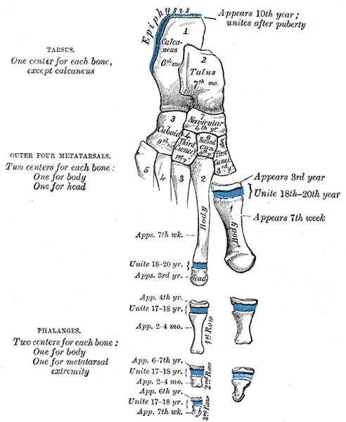
Figure 3Gray289
Long description
Plan of development or ossification of the foot.

Figure 3: Plan of development or ossification of the foot.
From top to bottom, the labels indicate the epiphysis, the reserve zone of chondrocytes, the zone of proliferation, the zone of hypertrophy, and the zone of calcified matrix. Below this lies the diaphysis, where osteoblasts are actively depositing bone on the calcified cartilage spicules.
Figure 4622 Longitudinal Bone Growth
Long description
Illustration from Anatomy & Physiology, Connexions Web site. http://cnx.org/content/col11496/1.6/, Jun 19, 2013.

Figure 4: Illustration from Anatomy & Physiology, Connexions Web site (http://cnx.org/content/col11496/1.6/, jun 19, 2013).
Limb formation begins with the emergence of limb buds on the lateral aspect of the embryo. The upper limb bud appears near the end of the fourth week of development, with the lower limb bud appearing shortly after[1]. By the sixth week, the distal ends of these buds expand into a paddle shape, which will become the hand or foot[1]. Constrictions form at the future wrist, ankle, elbow, and knee sites. Within the paddle, cell death creates separations between the growing digits
Rotation of the limbs occurs during the seventh week. The upper limb rotates laterally by 90 degrees, so that the palm faces anteriorly and the thumb points laterally[1]. In contrast, the lower limb undergoes a 90-degree medial rotation, bringing the big toe to the medial side[1]. Abnormalities in these processes can lead to congenital limb defects. For instance, failure of apoptosis may result in syndactyly (webbed digits), while errors in rotation can cause persistent medial or lateral deviation of the limbs.
Bone growth in length is driven by the epiphyseal plate, where chondrocytes proliferate and mature. The proliferative zone contains stacks of chondrocytes that divide via mitosis to replace those dying at the diaphyseal end. The zone of maturation and hypertrophy contains older, larger cells closer to the diaphysis. Longitudinal growth results from cellular division in the proliferative zone and maturation in the hypertrophic zone. Growth continues until early adulthood, when chondrocytes cease [2]
Once ossification is complete, epiphyseal plates fuse in a specific sequence. The lesser trochanter fuses first at the onset of puberty, around 11 years of age[1]. The greater trochanter follows approximately one year later[1]. The femoral head fuses between 14 and 17 years, while the distal condyles of the femur fuse last, between 16 and 19 years[1]. Radiologically, the cartilage of an epiphyseal plate is less dense than bone, appearing dark on X-rays[1].
The axial skeleton develops from somites, which are block-like mesoderm structures appearing by the fourth week alongside the notochord[1]. The sclerotome, the most medial part of the somite, consists of mesenchyme that forms the bones and cartilages of the body[1]. In the skull, bone formation differs from limb bones. At birth, the brain case and orbits are disproportionately large compared to the jaws and lower face[1]. This reflects the underdevelopment of the ma [3]
During early childhood, the face undergoes rapid growth. The mastoid process enlarges, and the two halves of the mandible and frontal bone fuse into single bones[1]. The paranasal sinuses enlarge, and the jaws expand as teeth appear[1]. These changes contribute to the significant enlargement of the face during childhood[1]. Understanding these developmental milestones helps clinicians assess normal craniofacial growth and identify conditions such as craniosynostosis or delayed dental eruption. [3] [2]
Ossification is the process by which bone tissue is created. It occurs via two primary mechanisms: intramembranous and endochondral ossification. Intramembranous ossification forms flat bones directly from mesenchymal tissue. Endochondral ossification replaces a hyaline cartilage model with bone. This process is responsible for the formation of long bones. [1]
DefinitionIntramembranous Ossification
The direct differentiation of mesenchymal cells into bone-producing cells, forming bone without a cartilage intermediate. [1]
DefinitionEndochondral Ossification
The replacement of a hyaline cartilage model with bone tissue, involving chondrocyte hypertrophy and vascular invasion. [1]
This process begins with the formation of an ossification centre within a fibrous membrane. Mesenchymal cells cluster and differentiate into osteoblasts, which secrete osteoid that later mineralises. [1]
Exam trapFlat Bone Formation
Flat bones of the skull and clavicle develop via intramembranous ossification, not endochondral. [2]
This mechanism involves the replacement of hyaline cartilage. It begins with the ossification centre in the diaphysis. Chondrocytes hypertrophy and the surrounding matrix calcifies. Blood vessels invade the calcified cartilage. Osteoblasts follow these vessels and deposit bone on cartilage remnants. Bone cell development and ossification ramps up and creates the primary ossification center. [2]
| Feature | Intramembranous | Endochondral |
|---|---|---|
| Precursor | Mesenchymal tissue | Hyaline cartilage |
| Primary Bones | Skull, clavicle | Long bones |
| Process | Direct mineralisation | Cartilage replacement |
Defects in ossification can lead to skeletal dysplasias. Rickets results from defective mineralisation of osteoid. This is often due to vitamin D deficiency. Osteomalacia is the adult equivalent of rickets. [2]
NoteVitamin D Role
- Intramembranous ossification forms flat bones directly.
- Endochondral ossification replaces cartilage with bone.
- The diaphysis ossifies before the epiphysis.
- Primary centres appear in the diaphysis.
- Secondary centres appear in the epiphysis.
- The epiphyseal plate allows longitudinal growth.
- The clavicle ossifies earliest in utero.
- Osteoblasts build bone; osteoclasts resorb it.
- Osteocytes maintain mature bone tissue.
- Osteoid is the unmineralised organic matrix.
- Rickets affects children; osteomalacia affects adults.
- Vitamin D deficiency causes soft bones.
Students frequently confuse the timeline and mechanisms of skeletal development, leading to errors in both anatomy and clinical scenarios. A primary error is assuming that bone formation begins with direct ossification of mesenchyme in the limbs. In reality, the bones of the appendicular skeleton arise from embryonic mesenchyme. The initial stage involves the formation of hyaline cartilage models. During the sixth week, mesenchyme within the limb bud begins to differentiate into hyaline cartilage, forming models for future bones. Students often overlook that this cartilaginous template is essential before ossification can occur. [1] [2]
Another frequent mistake involves the orientation of the limbs. It is incorrect to assume the limbs develop in their final adult position. During the seventh week, the upper limbs rotate laterally and the lower limbs rotate medially, bringing the limbs into their final positions. Failing to account for this rotation leads to confusion regarding the anatomical position of structures such as the biceps brachii and the gluteal muscles. [1]
Students also misinterpret the role of the apical ectodermal ridge. It is not merely a structural cap but a critical signalling centre. The apical ectodermal ridge, located at the end of the limb bud, stimulates growth and elongation of the limb. Without this ridge, the proximal-to-distal axis development halts. The ridge stimulates the underlying mesenchyme to rapidly proliferate, producing the outgrowth of the developing limb. [1]
A significant trap relates to the separation of digits. Many believe fingers form as distinct entities from the start. However, during the sixth week, the distal end of the limb bud becomes paddle-shaped, and selective cell death separates the developing fingers and toes. This process of apoptosis is crucial; its failure results in syndactyly. [1]
Regarding skull development, students often confuse fontanelles with sutures. While both involve fibrous tissue, their functions and timelines differ. The largest fontanelle is located on the anterior head, at the junction of the frontal and parietal bones. These fontanelles decrease in size and disappear by age 2. In contrast, the skull bones remained separated from each other at the sutures, which contain dense fibrous connective tissue that unites the adjacent bones. The connective tissue of the sutures allows for continued growth of the skull bones as the brain enlarges during childhood growth. Confusing the closure of fontanelles with the fusion of sutures is a common error. [3]
Finally, students misunderstand the cellular transition in bone formation. It is not instantaneous. The osteoblasts secrete osteoid, uncalcified matrix, which calcifies (hardens) within a few days as mineral salts are deposited on it, thereby entrapping the osteoblasts within[1]. This transition is vital for maintaining bone matrix. [2]
Exam trapCartilage vs Bone
Remember that limbs do not ossify directly. They first form hyaline cartilage models during the sixth week.
DefinitionApical Ectodermal Ridge
A thickened ectoderm at the limb bud tip that stimulates mesenchymal proliferation and limb elongation.
Understanding bone development is critical at the bedside, in the operating theatre, on imaging, and in examinations. At the bedside, knowledge of fontanelles and sutures is essential for assessing infant head growth. The connective tissue of the sutures allows for continued growth of the skull bones as the brain enlarges during childhood growth. Palpating the anterior fontanelle helps clinicians assess intracranial pressure and hydration status. Since the fontanelles decrease in size and disappear by age 2, their persistence beyond this age warrants investigation for conditions like hypothyroidism or rickets.
In the operating theatre, awareness of limb rotation is vital for surgical approaches and fracture reduction. During the seventh week, the upper limbs rotate laterally and the lower limbs rotate medially, bringing the limbs into their final positions. This rotation dictates the anatomical orientation of muscles and nerves. For instance, the lateral rotation of the upper limb places the biceps brachii anteriorly. Misunderstanding this can lead to errors in identifying neurovascular bundles during surgery.
On imaging, recognising the stages of ossification helps in diagnosing developmental disorders. The process begins with mesenchymal condensation followed by cartilage model formation. During the sixth week, mesenchyme within the limb bud begins to differentiate into hyaline cartilage, forming models for future bones. Radiologists look for the appearance of ossification centres, which appear after the cartilage model is established. The separation of digits relies on selective cell death. During the sixth week, the distal end of the limb bud becomes paddle-shaped, and selective cell death separates the developing fingers and toes. Failure of this process is visible on prenatal ultrasound or postnatal X-rays.
In examinations, students must accurately describe the sequence of events. The initial limb bud consists of mesenchyme covered by ectoderm. Initially, the limb buds consist of a core of mesenchyme covered by a layer of ectoderm. The apical ectodermal ridge forms at the end of this ectoderm. The ectoderm at the end of the limb bud thickens to form a narrow crest called the apical ectodermal ridge. This ridge drives proximal-to-distal growth. As the limb bud elongates, cells located farther from the apical ectodermal ridge slow their rates of cell division and begin to differentiate. This gradient explains why proximal structures form before distal ones. [1]
Clinical pearlFontanelle Closure
The anterior fontanelle typically closes by age 2, but sutures remain patent for skull growth.
NoteLimb Rotation
Upper limbs rotate laterally; lower limbs rotate medially during the seventh week.
Limb Development Sequence
- Week 4Limb buds appear from mesenchyme
- Week 4-5Apical ectodermal ridge forms
- Question: Ridge active?
Yes
- Mesenchyme proliferates
No
- Growth halts
- Week 6Cartilage models form
- Week 6Digit separation via apoptosis
- Week 7Limb rotation occurs
- Limb buds appear at the end of the fourth week.
- The apical ectodermal ridge stimulates limb elongation.
- Hyaline cartilage models form during the sixth week.
- Digit separation occurs via selective cell death in the sixth week.
- Upper limbs rotate laterally and lower limbs medially in the seventh week.
- Osteoblasts secrete osteoid which calcifies within a few days[1].
- The largest fontanelle is at the junction of frontal and parietal bones.
- Fontanelles disappear by age 2, but sutures allow continued growth.
Answer each one in your head first, then open the answer.
What structure stimulates the underlying mesenchyme to rapidly proliferate in the limb bud?
Show answerHide answer
Apical ectodermal ridgeDuring which week do the upper limbs rotate laterally?
Show answerHide answer
Seventh weekWhat type of tissue forms the initial models for future bones in the limbs?
Show answerHide answer
Hyaline cartilageWhere is the largest fontanelle located?
Show answerHide answer
Junction of the frontal and parietal bonesWhat happens to osteoblasts once they are entrapped in calcified matrix?
Show answerHide answer
They become osteocytes
- Betts JG, Young KA, Wise JA, et al. Development of the Appendicular Skeleton. OpenStax, Anatomy and Physiology 2e (CC BY-NC-SA 4.0). Accessed .
- Betts JG, Young KA, Wise JA, et al. Bone Formation and Development. OpenStax, Anatomy and Physiology 2e (CC BY-NC-SA 4.0). Accessed .
- Betts JG, Young KA, Wise JA, et al. Embryonic Development of the Axial Skeleton. OpenStax, Anatomy and Physiology 2e (CC BY-NC-SA 4.0). Accessed .
- Betts JG, Young KA, Wise JA, et al. Development of Joints. OpenStax, Anatomy and Physiology 2e (CC BY-NC-SA 4.0). Accessed .
Figure credits
- Figure 1: OpenStax College, via Wikimedia Commons, CC BY 3.0, CC BY 3.0.
- Figure 2: OpenStax College, via Wikimedia Commons, CC BY 3.0, CC BY 3.0.
- Figure 3: Henry Vandyke Carter, via Wikimedia Commons, Public domain, Public domain.
- Figure 4: OpenStax College, via Wikimedia Commons, CC BY 4.0, CC BY 4.0.
Educational content for medical students and doctors. It is not a substitute for clinical judgement or local guidelines.