Embryonic folding
Embryonic folding transforms the flat embryonic disc into a cylindrical embryo with distinct body regions.
Embryonic development occurs in distinct stages, with the embryonic period spanning weeks 3-8 post-fertilization. During this time, the flat embryonic disc undergoes significant transformations through folding processes. The embryo folds laterally and at both ends, forming a C-shape with distinct head and tail regions. This folding creates a tube-like structure called the primitive gut, which is lined by endoderm. [1]
The process of embryonic folding begins during the third week and continues through the eighth week of development. By the end of the eighth week, all major organ systems are established in rudimentary form. The folding essentially envelops a portion of the yolk sac, which protrudes with the umbilical cord from what will become the abdomen. The amniotic sac, previously sitting atop the flat embryo, envelops the embryo as it folds. [1]
Somites, which are block-like structures formed from paraxial mesoderm, appear on either side of the neural tube. These somites eventually differentiate into the axial skeleton, skeletal muscle, and dermis. The lateral plate mesoderm contributes to the formation of the intraembryonic coelom, which is a fluid-filled space that will house developing organs. [1]
Organogenesis, the process of organ formation, occurs primarily during weeks four to eight of development. By the end of the embryonic period, all organ systems are structured in rudimentary form, though the organs themselves are either nonfunctional or only semi-functional. The critical period for organ development spans weeks 3-8, with specific structures having particularly sensitive windows during this time. For example, craniopharyngioma, a tumor derived from remnants of Rathke's pouch, may develop if issues occur during the formation of the pituitary gland during this critical period. [1]
The embryonic period is defined by the transformation of a simple bilaminar disc into a complex, three-dimensional organism through the process of folding. This stage is critical because it establishes the basic body plan and positions the internal organs within the protective confines of the amniotic cavity.
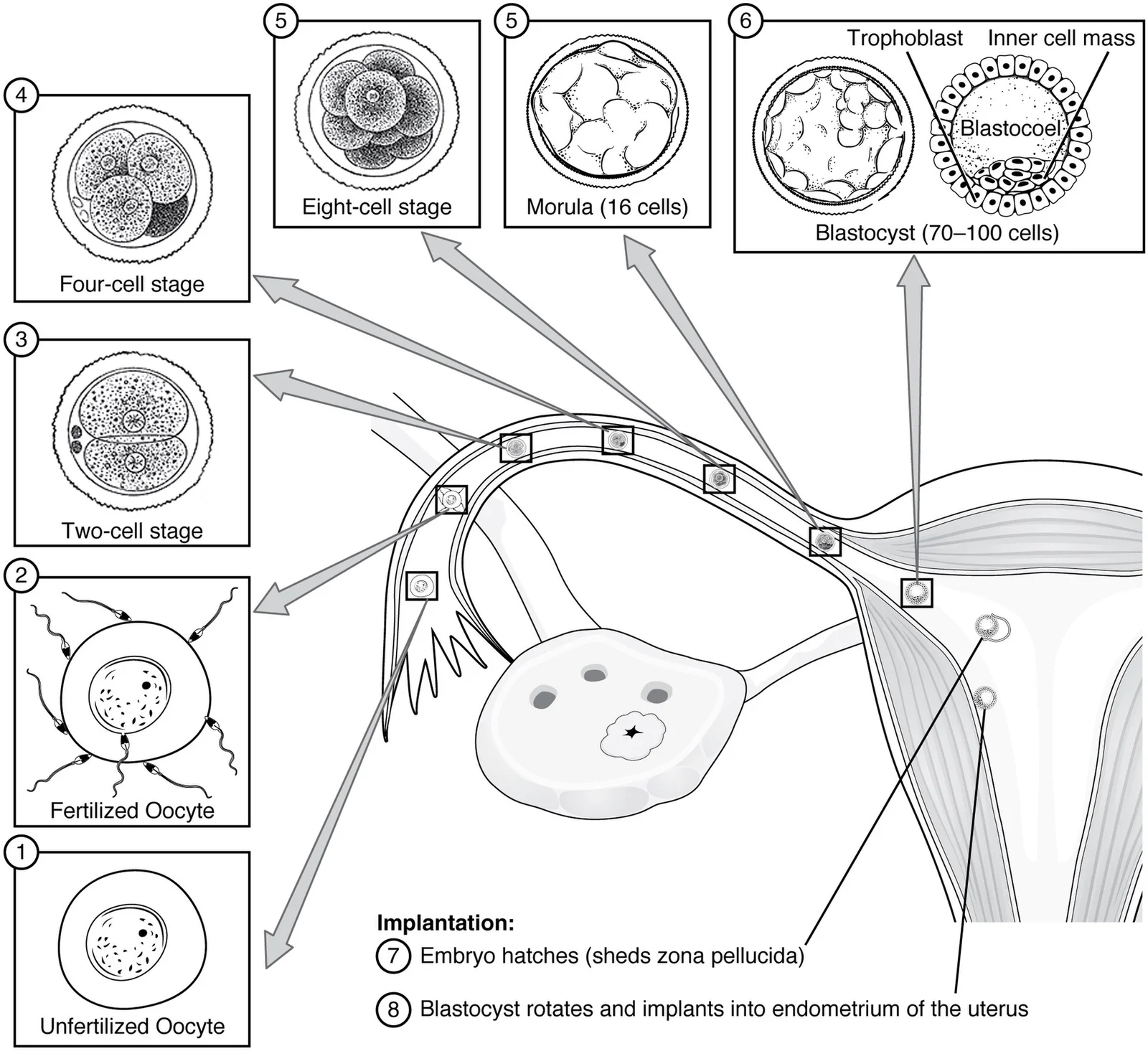
Figure 12904 Preembryonic Development-02
Long description
Ovulation, fertilization, pre-embryonic development, and implantation occur at specific locations within the female reproductive system in a time span of approximately 1 week.

Figure 1: Ovulation, fertilization, pre-embryonic development, and implantation occur at specific locations within the female reproductive system in a time span of approximately 1 week.
The head fold is prominent, bringing the oropharyngeal membrane and the developing heart tube ventrally. The tail fold brings the cloacal membrane to the ventral surface. The lateral body folds curve medially to close the ventral body wall. The amniotic cavity is now large and surrounds the embryo completely. The yolk sac is reduced in size and connected via the vitelline duct. The chorion surrounds the entire conceptus. The placenta is developing between the chorion and the uterine wall. The umbilical cord is forming, connecting the embryo to the placenta. The neural tube is closed in the cranial region. The heart is beating and looping. The limb buds are beginning to appear. The allantois is extending into the connecting stalk. The yolk sac is extra-embryonic. The amnion is extra-embryonic. The chorion is extra-embryonic. The embryo is intra-embryonic. The folding process brings the intra-embryonic coelom into the thoracic and abdominal cavities. The gut tube is formed from the foregut, midgut, and hindgut. The foregut is derived from the cranial part of the yolk sac. The midgut is connected to the yolk sac via the vitelline duct. The hindgut is distal to the vitelline duct. The folding is driven by the rapid growth of the neural tube and the liver. The neural tube grows faster than the surrounding mesoderm. This differential growth causes the embryo to fold. The head fold results from the growth of the brain and the heart. The tail fold results from the relative overgrowth of the neural tube compared to the tail region. The lateral folds result from the expansion of the intra-embryonic coelom. The folding process is essential for the formation of the body cavities. It also positions the gut tube within the abdominal cavity. The umbilical cord forms from the connecting stalk and the vitelline duct. The connecting stalk contains the allantois. The vitelline duct connects the midgut to the yolk sac. The umbilical arteries and vein run through the connecting stalk. The umbilical vein carries oxygenated blood from the placenta to the embryo. The umbilical arteries carry deoxygenated blood from the embryo to the placenta. The folding process is complete by the end of the fourth week. [1]
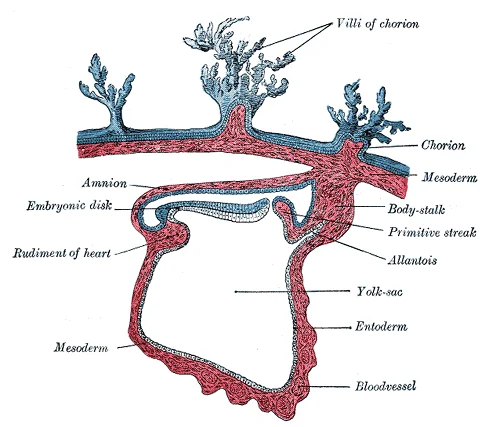
Figure 2Gray21
Long description
Section through the embryo which is represented in Fig. 17. (After Graf Spee.)

Figure 2: Section through the embryo which is represented in Fig (17).
DefinitionEmbryonic Folding
The process by which the flat bilaminar embryonic disc folds into a three-dimensional cylindrical embryo, establishing the body cavities and positioning the gut tube.
The inner mass of embryonic cells is totipotent during this stage, meaning that each cell has the potential to differentiate into any cell type in the human body. Totipotency lasts for only a few days before the cells’ fates are set as being the precursors to a specific lineage of cells. By the end of the embryonic period, the embryo is approximately 3 cm (1.2 in) from crown to rump and weighs approximately 8 g (0.25 oz). [1]
The axial skeleton begins to form during early embryonic development. However, growth, remodeling, and ossification (bone formation) continue for several decades after birth before the adult skeleton is fully formed. Knowledge of the developmental processes that give rise to the skeleton is important for understanding the abnormalities that may arise in skeletal structures. [2]
The bones of the skull arise from mesenchyme during embryonic development in two different ways. The first mechanism produces the bones that form the top and sides of the brain case. This involves the local accumulation of mesenchymal cells at the site of the future bone. These cells then differentiate directly into bone producing cells, which form the skull bones through the process of intramembranous ossification. As the brain case bones grow in the fetal skull, they remain separated from each other by large areas of dense connective tissue. [2]
The second mechanism for bone development in the skull produces the facial bones and floor of the brain case. This also begins with the localized accumulation of mesenchymal cells. However, these cells differentiate into cartilage cells, which produce a hyaline cartilage model of the future bone. As this cartilage model grows, it is gradually converted into bone through the process of endochondral ossification. This is a slow process and the cartilage is not completely converted to bone until the skull achieves its full adult size. [2]
Development of the vertebrae begins with the accumulation of mesenchyme cells from each sclerotome around the notochord. These cells differentiate into a hyaline cartilage model for each vertebra, which then grow and eventually ossify into bone through the process of endochondral ossification. As the developing vertebrae grow, the notochord largely disappears. However, small areas of notochord tissue persist between the adjacent vertebrae and this contributes to the formation of each intervertebral disc. [2]
Embryonic folding is a critical morphogenetic process that transforms the flat trilaminar embryonic disc into a cylindrical embryo. This transformation is driven by differential growth rates among the three germ layers and the expansion of specific structures, particularly the neural tube and the yolk sac. The process occurs simultaneously in two planes: cranial-caudal (head-to-tail) folding and lateral folding.
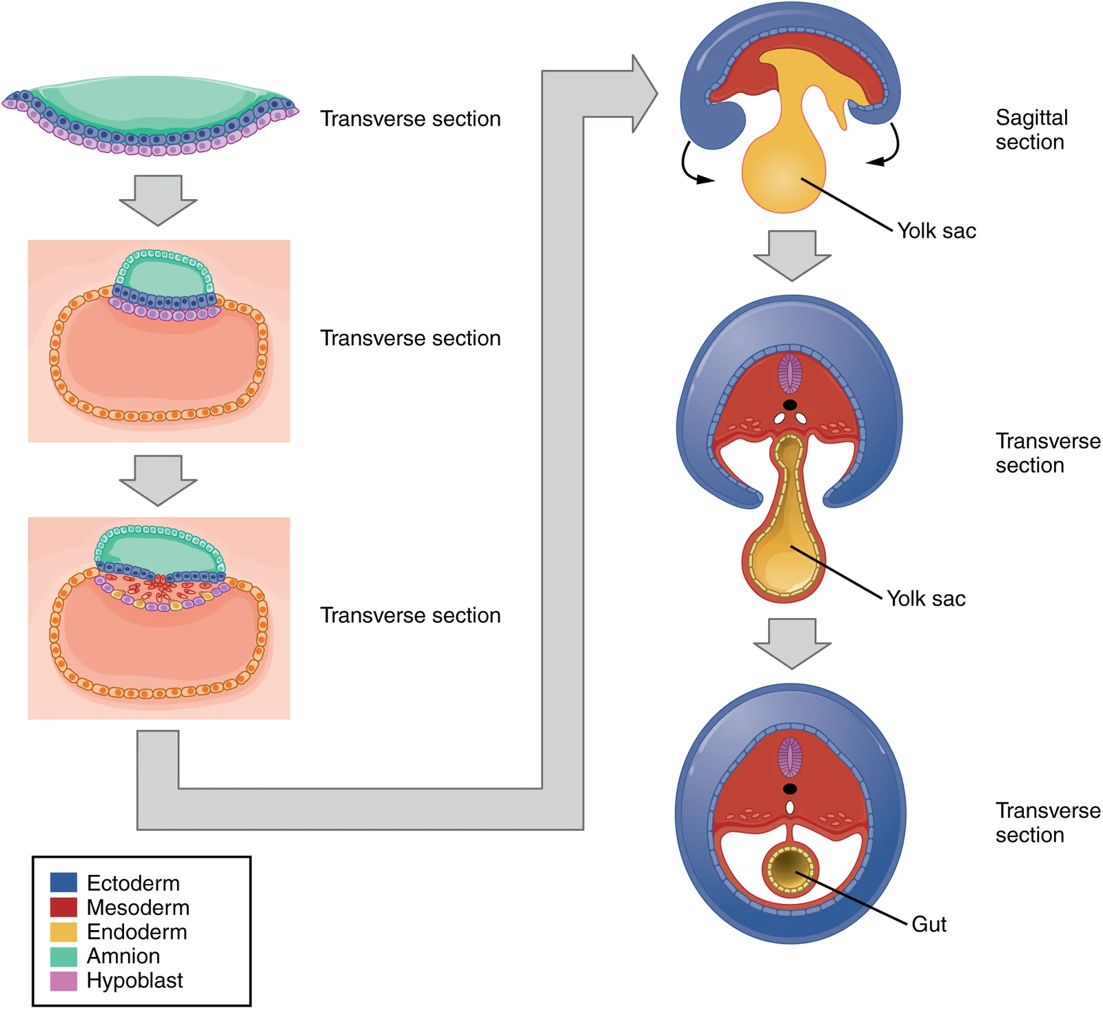
Figure 32913 Embryonic Folding
Long description
Embryonic folding converts a flat sheet of cells into a hollow, tube-like structure.

Figure 3: Embryonic folding converts a flat sheet of cells into a hollow, tube-like structure.
The central structure is the neural tube, which has closed and is positioned dorsally. Ventral to the neural tube is the heart field, which will bulge into the pericardial cavity. The chorionic cavity surrounds the embryo, separated from the yolk sac by the connecting stalk.
The molecular regulation of folding involves complex signalling pathways. Fibroblast growth factors (FGFs) and bone morphogenetic proteins (BMPs) play key roles in inducing and maintaining the germ layers. Wnt signalling is crucial for posterior development and tail folding. Shh (Sonic hedgehog) signalling from the notochord and floor plate influences ventral patterning. These signals coordinate the rapid growth of the neural tube, which pushes the head and tail regions forward and downward, initiating cranial and caudal folding.
Lateral folding is driven by the growth of the somatic mesoderm and the overlying ectoderm. As the lateral body walls fold ventrally, they meet at the midline, enclosing the gut tube. The yolk sac is partially incorporated into the embryo to form the primitive gut. The connecting stalk, which contains the allantois, becomes the umbilical cord. The amnion expands to surround the embryo, filling the chorionic cavity.
DefinitionEmbryonic Folding
The process by which the flat trilaminar embryonic disc folds into a cylindrical embryo, occurring in cranial-caudal and lateral planes, driven by differential growth and signalling pathways.
The outcome of embryonic folding is the establishment of the basic body plan. The head and tail folds bring the heart and foregut into the thoracic and abdominal cavities. The lateral folds close the ventral body wall, enclosing the intraembryonic coelom. The yolk sac is reduced to a small vestigial structure, while the allantois contributes to the formation of the urinary bladder and umbilical vessels.
| Structure | Role in Folding |
|---|---|
| Neural Tube | Rapid growth drives cranial and caudal folding |
| Yolk Sac | Incorporation into gut tube during lateral folding |
| Connecting Stalk | Forms umbilical cord, connects embryo to placenta |
| Amnion | Expands to surround embryo, fills chorionic cavity |
Disruptions in molecular signalling can lead to severe congenital anomalies. Defects in neural tube closure can result in spina bifida or anencephaly. Abnormal lateral folding can cause gastroschisis or omphalocele, where abdominal contents fail to be properly enclosed. Understanding these signalling pathways is essential for diagnosing and managing such conditions.
The placenta, formed from the chorion and decidua basalis, provides essential support during this period. The chorionic villi burrow into the endometrium, establishing maternal-fetal exchange. This interaction is regulated by trophoblast proliferation and vascular remodelling, ensuring adequate nutrient and oxygen supply to the rapidly growing embryo. [1]
Embryonic folding is the critical morphogenetic process that transforms the flat, trilaminar embryonic disc into a cylindrical embryo. This transformation is driven by the rapid growth of the neural tube and somites, which outpace the expansion of the surrounding extraembryonic mesoderm. As the head and tail folds occur, the ventral aspect of the embryo is drawn inward, while the dorsal aspect remains relatively open. This mechanical reshaping brings the lateral body walls together in the midline, effectively enclosing the primitive gut tube within the abdominal cavity. The process also establishes the definitive body plan, positioning the heart, lungs, and future abdominal organs in their correct anatomical relationships.
The central structure is the developing gut tube, which is suspended by the dorsal mesentery. Lateral to the gut tube are the lateral body folds, which have moved medially to close the ventral body wall. The amniotic cavity is large and surrounds the embryo, while the chorionic cavity is reduced due to the folding process.
The derivatives of embryonic folding are diverse and include structures derived from the endoderm, mesoderm, and ectoderm. The endoderm forms the epithelial lining of the gastrointestinal and respiratory tracts. The mesoderm contributes to the connective tissue, muscle, and vascular components of these organs. The ectoderm gives rise to the nervous system and epidermis. Understanding these derivatives is essential for correlating embryological anomalies with clinical presentations. [1]
| Embryonic Structure | Adult Derivative |
|---|---|
| Foregut | Pharynx, oesophagus, stomach, proximal duodenum, liver, pancreas |
| Midgut | Distal duodenum, jejunum, ileum, caecum, ascending colon, proximal 2/3 of transverse colon |
| Hindgut | Distal 1/3 of transverse colon, descending colon, sigmoid colon, rectum, upper anal canal |
| Yolk Sac | Vitelline duct (obliterates), primitive germ cells |
| Allantois | Urachus (obliterates), median umbilical ligament |
The foregut derivatives are supplied by the coeliac trunk, the midgut by the superior mesenteric artery, and the hindgut by the inferior mesenteric artery. This vascular supply is a direct consequence of the folding process, which aligns the gut tube with the developing cardiovascular system. The dorsal mesentery suspends the gut from the posterior abdominal wall, allowing for mobility and growth. The ventral mesentery is present only in the foregut region, forming the lesser omentum and falciform ligament.
Embryonic folding is not merely a change in shape but a fundamental reorganisation of tissue layers that establishes the adult body plan.
The process is complete by the end of the fourth week. At this stage, the embryo is recognisably human, with distinct head, trunk, and tail regions. The umbilical cord forms from the connecting stalk and the yolk sac, providing a conduit for blood vessels and the vitelline duct. Any disruption in this process can lead to serious congenital anomalies, such as gastroschisis or omphalocele, where the abdominal contents fail to return to the body cavity or close properly.
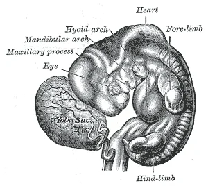
Figure 4Gray23
Long description
Human embryo from thirty-one to thirty-four days. (His.)

Figure 4: Human embryo from thirty-one to thirty-four days (His.).
Figure 5Gray22
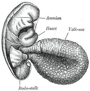
Long description
Human embryo of 2.6 mm. (His.). Labelled illustration from Wikimedia Commons, used under its open licence.

Figure 5: Human embryo of 2.6 mm (His.).
DefinitionEmbryonic Folding
The process by which the flat trilaminar embryonic disc is transformed into a cylindrical embryo through head and tail folding, enclosing the gut tube and establishing the body cavity.
Embryonic folding is the critical morphogenetic event that transforms the flat, trilaminar embryonic disc into a cylindrical embryo with a defined head, tail, and lateral body walls. This process occurs primarily during the fourth week of development, coinciding with the rapid growth of the neural tube and the formation of the heart tube. Failure of proper folding leads to a spectrum of congenital anomalies, ranging from minor structural defects to lethal malformations. Understanding these anomalies requires tracing the movement of the gut tube, the closure of the body wall, and the positioning of the heart.
At the cranial end, the head fold brings the oropharyngeal membrane and the heart field ventrally. The heart, initially dorsal to the foregut, swings ventrally into the thoracic cavity. The midgut remains connected to the yolk sac via the vitelline duct, which lies outside the body cavity initially. At the caudal end, the tail fold brings the cloacal membrane ventrally. The lateral body folds move medially to close the ventral body wall. The amniotic cavity expands to surround the embryo completely. The yolk sac is reduced and incorporated into the primitive gut. The allantois extends into the connecting stalk. The placenta remains attached via the umbilical cord. The relationship between the heart and the foregut is key; the heart must move ventrally to allow the lungs to develop in the thorax. The midgut herniation is a normal part of this process, occurring as the abdominal cavity is too small to contain the rapidly growing gut.
The clinical significance of understanding embryonic folding extends to surgical correction and genetic counselling. Defects in folding are often associated with chromosomal abnormalities, such as Trisomy 18 or Trisomy 13, necessitating comprehensive genetic testing when structural anomalies are detected. Early detection allows for multidisciplinary planning, including potential fetal surgery or postnatal repair. For instance, spina bifida may require in-utero repair to preserve neurological fun [1]
Cranial folding errors can lead to holoprosencephaly or exencephaly. Exencephaly, where the brain is exposed due to failure of the cranial vault to close, is a severe defect. The oropharyngeal membrane must rupture at the correct time; premature rupture can lead to facial clefts. The cloacal membrane must also rupture properly; failure can result in imperforate anus or persistent cloaca.
The heart's position is also dependent on folding. If the heart fails to move ventrally correctly, it may remain dorsal, leading to severe cardiac malformations. The connection between the heart and the foregut is mediated by the transverse pericardial sinus, which forms as the heart folds. Errors here can lead to persistent truncus arteriosus or other conotruncal defects.
The umbilical cord's structure is also a product of folding. It contains two umbilical arteries and one umbilical vein, surrounded by Wharton’s jelly. Abnormalities in the number of vessels, such as a single umbilical artery, are markers for other congenital anomalies. The vitelline duct normally obliterates; persistence can lead to Meckel’s diverticulum, a true diverticulum containing all layers of the gut wall.
Clinical pearlFolding Principle
Embryonic folding converts a 2D disc into a 3D tube, integrating the gut, heart, and body wall. Errors in this process are catastrophic and multifaceted.
The ribs and sternum also develop from mesenchyme. The ribs initially develop as part of the cartilage model for each vertebra, but in the thorax region, the rib portion separates from the vertebra by the eighth week. The cartilage model of the rib then ossifies, except for the anterior portion, which remains as the costal cartilage. The sternum initially forms as paired hyaline cartilage models on either side of the anterior midline, beginning during the fifth week of development. The cartilage models of the ribs become attached to the lateral sides of the developing sternum. Eventually, the two halves of the cartilaginous sternum fuse together along the midline and then ossify into bone. The manubrium and body of the sternum are converted into bone first, with the xiphoid process remaining as cartilage until late in life. [2]
| Structure | Developmental Event | Timing |
|---|---|---|
| Sternum | Paired hyaline cartilage models form | Fifth week |
| Ribs | Separate from vertebrae | Eighth week |
| Sternum | Two halves fuse at midline | Post-fifth week |
| Xiphoid | Remains cartilage | Late in life |
Embryonic folding is a critical morphogenetic process occurring during the fourth week of development, transforming the flat trilaminar embryonic disc into a three-dimensional cylindrical embryo. This complex movement involves simultaneous cephalocaudal and lateral folding, driven by the rapid growth of the neural tube and somites, particularly within the developing brain and spinal cord. The cephalocaudal fold results from the disproportionate growth of the forebrain and heart tube, causing the cranial end of the embryo to bend ventrally. Consequently, the oropharyngeal membrane comes to lie at the future ventral surface of the head, while the yolk sac is partially incorporated into the developing gut.
DefinitionCephalocaudal Folding
The bending of the embryo along its long axis due to rapid growth of the neural plate and somites, establishing the head and tail regions.
Lateral folding occurs as the lateral plate mesoderm splits into somatic and splanchnic layers, and the embryo's sides fold ventrally towards the midline. This process brings the lateral edges of the embryonic disc together, enclosing a portion of the yolk sac to form the primitive gut tube.
Prenatal diagnosis of abnormalities related to defective folding relies heavily on ultrasound imaging and maternal serum screening. Elevated levels of alpha-fetoprotein (AFP) in maternal serum are a key biochemical marker for open neural tube defects and ventral wall defects. Ultrasound can visualize structural anomalies such as exencephaly, spina bifida, or abdomin [1]
| Defect | Primary Cause | Diagnostic Marker |
|---|---|---|
| Gastroschisis | Failure of lateral folding | Normal AFP |
| Omphalocele | Failure of lateral folding | Elevated AFP |
| Spina Bifida | Failure of neural tube closure | Elevated AFP |
| Exencephaly | Cephalocaudal folding defect | Elevated AFP |
Prenatal diagnosis of abnormalities related to defective folding relies heavily on ultrasound imaging and maternal serum screening. Elevated levels of alpha-fetoprotein (AFP) in maternal serum are a key biochemical marker for open neural tube defects and ventral wall defects. Ultrasound can visualize structural anomalies such as exencephaly, spina bifida, or abdomin
Clinical pearlAFP Specificity
While AFP is elevated in open neural tube defects, it remains normal in closed defects like meningocele, distinguishing it from open myelomeningocele.
In summary, embryonic folding is a fundamental event that establishes the basic body plan. Disruptions in this process lead to significant congenital anomalies detectable through modern prenatal screening. Understanding the mechanisms of folding provides the foundation for recognizing and managing these clinical conditions, ensuring optimal outcomes for affected pregnancies. The integration of anatomical knowledge with diagnostic techniques remains essential for medical practitioners managing prenatal care.
The skull undergoes significant morphological changes from birth through childhood, driven by functional demands and growth patterns. Understanding these changes is critical for interpreting paediatric imaging and clinical presentations.
At birth, the neurocranium is disproportionately large compared to the viscerocranium. This reflects the relative underdevelopment of the maxilla and mandible, which lack teeth, and the small sizes of the paranasal sinuses and nasal cavity. The brain case and orbits of the skull are disproportionally large compared to the bones of the jaws and lower face. This disproportion is a normal physiological state, not a pathological one. [2]
Clinical pearlNeurocranium dominance
The brain case and orbits of the skull are disproportionally large compared to the bones of the jaws and lower face.
The underdevelopment of the facial skeleton is primarily due to the absence of dentition. The jaws also expand as the teeth begin to appear. Without teeth, the maxilla and mandible lack the structural support and growth stimuli provided by occlusion. Consequently, the paranasal sinuses and nasal cavity remain small at birth. [2]
During early childhood, specific skeletal events occur in a predictable sequence. The mastoid process enlarges. This enlargement is associated with the development of the sternocleidomastoid muscle attachment and the pneumatization of the mastoid air cells.
Exam trapMastoid development
Do not assume the mastoid process is fully developed at birth. During early childhood, the mastoid process enlarges.
Fusion of cranial bones is another key developmental milestone. The frontal bone consists of two halves at birth, which fuse later in childhood. [2]
These changes all contribute to the rapid growth and enlargement of the face during childhood. The expansion of the jaws is closely linked to dental eruption. As the teeth begin to appear, the alveolar processes develop, increasing the vertical height of the face. [2]
DefinitionViscerocranium growth
The jaws also expand as the teeth begin to appear. This expansion is driven by the need to accommodate the developing dentition.
The paranasal sinuses enlarge during childhood. This pneumatization continues well into adolescence. The maxillary and ethmoid sinuses are present at birth but are very small. The frontal and sphenoid sinuses develop later.
Clinical correlation: The large head size in infants is normal. However, disproportionate growth may indicate pathology. For example, hydrocephalus causes excessive enlargement of the neurocranium. Conversely, micrognathia results in a small mandible.
| Structure | Change | Timing |
|---|---|---|
| Mastoid process | Enlarges | Early childhood |
| Mandible halves | Fuse | Early childhood |
| Frontal bone halves | Fuse | Early childhood |
| Jaws | Expand | With teeth eruption |
| Paranasal sinuses | Enlarge | Early childhood |
Remember that the relative underdevelopment of the maxilla and mandible is due to the lack of teeth. This is a fundamental concept in paediatric dentistry and orthodontics. The small sizes of the paranasal sinuses and nasal cavity also reflect this lack of functional demand.
As the child grows, the face becomes more prominent. The chin projects forward, and the forehead recedes slightly. These changes are part of the normal maturation of the skull.
NoteGrowth pattern
These changes all contribute to the rapid growth and enlargement of the face during childhood. This growth is not uniform but follows specific patterns.
In summary, the skull transforms from a predominantly neurocranial structure to a more balanced neurovisceral structure. The key drivers are dental eruption and sinus pneumatization. The fusion of the mandible and frontal bones completes the basic skeletal framework.
Students frequently confuse the structural components of the early conceptus with the subsequent embryonic layers. A primary error is assuming that the trophoblast contributes to the embryo proper. The trophoblast forms the outer shell and develops into the chorionic sac and the fetal portion of the placenta, not the embryo itself[1]. The embryo arises from the inner cell mass, which differentiates into the three germ layers.
Another frequent mistake involves the timing and mechanism of implantation. Students often believe the blastocyst implants while still enclosed in the zona pellucida. In reality, the trophoblast excretes enzymes that degrade the zona pellucida in a process called “hatching,” allowing the conceptus to break free before adhering to the endometrium[1]. Implantation only succeeds after this hatching event.
There is also confusion regarding the function of the syncytiotrophoblast. It is not merely a protective layer but a multinucleated body that digests endometrial cells to firmly secure the blastocyst to the uterine wall[1]. This invasive behaviour is crucial for establishing the maternal-fetal interface. Students must distinguish this from the cytotrophoblast, which remains cellular and mitotically active.
A significant trap is misattributing the source of human chorionic gonadotropin (hCG). The hormone is secreted by the trophoblast, specifically the syncytiotrophoblast, not by the embryo or the corpus luteum[1]. Its function is to direct the corpus luteum to survive and continue producing progesterone and estrogen, thereby suppressing menses. This hormonal support is necessary for creating an environment suitable for the developing embryo[1].
Students often overlook the complete timeline of implantation. It is incorrect to state that implantation is immediate upon contact. Implantation is complete by the middle of the second week[1]. Understanding this two-week window is critical for correlating clinical signs with developmental stages.
Another common error is conflating the germ layers with their derivatives. The ectoderm forms the central and peripheral nervous systems, sensory organs, epidermis, hair, and nails[1]. The mesoderm becomes the skeleton, muscles, connective tissue, heart, blood vessels, and kidneys[1]. The endoderm forms the epithelial lining of the gastrointestinal tract, liver, and pancreas, as well as the lungs[1]. Mixing these lineages leads to incorrect anatomical predictions.
Finally, students may ignore the clinical implications of placental permeability. The chorionic villi provide ample surface area for exchange, but this permeability extends to lipid-soluble fetotoxic substances such as alcohol, nicotine, and barbiturates[1]. Alcohol consumption can result in fetal alcohol spectrum disorders, including organ and facial malformations, cognitive and behavioral disorders[1]. Recognizing these risks is part of understanding placental function.
This knowledge is applied at the bedside through the detection of hCG. Because hCG accumulates in the maternal bloodstream and is excreted in the urine, its presence confirms pregnancy[1]. Clinicians use this to monitor the early survival of the conceptus. The hormone’s role in maintaining the corpus luteum is vital for sustaining the pregnancy until the placenta takes over steroidogenesis.
In the operating theatre, understanding the invasion of the syncytiotrophoblast is critical. The fusion of trophoblast cells and their digestive capacity explain the risk of placenta accreta if the decidua basalis is deficient. Surgeons must be aware that the placenta is not a passive organ but an invasive structure anchored by enzymatic digestion of the endometrium[1].
On imaging, ultrasound relies on the distinction between the chorionic sac and the embryo. The chorionic sac develops from the trophoblast[1]. Early scans identify the gestational sac within the uterine cavity. Later, the differentiation of germ layers allows for the identification of specific organ systems. For instance, the development of the neural tube from the ectoderm is a key milestone visible in mid-trimester scans[1].
In examinations, students must accurately map germ layer derivatives. Questions often test the origin of specific tissues. The skeleton and muscles derive from mesoderm[1]. The epidermis and nervous system from ectoderm[1]. The gut lining from endoderm[1]. Precision in these associations is required for high marks.
The fontanelles and sutures are also relevant in clinical practice. The largest fontanelle is located on the anterior head, at the junction of the frontal and parietal bones. These structures decrease in size and disappear by age 2. However, the sutures remain, containing dense fibrous connective tissue that allows for continued skull growth as the brain enlarges. Palpation of these areas helps assess intracranial pressure and developmental progress. [2]
The umbilical blood vessels are another practical focus. Fetal mesenchymal cells differentiate into blood vessels, including the three umbilical blood vessels that connect the embryo to the developing placenta[1]. Understanding this connection is essential for interpreting cord abnormalities and fetal circulation patterns.
The permeability of the placenta informs clinical advice. Pregnant people should avoid fetotoxic substances because the villi allow two-way exchange of substances[1]. This includes pathogens and drugs. Clinicians counsel patients on avoiding alcohol to prevent fetal alcohol spectrum disorders, which include facial malformations and cognitive disorders[1].
Placental Development and Function
- FertilizationZygote forms
- CleavageBlastomeres form; volume constant
- BlastocystTrophoblast (outer) and Inner Cell Mass
- Question: Hatching?
Yes
- Zona Pellucida DegradedEnzymes excreted
No
- No ImplantationConceptus remains enclosed
- ImplantationSyncytiotrophoblast digests endometrium
- hCG SecretionMaintains Corpus Luteum
- Placental FormationVilli develop; Exchange occurs
- Clinical MonitoringDetect hCG; Ultrasound anatomy
The trophoblast forms the chorionic sac and fetal placenta, not the embryo. Hatching involves the degradation of the zona pellucida before implantation. The syncytiotrophoblast digests endometrial cells to secure the blastocyst. hCG is secreted by the trophoblast to maintain the corpus luteum. Implantation is complete by the middle of the second week. Ectoderm forms the nervous system and epidermis. Mesoderm forms the skeleton, muscles, and kidneys. Endoderm forms the gut lining, liver, and lungs.
Answer each one in your head first, then open the answer.
Which cells form the outer shell of the blastocyst?
Show answerHide answer
TrophoblastsWhat is the process called when the conceptus breaks free of the zona pellucida?
Show answerHide answer
HatchingWhich hormone is secreted by the trophoblast to maintain the corpus luteum?
Show answerHide answer
Human chorionic gonadotropin (hCG)When is implantation complete?
Show answerHide answer
By the middle of the second weekWhich germ layer gives rise to the skeleton and muscles?
Show answerHide answer
Mesoderm
- Betts JG, Young KA, Wise JA, et al. Embryonic Development. OpenStax, Anatomy and Physiology 2e (CC BY-NC-SA 4.0). Accessed .
- Betts JG, Young KA, Wise JA, et al. Embryonic Development of the Axial Skeleton. OpenStax, Anatomy and Physiology 2e (CC BY-NC-SA 4.0). Accessed .
Figure credits
- Figure 1: OpenStax College, via Wikimedia Commons, CC BY 3.0, CC BY 3.0. resized.
- Figure 2: Henry Vandyke Carter, via Wikimedia Commons, Public domain, Public domain.
- Figure 3: OpenStax College, via Wikimedia Commons, CC BY 3.0, CC BY 3.0. resized.
- Figure 4: Henry Vandyke Carter, via Wikimedia Commons, Public domain, Public domain.
- Figure 5: Henry Vandyke Carter, via Wikimedia Commons, Public domain, Public domain.
Educational content for medical students and doctors. It is not a substitute for clinical judgement or local guidelines.