Placental development
Embryological formation of extra-embryonic membranes and the placenta from weeks 2 to 16.
During the second week of development, with the embryo implanted in the uterus, cells within the blastocyst start to organize into layers. Some grow to form the extra-embryonic membranes needed to support and protect the growing embryo: the amnion, the yolk sac, the allantois, and the chorion. At the beginning of the second week, the cells of the inner cell mass form into a two-layered disc of embryonic cells, and a space—the amniotic cavity—opens up between it and the trophoblast. Cells from th [1]
On the ventral side of the embryonic disc, opposite the amnion, cells in the lower layer of the embryonic disk (the hypoblast) extend into the blastocyst cavity and form a yolk sac. The yolk sac supplies some nutrients absorbed from the trophoblast and also provides primitive blood circulation to the developing embryo for the second and third week of development. When the placenta takes over nourishing the embryo at approximately week 4, the yolk sac has been greatly reduced in size and its main function is to serve as the source of blood cells and germ cells (cells that will give rise to gametes). During week 3, a finger-like outpocketing of the yolk sac develops into the allantois, a primitive excretory duct of the embryo that will become part of the urinary bladder. Together, the stalks of the yolk sac and allantois establish the outer structure of the umbilical cord. [1]
The last of the extra-embryonic membranes is the chorion, which is the one membrane that surrounds all others. The development of the chorion will be discussed in more detail shortly, as it relates to the growth and development of the placenta. The maternal portion of the placenta develops from the deepest layer of the endometrium, the decidua basalis. To form the embryonic portion of the placenta, the syncytiotrophoblast and the underlying cells of the trophoblast (cytotrophoblast cells) begin to proliferate along with a layer of extraembryonic mesoderm cells. These form the chorionic membrane, which envelops the entire conceptus as the chorion. The chorionic membrane forms finger-like structures called chorionic villi that burrow into the endometrium like tree roots, making up the fetal portion of the placenta. [1]
The placenta develops throughout the embryonic period and during the first several weeks of the fetal period; placentation is complete by weeks 14–16. As a fully developed organ, the placenta provides nutrition and excretion, respiration, and endocrine function. It receives blood from the fetus through the umbilical arteries. Nutrients and oxygen are transferred from maternal blood surrounding the villi through the capillaries and into the fetal bloodstream. Some substances move across the place [1]
Two umbilical arteries carry oxygen-depleted fetal blood, including wastes and carbon dioxide, to the placenta. Remnants of the umbilical arteries remain in the adult. [2]
DefinitionPlacenta
A circulatory organ unique to pregnancy that develops jointly from the embryo and uterine wall structures to fill the growing embryo's need for nutrients and gas exchange.
DefinitionDecidua basalis
The maternal portion of the placenta, developing from the deepest layer of the endometrium.
DefinitionAmniotic fluid
Early fluid is a filtrate of maternal plasma; from the eighth week, fetal urine adds to the volume.
NoteTwinning
Identical twins arise from the splitting of the inner cell mass; fraternal twins from two separate zygotes.
NoteTwin-twin transfusion
A complication in monochorionic twins where vascular connections in the shared placenta cause unequal blood flow.
DefinitionUmbilical cord
Surrounded by the amnion, with spaces filled with Wharton’s jelly, a mucous connective tissue.
DefinitionUrachus
Remnant of the allantois, a primitive excretory duct that becomes part of the urinary bladder.
DefinitionVitelline duct
Remnant of the yolk sac stalk, connecting the midgut to the yolk sac during early development.
Figure 1Gray28
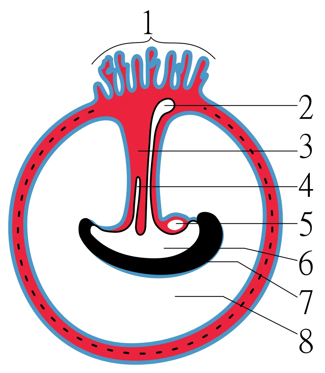
Long description
Diagram illustrating a later stage in the development of the umbilical cord. 1 Placental villi 2 Yolk-sac 3 Umbilical cord 4 Allantois 5 Heart 6 Digestive tube 7 Embryo 8 Amniotic cavity

Figure 1: Diagram illustrating a later stage in the development of the umbilical cord.
The period required for full development in utero is termed gestation. This period is subdivided into distinct stages. The first two weeks are the pre-embryonic stage, characterized by rapid cell division and migration. From week 3 to week 8, the developing human is an embryo. During this embryonic phase, the embryo implants within the uterine body in most cases. However, in one to two percent of cases, implantation occurs outside the uterus or in a location that creates complications. For instance, if implantation occurs in the inferior portion of the uterus, the placenta may grow over the cervical opening, a condition known as placenta previa. [1]
From the ninth week until birth, the developing human is a fetus. This 30-week period is marked by continued cell growth and differentiation. These processes fully develop the structures and functions of the immature organ systems formed during the embryonic period. The completion of fetal development results in a newborn capable of survival outside the womb, although still immature in many ways. [3] [1]
DefinitionGestation
The period of time required for full development of a fetus in utero, subdivided into distinct gestational periods.
DefinitionEmbryo
A developing human referred to during weeks 3–8 of gestation, characterized by cell division, migration, and differentiation.
DefinitionFetus
A developing human referred to from the ninth week of gestation until birth.
Figure 2Gray39
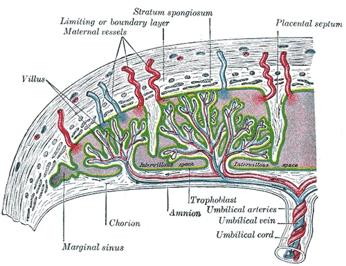
Long description
Scheme of placental circulation. Labelled illustration from Wikimedia Commons, used under its open licence.

Figure 2: Scheme of placental circulation.
The molecular regulation of placental development begins with the critical process of implantation, which occurs at the end of the first week of development. The blastocyst comes into contact with the uterine wall and adheres to it via trophoblast cells, embedding itself in the uterine lining. This process marks the end of the pre-embryonic stage and is initiated when the blastocyst typically implants in the fundus of the uterus or on the posterior wall. However, implantation failure is common, with 50-75 percent of blastocysts failing to implant when the endometrium is not fully developed. [1]
During the third week of development, gastrulation transforms the two-layered embryonic disc into a three-layered structure through cell migration and differentiation. The primitive streak forms along the dorsal surface of the epiblast, with a node at its caudal end emitting growth factors that direct cell migration. These migrating cells give rise to the three germ layers: the endoderm (adjacent to the yolk sac), the mesoderm (middle layer), and the ectoderm (remaining epiblast cells). [1]
The mesodermal cells further differentiate into angioblasts and pluripotent stem cells, which form blood islands throughout the embryonic disc. These blood islands develop vessel lumens, with endothelial linings arising from angioblasts, while surrounding mesenchymal cells contribute to the smooth muscle and connective tissue layers of the developing vessels. Simultaneously, the pluripotent stem cells begin forming the formed elements of blood. [2]
The fetal circulatory system integrates with the placenta via the umbilical cord, allowing the fetus to receive oxygen and nutrients. This connection is established through umbilical arteries that carry deoxygenated blood and waste products from the fetus to the placenta, where exchange occurs with maternal circulation. The specialized fetal circulatory system includes three key shunts—the ductus venosus, foramen ovale, and ductus arteriosus—that allow blood to bypass the liver and pulmonary circuit until birth. [3] [2]
DefinitionImplantation
The process by which the blastocyst adheres to and embeds itself in the uterine lining via trophoblast cells, marking the end of the pre-embryonic stage.
DefinitionGastrulation
The process during which the two-layered embryonic disc becomes three-layered through cell migration, establishing the endoderm, mesoderm, and ectoderm.
The placenta is a composite organ derived from both embryonic and maternal tissues, serving as the primary interface for nutrient, waste, and gas exchange. Its formation begins shortly after fertilization, with distinct cellular lineages establishing the structural foundation.
Approximately 3 days after fertilization, a 16-cell conceptus reaches the uterus. The cells compact into a solid mass known as the morula, resembling a "little mulberry". As it floats freely in the uterus, the conceptus divides into a ball of approximately 100 cells, consuming uterine milk while the endometrial lining thickens. Fluid secretion creates a cavity called the blastocoel, defining the blastocyst stage. [1]
Figure 3Placental cotyledon
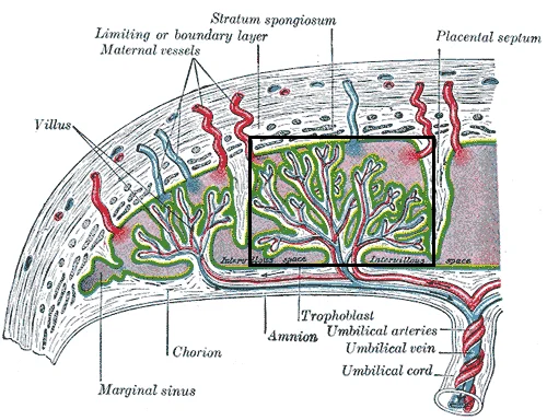
Long description
Derivative of File:Gray39.png Structure of the placenta , with a placental cotyledon marked in rectangle.

Figure 3: Derivative of File:Gray39.png Structure of the placenta , with a placental cotyledon marked in rectangle.
The cells forming the outer shell are called trophoblasts. These cells develop into the chorionic sac and the fetal portion of the placenta. The placenta functions as the organ of nutrient, waste, and gas exchange between the pregnant person and the developing offspring. [1]
Totipotency lasts for only a few days before the cells’ fates are set as precursors to specific lineages. [1]
The placenta is permeable to lipid-soluble fetotoxic substances, including alcohol, nicotine, barbiturates, antibiotics, and certain pathogens. Although blood cells are not exchanged, the chorionic villi provide ample surface area for two-way exchange. The rate of exchange increases throughout gestation as the villi become thinner and increasingly branched. [1]
The embryo folds laterally and at either end, forming a C-shape. The embryo envelops a portion of the yolk sac, which protrudes with the umbilical cord from what will become the abdomen. Blood vessels form from the embryonic mesoderm[1]. The placenta supplies blood rich in oxygen and nutrients via the umbilical vein and removes wastes via the umbilical arteries[1]. The umbilical vein carries oxygenated blood to the fetal heart, bypassing the immature liver via the ductus venosus shunt[1]. [2][3]
| Embryonic Structure | Adult/Functional Derivative |
|---|---|
| Trophoblasts | Chorionic sac and fetal portion of the placenta |
| Inner Cell Mass | The embryo |
| Embryonic Mesoderm | Blood vessels and blood elements[1] |
| Yolk Sac | Part of the umbilical cord structure |
Placental development is the foundation for fetal viability, yet disruptions during this intricate process can lead to significant congenital anomalies. Understanding these defects requires tracing their origins back to the earliest stages of embryogenesis. Within the first 8 weeks of gestation, a developing embryo establishes the rudimentary structures of all of its organs and tissues from the ectoderm, mesoderm, and endoderm. This critical period, known as organogenesis, is when the placenta and its associated membranes form. Any error in the formation of the chorionic villi or the amniotic cavity during this window can result in structural abnormalities that persist throughout pregnancy. [1]
DefinitionAmniotic Fluid Dynamics
Amniotic fluid volume is a key indicator of fetal health, regulated by fetal swallowing and urine output, with deviations indicating potential anomalies.
The volume of amniotic fluid is tightly regulated. Normally, the fetus moves freely within this fluid and can prepare for swallowing and breathing out of the uterus. This dynamic equilibrium is crucial. Polyhydramnios, an excess of amniotic fluid, often arises when the fetus cannot swallow effectively, preventing the normal removal of fluid. Conversely, oligohydramnios, a deficiency of fluid, may result from impaired fetal urine production or rupture of the membranes. These conditions are not merely numerical abnormalities but reflect underlying physiological failures in the placental-fetal unit. [1]
Figure 4Gray26
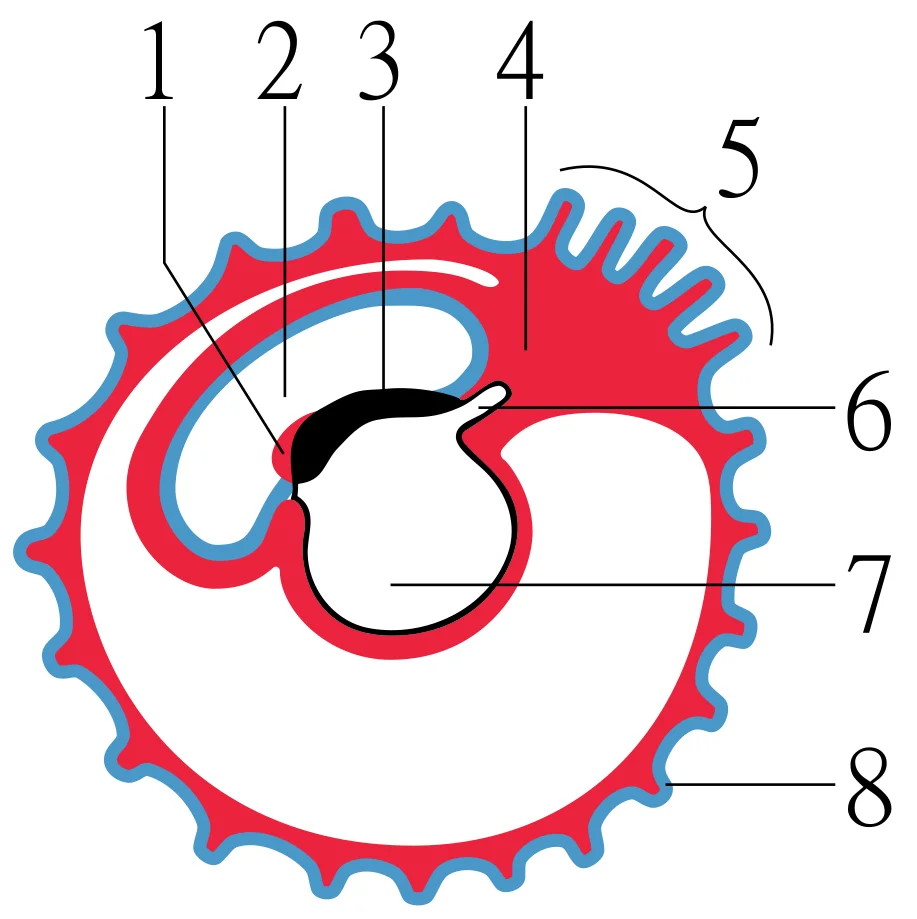
Long description
Diagram showing later stage of allantoic development with commencing constriction of the yolk-sac. 1 Heart 2 Amniotic cavity 3 Embryo 4 Body-stalk 5 Placental villi 6 Allantois 7 Yolk-sac 8 Chorion

Figure 4: Diagram showing later stage of allantoic development with commencing constriction of the yolk-sac.
In the context of polyhydramnios, the fluid space appears distended, pushing the fetal membranes against the uterine wall. In oligohydramnios, the fluid space is reduced, allowing the fetal membranes to adhere closely to the fetus. The umbilical cord connects the fetus to the placenta, serving as the lifeline for nutrient and gas exchange.
| Condition | Definition | Primary Embryological/Physiological Basis |
|---|---|---|
| Polyhydramnios | Excess amniotic fluid | Impaired fetal swallowing or increased fetal urine output |
| Oligohydramnios | Deficient amniotic fluid | Impaired fetal urine production or membrane rupture |
| Structure | Origin | Course | Relations | Termination |
|---|---|---|---|---|
| Chorionic Villi | Trophoblast | Extend into intervillous space | Surrounded by maternal blood | Fuse with decidua basalis |
| Amniotic Cavity | Epiblast | Surrounds the embryo | Bounded by amnion | Fuses with chorion at 12 weeks |
Exam trapSurfactant Timing
Surfactant production is not complete until late gestation; premature birth before this stage leads to respiratory distress, not a placental anomaly per se, but a consequence of incomplete organogenesis.
While placental anomalies are structural, their functional consequences can mirror those of other developmental delays. For instance, inadequate surfactant production in premature newborns may result in respiratory distress syndrome. Although this is primarily a pulmonary issue, it highlights the importance of timing in organogenesis. The fetus at 30 weeks measures 28 cm (11 in) from crown to rump and exhibits the approximate body proportions of a full-term newborn. However, the lungs and other systems may not be fully mature. This underscores that while the placenta provides the environment, the fetus must complete its own organogenesis within that timeframe. Anomalies in placental development can compromise this environment, leading to secondary effects on fetal growth and organ maturation. [3]
The placenta is a transient organ essential for fetal survival, and its development is closely linked to several critical clinical conditions. Understanding the embryological timeline allows clinicians to anticipate complications arising from abnormal implantation or vascular formation.
Normal implantation occurs in the upper posterior or anterior wall of the uterine body. When the blastocyst implants in the lower uterine segment, it may invade the myometrium, leading to placenta accreta spectrum disorders. This condition is characterised by the absence of the decidua basalis, causing the chorionic villi to attach directly to the myometrium. The risk increases significantly with previous caesarean sections.
Exam trapPlacenta Accreta
Placenta accreta is defined as abnormal adherence of the placenta to the uterine wall due to defective decidua basalis.
Another critical anomaly is placenta praevia, where the placenta implants over or near the internal cervical os. This condition presents with painless vaginal bleeding in the second or third trimester. Diagnosis is typically confirmed via ultrasound. The classification depends on the relationship between the placental edge and the cervical os.
| Type | Description |
|---|---|
| Type 1 | Low-lying placenta, edge within 2 cm of os but not covering it. |
| Type 2 | Marginal praevia, edge reaches the margin of the os. |
| Type 3 | Complete praevia, placenta covers the internal os completely. |
| Type 4 | Complete praevia, placenta covers the internal os completely with myometrial invasion. |
The umbilical cord normally contains two arteries and one vein. A single umbilical artery is the most common vascular anomaly, occurring in approximately 1% of singleton pregnancies. This finding is associated with an increased risk of congenital anomalies, particularly renal and cardiac defects. Therefore, the detection of a single umbilical artery should prompt a detailed anatomical survey.
NoteSingle Umbilical Artery
A single umbilical artery is found in 1% of singleton pregnancies and is associated with congenital anomalies.
Cord entanglement, or nuchal cord, occurs when the umbilical cord wraps around the fetal neck. This is common, occurring in about 30% of deliveries. While often benign, tight entanglement can lead to fetal distress during labour due to compression of the vessels.
Prenatal diagnosis relies heavily on ultrasound imaging. The first-trimester scan assesses viability and dating. The nuchal translucency measurement is a key screening tool for chromosomal abnormalities.
DefinitionNuchal Translucency
Nuchal translucency is the sonographic appearance of fluid accumulation in the subcutaneous tissue behind the fetal neck.
In the second trimester, the anatomy scan evaluates fetal structures and placental location. Doppler ultrasound may be used to assess uteroplacental blood flow in high-risk pregnancies, such as those with pre-eclampsia or intrauterine growth restriction. [1]
| Finding | Clinical Significance |
|---|---|
| Early diastolic notch | Increased resistance, associated with pre-eclampsia. |
| Absent end-diastolic flow | Severe placental insufficiency, high risk of fetal demise. |
| Reversed end-diastolic flow | Critical placental failure, requires immediate delivery. |
The placenta also serves as a source of fetal cells for non-invasive prenatal testing. Cell-free fetal DNA in maternal blood can be analysed for common aneuploidies such as trisomy 21, 18, and 13. [1]
Clinical pearlNIPT
Non-invasive prenatal testing uses cell-free fetal DNA from maternal blood to screen for common aneuploidies.
The neural tube rests atop the notochord, a mesoderm-derived rod, which eventually becomes the nucleus pulposus of intervertebral discs. [1]
Block-like structures called somites form on either side of the tube. [1]
During the fourth and fifth weeks, the anterior neural tube dilates. [1]
This angiogenesis—the creation of new blood vessels from existing ones—continues as needed throughout life as we grow and develop. [2]
Sexual differentiation does not begin until the fetal period. This process occurs specifically during weeks 9 to 12 of gestation. Prior to this, embryonic males and females are morphologically identical. Genetic sex is distinguishable, but physical structures are not. Bipotential gonads are present and can develop into either male or female organs. These gonads connect to the cloaca via Müllerian and Wolffian ducts. The cloaca serves as an extension of the primitive gut. Several coordinated even [3]
Students frequently confuse the timing of implantation with the formation of the blastocyst. A common error is assuming that the conceptus implants immediately upon entering the uterine cavity. In reality, the conceptus must first undergo "hatching" to break free of the zona pellucida before it can adhere to the endometrium[1]. This hatching process is a prerequisite for implantation, not a concurrent event. Another frequent mistake is attributing the maintenance of the corpus luteum directly to the developing embryo's metabolic needs. It is specifically the syncytiotrophoblast that secretes human chorionic gonadotropin (hCG) to signal the corpus luteum to survive and continue producing progesterone and estrogen[1]. Without this hormonal signal, menses would resume, and the pregnancy would not be sustained.
A significant conceptual trap involves the functional status of fetal organs during gestation. Students often assume that the fetal lungs are functional from early in development. However, the fetal lungs are nonfunctional during gestation, and the circulation is designed to bypass them entirely. Blood is shunted through the foramen ovale and the ductus arteriosus to avoid the high-resistance pulmonary circuit. This physiological arrangement ensures that only a small volume of blood passes through the immature pulmonary circuit, which has only minor metabolic requirements. Confusing fetal circulation with adult circulation leads to errors in understanding congenital heart defects. [2] [1][3]
Similarly, the production of surfactant, which is critical for postnatal lung expansion, begins during the period characterized by rapid weight gain, not ea
Finally, students often misinterpret the measurements used to describe fetal size. It is crucial to distinguish between crown-to-rump length and head-to-toe length. Immediately before birth, the average crown-to-rump length is 35.5–40.5 cm (14–16 in). However, once born, the newborn is no longer confined to the fetal position, so subsequent measurements are made from head-to-toe instead of from crown-to-rump. The average length at birth is approximately 51 cm (20 in). Using the wrong metric can lead to significant errors in growth chart interpretation. [3]
Exam trapImplantation Sequence
Implantation is not immediate; the conceptus must first hatch from the zona pellucida before adhering to the endometrium[1].
DefinitionFetal Circulation Bypass
The fetal lungs are nonfunctional, so blood bypasses them via the foramen ovale to avoid high-resistance pulmonary circuits. [3]
In clinical practice, understanding placental and embryological development is essential for interpreting diagnostic tests and managing pregnancy complications. The detection of human chorionic gonadotropin (hCG) in the maternal bloodstream and urine is the basis for pregnancy tests[1]. Clinicians rely on the fact that hCG accumulates in the maternal bloodstream due to secretion by the syncytiotrophoblast[1]. This hormone is necessary for creating an environment suitable for the developing embryo by directing the corpus luteum to survive[1]. Recognizing the source and function of hCG helps in diagnosing ectopic pregnancies or gestational trophoblastic disease, where hCG levels may be abnormal.
On imaging, knowledge of fetal anatomy and circulation is critical. Ultrasound examinations assess fetal growth using specific measurements. Clinicians measure crown-to-rump length in the first trimester and later use head circumference and abdominal circumference. Understanding that the average crown-to-rump length is 35.5–40.5 cm (14–16 in) immediately before birth helps in dating pregnancies and identifying growth restriction. Additionally, imaging can detect structural abnormalities related to the bypass of fetal circulation. For instance, the foramen ovale and ductus arteriosus are key structures that close after birth. Failure of these shunts to close properly can lead to congenital heart defects, which are identified through echocardiography.
In the operating theatre, particularly during caesarean sections or emergency deliveries, awareness of fetal development stages informs surgical decisions. For example, the presence of vernix caseosa, a waxy, protective substance coating the skin, is noted during delivery. This substance protects and moisturizes the skin and may provide lubrication during childbirth. Its presence indicates a fetus that has reached a certain stage of development, typically after week 16. Similarly, the absence of [3]
In examinations, students are tested on the precise timing of developmental milestones. Questions may ask about the onset of quickening, which occurs during approximately weeks 16–20. Recognizing that quickening corresponds to powerful limb movements helps in dating pregnancies clinically. Another common exam topic is the production of surfactant, which begins during weeks 21–30. Understanding that surfactant reduces surface tension in the lungs and assists proper lung expansion after birth is crucial for managing neonatal respiratory distress syndrome. Students must also know that the bone marrow completely takes over erythrocyte synthesis during weeks 21–30. This shift from hepatic to marrow-based hematopoiesis is a key developmental transition. [3]
Fetal Circulation Shunts
- Start: Fetal CirculationNonfunctional lungs require bypass
- Question: Blood from Right Atrium?
Yes
- Shunt: Foramen OvaleDirectly connects right and left atria
- Result: Blood enters Left AtriumAvoids pulmonary trunk
No
- Blood to Right VentriclePumped to pulmonary trunk
- Question: Blood in Pulmonary Artery?
Yes
- Shunt: Ductus ArteriosusDiverts blood into aorta
- Result: Blood enters AortaAvoids high-resistance lungs
No
- EndN/A
The conceptus undergoes hatching from the zona pellucida before implantation[1]. Syncytiotrophoblast secretes hCG to maintain the corpus luteum[1]. Implantation is complete by the middle of the second week[1]. Fetal lungs are nonfunctional; blood bypasses them via foramen ovale and ductus arteriosus. [3] Quickening occurs during approximately weeks 16–20. Lanugo covers skin during weeks 17–20 but is shed later. [3] Surfactant production begins during weeks 21–30. Average crown-to-rump length before birth is 35.5–40.5 cm (14–16 in).
Answer each one in your head first, then open the answer.
What process allows the conceptus to break free of the zona pellucida before implantation?
Show answerHide answer
HatchingWhich hormone is secreted by the syncytiotrophoblast to maintain the corpus luteum?
Show answerHide answer
Human chorionic gonadotropin (hCG)What is the primary function of the foramen ovale in fetal circulation?
Show answerHide answer
To directly connect the right and left atria and bypass the pulmonary trunkDuring which weeks does the fetus typically begin to feel quickening?
Show answerHide answer
Weeks 16–20What substance reduces surface tension in the lungs and assists proper lung expansion after birth?
Show answerHide answer
Surfactant
- Betts JG, Young KA, Wise JA, et al. Embryonic Development. OpenStax, Anatomy and Physiology 2e (CC BY-NC-SA 4.0). Accessed .
- Betts JG, Young KA, Wise JA, et al. Development of Blood Vessels and Fetal Circulation. OpenStax, Anatomy and Physiology 2e (CC BY-NC-SA 4.0). Accessed .
- Betts JG, Young KA, Wise JA, et al. Fetal Development. OpenStax, Anatomy and Physiology 2e (CC BY-NC-SA 4.0). Accessed .
Figure credits
Educational content for medical students and doctors. It is not a substitute for clinical judgement or local guidelines.