Fetal membranes
Embryonic membranes support early development before placental dominance.
By day 21 post-fertilization, the heart has developed enough to begin beating, and circulation patterns are clearly established by the fourth week of embryonic life. This early circulatory system is critical for su [1]
Figure 1Gray41
Long description
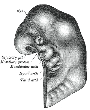
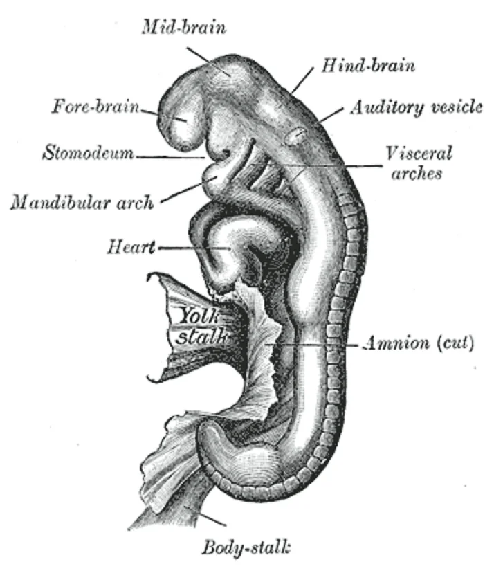
Head end of human embryo, about the end of the fourth week. (From model by Peter.)

Figure 1: Head end of human embryo, about the end of the fourth week (From model by Peter.).
The primary fetal membranes comprise the chorion, amnion, yolk sac, and allantois. The chorion is the outermost membrane that contributes jointly with uterine wall structures to form the placenta, a unique circulatory organ that develops to meet the growing embryo’s increasing requirements for nutrient and gas exchange. The amnion encloses the amniotic cavity. The yolk sac is an early site of haematopoiesis. The allantois is a small diverticulum that contributes to the umbilical cord structure.
Figure 2Gray37
Long description
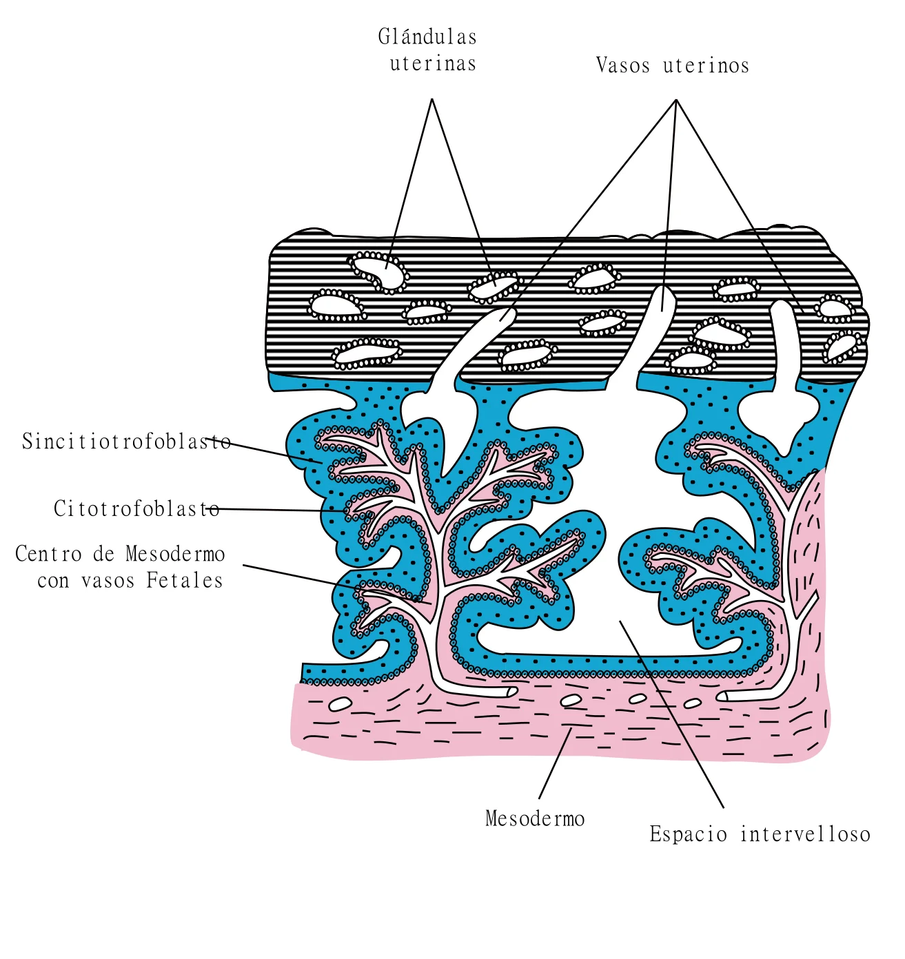
Secondary chorionic villi. Diagrammatic. (Modified from Bryce.)

Figure 2: Secondary chorionic villi (diagrammatic).
Figure 3Gray36
Long description
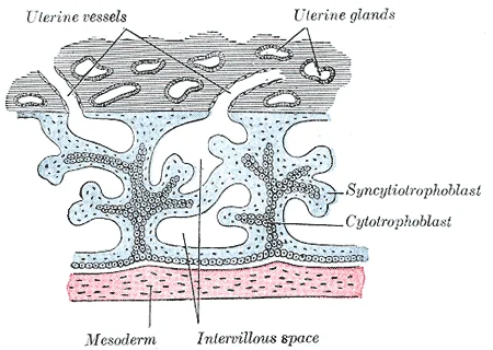
Primary chorionic villi. Diagrammatic. (Modified from Bryce.)

Figure 3: Primary chorionic villi (diagrammatic).
Figure 4Gray35
Long description
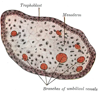
Transverse section of a chorionic villus.

Figure 4: Transverse section of a chorionic villus.
Figure 5Gray34
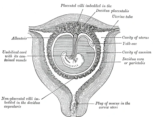
Long description
Sectional plan of the gravid uterus in the third and fourth month. (Modified from Wagner.)

Figure 5: Sectional plan of the gravid uterus in the third and fourth month (Modified from Wagner.).
Figure 6Gray33
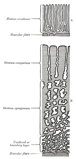
Long description
Diagrammatic sections of the uterine mucous membrane: A. The non-pregnant uterus. B. The pregnant uterus, showing the thickened mucous membrane and the altered condition of the uterine glands. (Kundrat and Engelmann.)

Figure 6: Diagrammatic sections of the uterine mucous membrane: A (the non-pregnant uterus).
Figure 7Gray32
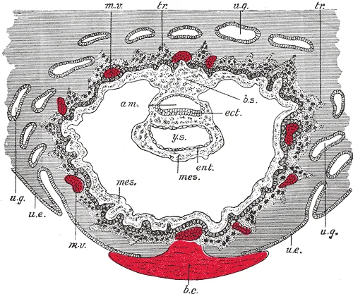
Long description
Section through ovum imbedded in the uterine decidua. Semidiagrammatic. (After Peters.) am. Amniotic cavity. b.c. Blood-clot. b.s. Body-stalk. ect. Embryonic ectoderm. ent. Entoderm. mes. Mesoderm. m.v. Maternal vessels. tr. Trophoblast. u.e. Uterine epithelium. u.g. Uterine glands. y.s. Yolk-sac.

Figure 7: Section through ovum imbedded in the uterine decidua (semidiagrammatic).
Figure 8Gray27
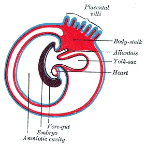
Long description
Diagram showing the expansion of amnion and delimitation of the umbilicus.

Figure 8: Diagram showing the expansion of amnion and delimitation of the umbilicus.
During the fetal period, which lasts from the ninth week of development until birth, these membranes support rapid growth and differentiation. The fetus grows from an embryo measuring approximately 3.3 cm (1.3 in) and weighing 7 g (0.25 oz) to an infant measuring approximately 51 cm (20 in) and weighing an average of approximately 3.4 kg (7.5 lbs). Between weeks 9 and 12, the brain continues to expand, the body elongates, and ossification continues. The fetus circulates amniotic fluid by swallow [2]
During approximately weeks 16–20, limb movements become more powerful, and the pregnant person may begin to feel quickening. Space restrictions typically force the growing fetus into the “fetal position,” with arms crossed and legs bent at the knees. Sebaceous glands coat the skin with vernix caseosa, a waxy, protective substance that moisturizes the skin and may provide lubrication during childbirth. A silky hair called lanugo covers the skin during weeks 17–20 but is shed as the fetus continues to grow. These membranes and their associated fluids create a stable environment for these complex morphological changes. [2]
DefinitionFetal Period
The stage of prenatal development from the ninth week until birth, characterised by growth and functional maturation of organs formed during the embryonic period.
Clinical pearlEarly Circulation
Blood vessel production in the yolk sac, chorion, and connecting stalk begins around day 15–16, preceding intra-embryonic circulation which starts approximately two days later. [1]
| Structure | Primary Function | Timeline Note |
|---|---|---|
| Chorion | Contributes to placenta formation | Active from early embryonic stage |
| Amnion | Encloses amniotic fluid | Protects embryo throughout gestation |
| Yolk Sac | Early blood cell and vessel production | Begins ~15-16 days post-fertilization |
| Allantois | Contributes to umbilical cord | Involved in early waste handling |
The formation of the fetal membranes and the associated circulatory infrastructure begins with the differentiation of the embryonic mesoderm. Together, these cells form masses known as blood islands scattered throughout the embryonic disc[1]. Spaces appear on the blood islands that develop into vessel lumens[1]. The endothelial lining of the vessels arise from the angioblasts within these islands[1]. Surrounding mesenchymal cells give rise to the smooth muscle and connective tissue layers of the vessels[1]. While the vessels are developing, the pluripotent stem cells begin to form the blood[1].
Figure 9Gray24

Long description
Diagram showing earliest observed stage of human ovum.

Figure 9: Diagram showing earliest observed stage of human ovum.
DefinitionAngioblasts
Cells derived from hemangioblasts that form the endothelial lining of blood vessels and contribute to the formation of blood islands.
This occurs because the many factors directing growth of nerves also stimulate blood vessels to follow a similar pattern[1]. Whether a given vessel develops into an artery or a vein is dependent upon local concentrations of signaling proteins[1]. Extensions known as vascular tubes eventually connect the vascular network[1].
The central structure is the developing heart tube, formed from two parallel tubes derived from mesoderm and lined with endothelium, which then fuse together. Surrounding the heart are the angioblasts forming the initial vascular network.
As the embryo grows within the mother’s womb, the placenta develops to supply blood rich in oxygen and nutrients via the umbilical vein and to remove wastes in oxygen-depleted blood via the umbilical arteries[1]. The placenta provides the fetus with necessary oxygen and nutrients via the umbilical vein. Remember that veins carry blood toward the heart. In this case, the blood flowing to the fetal heart is oxygenated because it comes from the placenta. The respiratory system is immature and cannot yet oxygenate blood on its own. From the umbilical vein, the oxygenated blood flows toward the inferior vena cava, all but bypassing the immature liver, via the ductus venosus shunt. The liver receives just a trickle of blood, which is all that it needs in its immature, semifunctional state. [2]
Blood flows from the inferior vena cava to the right atrium, mixing with fetal venous blood along the way. Three major shunts found in the fetus are the foramen ovale and ductus arteriosus, which divert blood from the pulmonary to the systemic circuit, and the ductus venosus, which carries freshly oxygenated blood high in nutrients to the fetal heart[1]. A shunt is an anatomical (or sometimes surgical) diversion that allows blood flow to bypass immature organs such as the lungs and liver until childbirth. [2]
As you will recall, a developing human is called a fetus from the ninth week of gestation until birth. The completion of fetal development results in a newborn who, although still immature in many ways, is capable of survival outside the womb. [2]
The fetus continues to lay down subcutaneous fat from week 31 until birth. The added fat fills out the hypodermis, and the skin transitions from red and wrinkled to soft and pink. Lanugo is shed, and the nails grow to the tips of the fingers and toes. Immediately before birth, the average crown-to-rump length is 35.5–40.5 cm (14–16 in), and the fetus weighs approximately 2.5–4 kg (5.5–8.8 lbs). Once born, the newborn is no longer confined to the fetal position, so subsequent measurements are made from head-to-toe instead of from crown-to-rump. At birth, the average length is approximately 51 cm (20 in). [2]
DefinitionFetal Membranes
The fetal membranes consist of the amnion and chorion, which enclose the amniotic cavity and protect the developing embryo, providing a stable environment for growth and movement.
It forms the innermost layer of the gestational sac. The chorion is derived from the trophoblast and extra-embryonic mesoderm, forming the outer layer. The structural integrity of these membranes is essential for maintaining the amniotic fluid volume and protecting the fetus from mechanical injury and infection.
The outermost layer is the chorion, which interfaces with the maternal decidua basalis. Beneath the chorion lies the amnion, which directly encloses the amniotic fluid. The interface between the amnion and chorion is a potential space that typically fuses.
The metabolic support of the fetal membranes is linked to the overall fetal circulation. This blood then moves into the left ventricle, where it is pumped into the aorta. Some of this blood moves through the coronary arteries into the myocardium, and some moves through the carotid arteries to the brain. This efficient shunting ensures that oxygen-rich blood is prioritised for the brain and heart, supporting the high metabolic activity of the developing tissues, including the membranes. [2]
NoteClinical Significance
Understanding the link between circulation and tissue support is vital, as compromised blood flow can affect membrane integrity and function.
Molecular regulation involves growth factors and cytokines that control cell proliferation and apoptosis. Key pathways include those regulating extracellular matrix production and degradation. An imbalance in these pathways can lead to weakening of the membranes. For example, increased activity of matrix metalloproteinases can degrade collagen, reducing tensile strength. Conversely, tissue inhibitors of metalloproteinases help maintain structural integrity.
| Feature | Amnion | Chorion |
|---|---|---|
| Origin | Epiblast and extra-embryonic mesoderm | Trophoblast and extra-embryonic mesoderm |
| Position | Innermost layer | Outermost layer |
| Function | Protects embryo, allows movement | Forms placenta, nutrient exchange |
| Cell Type | Cuboidal epithelium | Syncytiotrophoblast and cytotrophoblast |
The development of the fetal membranes is a critical process where embryonic structures, initially primitive and nonfunctional, mature sufficiently to ensure the newborn can survive in the outside world. This transformation involves the differentiation of extraembryonic membranes that provide protection, nutrition, and waste removal for the developing conceptus. Understanding these derivatives is essential for recognizing congenital anomalies that arise from their incomplete obliteration or persistence. [2]
| Embryonic Structure | Adult Derivative | Clinical Significance |
|---|---|---|
| Amnion | Amniotic sac and part of the placenta | Forms the innermost protective layer |
| Chorion | Chorionic plate and placental tissue | Forms the outer protective layer |
| Yolk sac | Vitelline duct (obliterates) | Persistence causes Meckel's diverticulum |
| Allantois | Urachus (obliterates) | Persistence causes urachal cysts or fistulas |
| Umbilical vessels | Ligamentum teres and ligaments | Fibrous remnants of fetal circulation |
Surrounding the amnion is the chorion, which is the outermost membrane and contributes significantly to the formation of the placenta. In normal development, this duct obliterates, but if it persists, it can lead to Meckel's diverticulum, a common congenital anomaly of the ileum. Normally, the allantois regresses and forms the urachus, which becomes the median umbilical ligament. Failure of the urachus to obliterate can result in various anomalies, including urachal cysts or fistulas.
The amnion and chorion are the primary fetal membranes. The amnion is a thin, transparent membrane that encloses the amniotic cavity, which is filled with amniotic fluid. This fluid cushions the fetus, allowing for movement and lung development. The chorion is a thicker membrane that forms the fetal portion of the placenta. It interacts with the maternal decidua basalis to facilitate nutrient and gas exchange. Its connection to the midgut, the vitelline duct, normally closes by the fifth week of gestation. If it remains patent, it can lead to complications such as intestinal obstruction or inflammation.
Figure 10Gray38
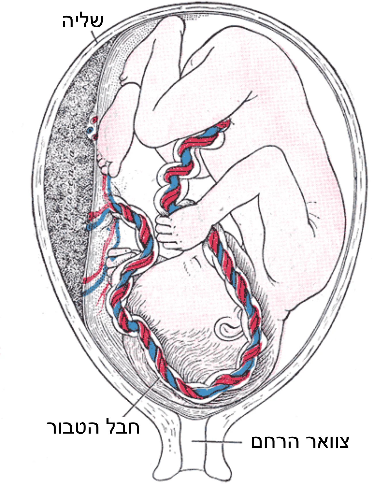
Long description
Fetus in utero, between fifth and sixth months.

Figure 10: Fetus in utero, between fifth and sixth months.
Figure 11Gray30
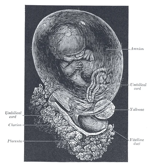
Long description
Fetus of about eight weeks, enclosed in the amnion. Magnified a little over two diameters. (Drawn from stereoscopic photographs lent by Prof. A. Thomson, Oxford.)

Figure 11: Fetus of about eight weeks, enclosed in the amnion.
It plays a role in early blood formation and waste removal. As development progresses, the allantois regresses, and its proximal part forms the urachus. The urachus connects the bladder to the umbilicus and normally obliterates to become the median umbilical ligament. If this obliteration is incomplete, it can result in a urachal cyst, sinus, or fistula, which may present with urinary discharge from the umbilicus or recurrent infections.
Figure 12Gray25
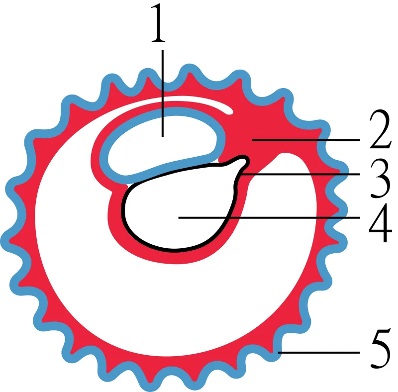
Long description
Diagram illustrating early formation of allantois and differentiation of body-stalk.

Figure 12: Diagram illustrating early formation of allantois and differentiation of body-stalk.
The umbilical cord is a vital structure that contains two umbilical arteries and one umbilical vein. These vessels are surrounded by Wharton's jelly, a gelatinous substance that protects the vessels from compression. The umbilical cord serves as the lifeline between the fetus and the placenta, ensuring the continuous supply of oxygen and nutrients. After birth, the umbilical vessels constrict and eventually fibrose, forming the round ligament of the liver (ligamentum teres) and the medial umbilical ligaments.
Figure 13Gray31
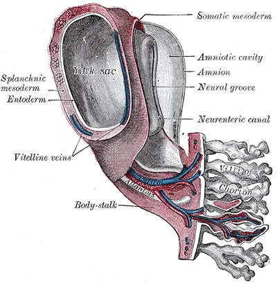
Long description
Model of human embryo 1.3 mm. long. (After Eternod.)

Figure 13: Model of human embryo 1.3 mm (long).
Congenital anomalies of the fetal membranes are relatively uncommon but important to recognize as they can significantly impact pregnancy outcomes and fetal well-being. These anomalies typically result from disruptions in the normal embryological development of the amnion, chorion, and placenta.
Figure 14Gray40
Long description
Embryo between eighteen and twenty-one days. (His.)

Figure 14: Embryo between eighteen and twenty-one days (His.).
DefinitionAmniotic band syndrome
A condition where strands of amniotic tissue wrap around parts of the fetus, causing constriction and potential tissue damage.
One of the most well-known anomalies is amniotic band syndrome, which occurs when the amniotic sac ruptures prematurely, allowing fetal parts to become entangled in the amnion strands. This can lead to congenital amputations, craniofacial defects, or body wall defects. [1]
Another anomaly is chorioangioma, a benign tumor of the placenta consisting of an overgrowth of chorionic tissue and blood vessels. These are found in approximately 1% of placentas and can range in size from a few millimeters to several centimeters. While most are asymptomatic, large chorioangiomas (>5 cm) can lead to polyhydramnios, fetal hydrops, or intrauterine growth restriction. [1]
| Anomaly | Embryological basis | Clinical significance |
|---|---|---|
| Amniotic band syndrome | Rupture of amnion after 4th week | Constriction defects, amputations |
| Chorioangioma | Overgrowth of chorionic tissue | Polyhydramnios, fetal hydrops if >5cm |
| Placental cysts | Degeneration of chorionic villi | Usually asymptomatic unless large |
| Membranous amnion | Failure of amnion to fuse with chorion | Increased risk of preterm rupture |
Placental cysts, which are filled with fluid, result from degeneration of chorionic villi and are typically found in 4-6% of placentas. Most are small and asymptomatic, but large cysts (>5 cm) may cause compression of adjacent structures or be associated with fetal anomalies. [1]
A rare anomaly is the membranous amnion, where the amnion fails to fuse with the chorion, resulting in a double-layered membrane. This condition is associated with an increased risk of preterm rupture of membranes and preterm birth. [1]
Adjacent to these, there are several placental cysts appearing as translucent, fluid-filled structures.
The integrity of the fetal membranes is paramount for maintaining a sterile intrauterine environment and facilitating gas exchange. The amnion and chorion form a protective sac that encloses the amniotic fluid, which cushions the fetus against mechanical trauma and allows for symmetrical growth. Clinically, the rupture of these membranes, known as the 'breaking of waters', is a critical event in obstetrics. It can occur spontaneously at term, signalling the onset of labour, or prematurely before the onset of labour, a condition termed preterm premature rupture of membranes (PPROM). The timing of rupture dictates the management strategy, as prolonged exposure to the vaginal flora increases the risk of chorioamnionitis, a serious infection of the fetal membranes and amniotic fluid.
The chorion frondosum, the villous portion of the chorion, interacts with the decidua basalis to form the placenta. The chorion laeve, the smooth portion, fuses with the decidua capsularis. Understanding this distinction is vital when interpreting ultrasound findings. For instance, a subchorionic haemorrhage involves bleeding between the chorion and the uterine wall, which can be visualised as an anechoic space on ultrasound.
Ultrasound is the primary modality for assessing fetal membrane integrity and amniotic fluid volume. The 'sludge' seen in the amniotic fluid on ultrasound, consisting of echogenic particles, is often indicative of intra-amniotic infection or meconium staining, requiring close monitoring for signs of fetal distress. [1]
When PPROM is diagnosed before 34 weeks of gestation, the primary goal is to prolong pregnancy to improve fetal lung maturity while preventing infection. Corticosteroids are administered to accelerate fetal lung development, typically given between 24 and 34 weeks of gestation. Antibiotic prophylaxis is standard to reduce the risk of chorioamnionitis and prolong the latency period. In cases of term PPROM, induction of labour is usually recommended to reduce the risk of neonatal sepsis, as the be [1]
1. Origin of the Amnion The amnion is derived from the epiblast layer of the bilaminar disc. It forms a fluid-filled cavity that encloses the embryo, providing a protective mechanical cushion and a stable thermal environment for development.
2. Origin of the Chorion The chorion is formed by the trophoblast and the extra-embryonic mesoderm. It is the outermost fetal membrane and is responsible for implantation into the uterine wall and the formation of the placenta.
3. Amniotic Fluid Volume Amniotic fluid volume increases progressively throughout pregnancy. By term, the volume is approximately 800 mL. This fluid allows for fetal movement, musculoskeletal development, and lung maturation.
4. Oligohydramnios Definition Oligohydramnios is defined as an amniotic fluid index (AFI) of less than 5 cm or a single deepest pocket of less than 2 cm. It is associated with fetal renal anomalies and pulmonary hypoplasia.
5. Polyhydramnios Definition Polyhydramnios is defined as an AFI greater than 24 cm or a single deepest pocket greater than 8 cm. Causes include fetal gastrointestinal obstructions, neurological defects, and maternal diabetes.
6. Umbilical Cord Structure The umbilical cord contains two umbilical arteries and one umbilical vein. The vessels are surrounded by Wharton’s jelly, a mucous connective tissue that protects the vessels from compression.
7. Umbilical Artery Function The umbilical arteries carry deoxygenated blood and waste products from the fetus to the placenta.
8. Umbilical Vein Function The umbilical vein carries oxygenated and nutrient-rich blood from the placenta to the fetus.
9. Velamentous Insertion In velamentous cord insertion, the umbilical vessels insert into the membranes rather than the placental disc.
10. Succenturiate Lobe A succenturiate lobe is an accessory placental lobe separate from the main placenta. It is connected by vessels running through the membranes, posing a risk of retained placental tissue and postpartum haemorrhage.
11. Chorioamniotic Separation Chorioamniotic separation is a detachment of the amnion from the chorion. It is often an incidental finding but can be associated with preterm premature rupture of membranes (PPROM) and infection.
12. Common Trap: Membrane Rupture A common trap is confusing the amnion and chorion in ultrasound imaging. The amnion is thin and often adherent to the chorion later in pregnancy, whereas the chorion is thicker and forms the fetal part of the placenta.
Clinical pearlKey Distinction
The amnion is derived from epiblast, while the chorion is derived from trophoblast and extra-embryonic mesoderm.
Exam trapVessel Count
Remember: Two arteries, one vein. The mnemonic "Artery, Artery, Vein" (AAV) helps recall the order from left to right in the cord.
Students frequently confuse the timeline of fetal development, particularly regarding the onset of lung maturity and the specific functions of the fetal membranes in relation to surfactant production. A primary error is assuming that the fetal membranes themselves produce surfactant. The membranes provide the structural and protective environment, but the surfactant is synthesised by the type II pneumocytes within the developing lung tissue itself. This distinction is critical because pathology related to respiratory distress stems from pulmonary insufficiency, not membrane failure.
Another frequent mistake involves the timing of myelination. Students often believe that the nervous system is fully functional or myelinated at birth. In reality, myelination is a protracted process that begins in the spinal cord during weeks 21–30 but is not completed until adolescence. This incomplete myelination explains why reflexes are primitive and why the nervous system continues to mature significantly after birth. It is essential to remember that while axons begin to be coated in electrically insulating glial cell sheaths during this period, efficient nervous system functioning is still developing. [2]
Students also frequently misinterpret the significance of weight gain in the third trimester. It is a common error to view weight gain solely as a marker of size. However, the rapid weight gain occurring in developmental weeks 21–30 is physiologically crucial for thermoregulation. This adipose tissue accumulation is the primary mechanism for maintaining a stable body temperature after birth. Without this specific period of weight gain, the newborn would struggle to prevent hypothermia due to the high surface-area-to-volume ratio. [2]
A further pitfall relates to the timeline of testicular descent. Students may assume that testes descend immediately upon formation. However, in male fetuses, the testes descend into the scrotum near the end of the period characterised by rapid weight gain and lung maturation. This specific timing is a key landmark for assessing male fetal development at approximately 30 weeks. [2]
Finally, students often overlook the specific measurement standards for fetal size at 30 weeks. It is incorrect to assume that a 30-week fetus is significantly smaller in proportion than a term newborn. At 30 weeks, the fetus measures 28 cm (11 in) from crown to rump and exhibits the approximate body proportions of a full-term newborn. The primary difference is that the 30-week fetus is much leaner, lacking the subcutaneous fat reserves of the term infant. Confusing size with proportionality is a common analytical error in clinical assessments. [2]
This knowledge is applied at the bedside when managing premature infants who present with respiratory difficulties. If a newborn born before the completion of surfactant production exhibits signs of respiratory distress, clinicians must recognise that inadequate surfactant production may result in respiratory distress syndrome. This understanding dictates the immediate management plan, which may include surfactant replacement therapy, supplemental oxygen, or maintenance in a continuous positive airway pressure (CPAP) chamber during their first days or weeks of life. Recognising the link between gestational age and surfactant capability is vital for initiating the correct supportive care. [2]
In the operating theatre and during neonatal examinations, understanding the timeline of myelination helps in interpreting neurological findings. Since the process of myelination is not completed until adolescence, clinicians should not expect mature neurological responses in preterm infants. This knowledge prevents the misdiagnosis of neurological deficits in infants whose nervous systems are simply immature rather than damaged. The axons of the spinal cord begin to be myelinated during weeks 21–30, but this is an ongoing process.
On imaging, such as ultrasound, the measurement of fetal length is used to estimate gestational age. A fetus measuring 28 cm (11 in) from crown to rump is consistent with a gestational age of 30 weeks. This metric, combined with the observation of body proportions, allows sonographers to confirm that the fetus exhibits the approximate body proportions of a full-term newborn. This is particularly useful for assessing growth restriction, as a fetus of this size should be leaner than a term infant but proportionally similar.
In examinations, students are often tested on the specific functions of fetal structures during the third trimester. Questions may ask about the substance that reduces surface tension in the lungs. The correct answer is surfactant, which assists proper lung expansion after birth. Another common question involves the timeline of eyelid development. By weeks 21–30, the eyelids are no longer fused and can be opened and closed. This is a distinct developmental milestone that helps differentiate betw [2]
Furthermore, clinical practice relies on understanding the role of bone marrow. By weeks 21–30, the bone marrow completely takes over erythrocyte synthesis. This shift is important for interpreting haematological parameters in newborns. A failure of this transition or a premature birth before this takeover is complete can lead to anaemia or other blood disorders. The rapid weight gain during this period is also monitored to ensure the fetus is preparing for extra-uterine life, specifically for temperature regulation. [2]
Management of Preterm Respiratory Distress
- Identify Preterm NewbornGestational age < 30 weeks or risk factors
- Question: Respiratory Distress Present?
Yes
- Assess Surfactant StatusInadequate production likely
- Question: Severity?
Moderate/Severe
- Initiate TherapySurfactant replacement therapy
Mild
- Supportive CareSupplemental oxygen or CPAP
No
- MonitorObserve for delayed onset
Developmental weeks 21–30 are characterised by rapid weight gain for thermoregulation. The bone marrow completely takes over erythrocyte synthesis during this period. Axons of the spinal cord begin to be myelinated, a process completed in adolescence. The fetus grows eyelashes and the eyelids can be opened and closed. [2] The lungs begin producing surfactant to reduce surface tension and assist expansion. [2] Inadequate surfactant may result in respiratory distress syndrome in premature newborns. Management may include surfactant replacement therapy, supplemental oxygen, or CPAP. At 30 weeks, the fetus measures 28 cm (11 in) and has term-like proportions.
Answer each one in your head first, then open the answer.
What substance reduces surface tension in the lungs?
Show answerHide answer
SurfactantWhen is myelination of the nervous system completed?
Show answerHide answer
AdolescenceWhat is the crown-to-rump length of a 30-week fetus?
Show answerHide answer
28 cm (11 in)What therapy is used for inadequate surfactant production?
Show answerHide answer
Surfactant replacement therapyWhat takes over erythrocyte synthesis by weeks 21–30?
Show answerHide answer
Bone marrow
- Betts JG, Young KA, Wise JA, et al. Development of Blood Vessels and Fetal Circulation. OpenStax, Anatomy and Physiology 2e (CC BY-NC-SA 4.0). Accessed .
- Betts JG, Young KA, Wise JA, et al. Fetal Development. OpenStax, Anatomy and Physiology 2e (CC BY-NC-SA 4.0). Accessed .
Figure credits
- Figure 1: Henry Vandyke Carter, via Wikimedia Commons, Public domain, Public domain.
- Figure 2: Henry Vandyke Carter, via Wikimedia Commons, Public domain, Public domain.
- Figure 3: Henry Vandyke Carter, via Wikimedia Commons, Public domain, Public domain.
- Figure 4: Henry Vandyke Carter, via Wikimedia Commons, Public domain, Public domain.
- Figure 5: Henry Vandyke Carter, via Wikimedia Commons, Public domain, Public domain.
- Figure 6: Henry Vandyke Carter, via Wikimedia Commons, Public domain, Public domain.
- Figure 7: Henry Vandyke Carter, via Wikimedia Commons, Public domain, Public domain.
- Figure 8: Henry Vandyke Carter, via Wikimedia Commons, Public domain, Public domain.
- Figure 9: Henry Vandyke Carter, via Wikimedia Commons, CC0, CC0.
- Figure 10: Henry Vandyke Carter, via Wikimedia Commons, Public domain, Public domain.
- Figure 11: Henry Vandyke Carter, via Wikimedia Commons, Public domain, Public domain.
- Figure 12: Henry Vandyke Carter, via Wikimedia Commons, CC0, CC0.
- Figure 13: Henry Vandyke Carter, via Wikimedia Commons, Public domain, Public domain.
- Figure 14: Henry Vandyke Carter, via Wikimedia Commons, Public domain, Public domain.
Educational content for medical students and doctors. It is not a substitute for clinical judgement or local guidelines.