Cardiovascular development
Embryonic heart development from tube formation to fetal circulation shunts.
The human heart is the first functional organ to develop, beginning to beat and pump blood around day 21 or 22, a mere three weeks after fertilization. This early activity is vital for distributing blood through vessels and facilitating the exchange of nutrients, oxygen, and wastes. Initially, the heart exists as two parallel tubes derived from mesoderm and lined with endothelium, which then fuse together. These structures include the truncus arteriosus, bulbus cordis, primitive ventricle, primi [1][3]
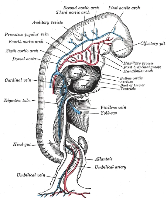
Figure 1Gray472
Long description
Profile view of a human embryo estimated at twenty or twenty-one days old. (After His.)

Figure 1: Profile view of a human embryo estimated at twenty or twenty-one days old (After His.).
As the primitive heart tube elongates, it begins to fold within the pericardium, eventually forming an S shape. This process occurs between days 23 and 28, placing chambers and major vessels into an alignment similar to the adult heart. The internal septa begin to form about day 28, separating the heart into the atria and ventricles. Partitioning of the atria and ventricles by the interatrial septum, interventricular septum, and atrioventricular septum is complete by the end of the fifth week. The atrioventricular valves form between weeks five and eight, and the semilunar valves form between weeks five and nine. [3]
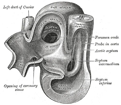
Figure 2Gray468
Long description
Same heart as in Fig. 467, opened on right side. (From model by His.)

Figure 2: Same heart as in Fig (467, opened on right side).
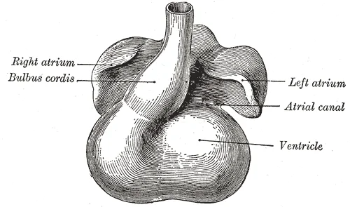
Figure 3Gray466
Long description
Heart showing expansion of the atria. (Drawn from Ecker-Zeigler model.)

Figure 3: Heart showing expansion of the atria (Drawn from Ecker-Zeigler model.).
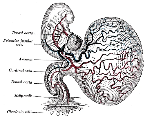
Figure 4Gray459
Long description
Human embryo of about fourteen days, with yolk-sac. (After His.)

Figure 4: Human embryo of about fourteen days, with yolk-sac (After His.).
The fetal period lasts from the ninth week of development until birth. During this time, the circulatory system becomes specialized, including three shunts: the ductus venosus, the foramen ovale, and the ductus arteriosus. These shunts enable the fetus to bypass the semifunctional liver and pulmonary circuit until after childbirth. The placenta provides oxygen and nutrients via the umbilical vein. Blood flows from the umbilical vein toward the inferior vena cava, bypassing the immature liver via the ductus venosus shunt. [1][2][3]
Figure 5Gray502
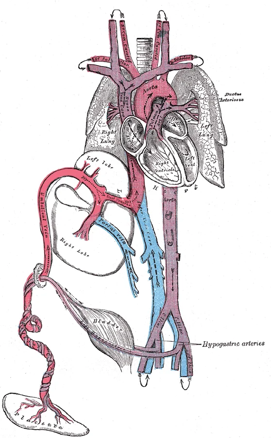
Long description
Plan of the fetal circulation. In this plan the figured arrows represent the kind of blood, as well as the direction which it takes in the vessles. Thus—arterial blood is figured >—>; venous blood, >—>; mixed (arterial and venous) blood, >—>.

Figure 5: Plan of the fetal circulation.
Figure 6Gray480
Long description
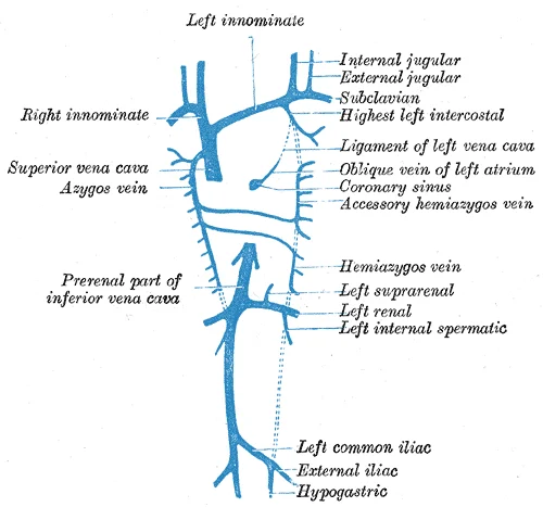
Diagram showing completion of development of the parietal veins.

Figure 6: Diagram showing completion of development of the parietal veins.
Figure 7Gray479
Long description
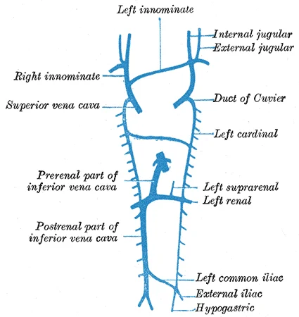
Diagram showing development of main cross branches between jugulars and between cardinals.

Figure 7: Diagram showing development of main cross branches between jugulars and between cardinals.
Figure 8Gray478
Long description
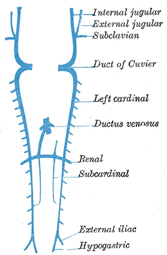
Scheme showing early stages of development of the inferior vena cava.

Figure 8: Scheme showing early stages of development of the inferior vena cava.
Figure 9Gray458
Long description
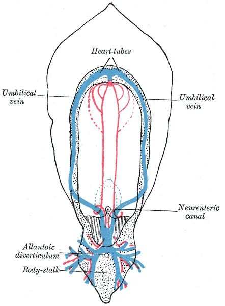
Diagram of the vascular channels in a human embryo of the second week. (After Eternod.) The red lines are the dorsal aortæ continued into the umbilical arteries. The red dotted lines are the ventral aortæ, and the blue dotted lines the vitelline veins.

Figure 9: Diagram of the vascular channels in a human embryo of the second week.
Management depends on the type and severity of the defect. Many simple defects, such as small ASDs or VSDs, may only require observation. Complex defects, such as tetralogy of Fallot, require surgical correction. [1]
DefinitionFetal Shunt
An anatomical diversion that allows blood flow to bypass immature organs such as the lungs and liver until childbirth.
| Structure | Timeline |
|---|---|
| Heart beating | Day 21 or 22 |
| Cardiac looping | Days 23–28 |
| Septation complete | End of fifth week |
| AV valves form | Weeks five–eight |
| Semilunar valves form | Weeks five–nine |
Fetal Circulation Pathway
- PlacentaOxygen and nutrients
- Umbilical VeinCarries oxygen-rich blood
- Question: Ductus Venosus?
Yes
- Bypasses LiverTo Inferior Vena Cava
No
- LiverReceives trickle of blood
- Inferior Vena CavaMixes with fetal venous blood
- Right AtriumBlood enters heart
The ventricle is the large, central chamber, while the atrium lies further caudal. The labels 'valve development' point to the endocardial cushions within the atrioventricular canal, indicating the site where the future atrioventricular valves will form. Understanding this spatial arrangement is essential, as the looping process brings the future atria and ventricles into their correct anatomical positions relative to the great vessels. [2]
Figure 10Gray488
Long description
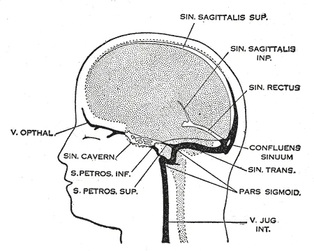
Profile drawings of the dural veins showing principal stages in their development in human embryos from 4 mm. to birth. It is of particular interest to notice their adaptation to the growth and changes in the form of the central nervous system. Fig. 481, 4 mm.; Fig. 482, 14 mm.; Fig. 483, 18 mm.; Fi

Figure 10: Profile drawings of the dural veins showing principal stages in their development in human embryos from 4 mm (to birth).
Figure 11Gray477
Long description
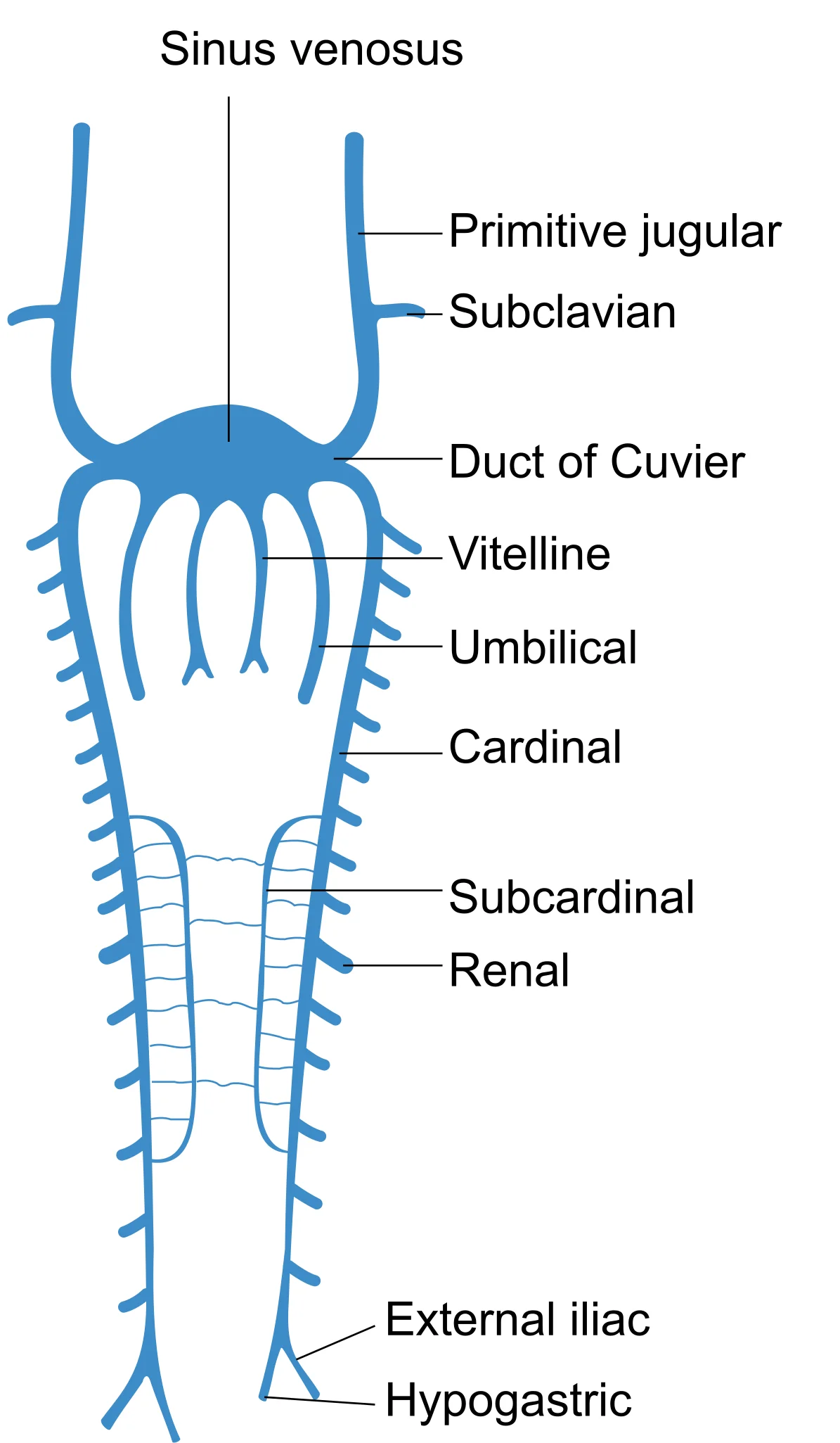
Scheme of arrangement of parietal veins.

Figure 11: Scheme of arrangement of parietal veins.
The cardiovascular system is among the first to function in the embryo. By day 21 post-fertilization, the heart has developed enough to begin beating[1]. This early function is critical for survival, as the circulatory system must supply growing tissues with nutrients and gases and remove waste products[1]. Blood vessel development begins in structures outside the embryo proper, such as the yolk sac, chorion, and connecting stalk, starting about 15 to 16 days following fertilization[1]. Development within the embryo itself begins approximately 2 days later[1]. [2][3]
Figure 12Gray475
Long description
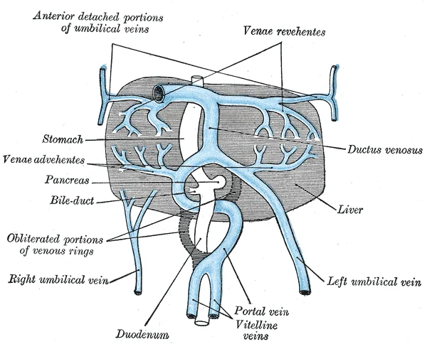
The liver and the veins in connection with it, of a human embryo, twenty-four or twenty-five days old, as seen from the ventral surface. (After His.)

Figure 12: The liver and the veins in connection with it, of a human embryo, twenty-four or twenty-five days old, as seen from the ventral surface (After His.).
Figure 13Gray467
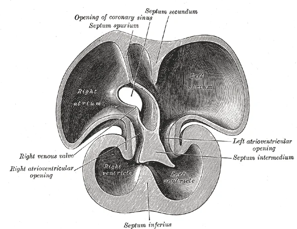
Long description
Interior of dorsal half of heart of human embryo of about thirty-five days. (From model by His.)

Figure 13: Interior of dorsal half of heart of human embryo of about thirty-five days (From model by His.).
Figure 14Gray465
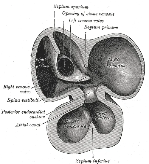
Long description
Interior of dorsal half of heart from a human embryo of about thirty days. (From model by His.)

Figure 14: Interior of dorsal half of heart from a human embryo of about thirty days (From model by His.).
Figure 15Gray464
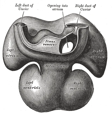
Long description
Dorsal surface of heart of human embryo of thirty-five days. (From model by His.)

Figure 15: Dorsal surface of heart of human embryo of thirty-five days (From model by His.).
Figure 16Gray463
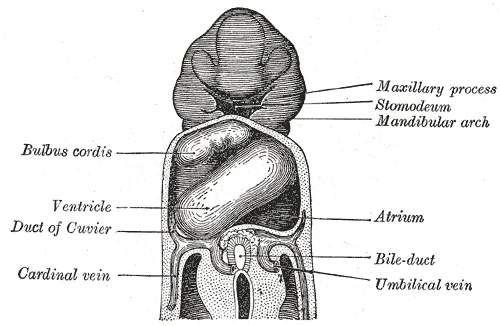
Long description
Heart of human embryo of about fifteen days. (Reconstruction by His.)

Figure 16: Heart of human embryo of about fifteen days (Reconstruction by His.).
Figure 17Gray462
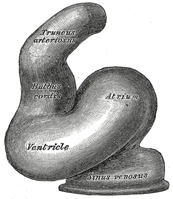
Long description
Heart of human embryo of about fourteen days. (From model by His.)

Figure 17: Heart of human embryo of about fourteen days (From model by His.).
The heart forms from mesoderm around 18 to 19 days after fertilization[1]. Mesoderm is one of the three primary germ layers that differentiates early in development[1]. The heart begins to develop near the head of the embryo in a region known as the cardiogenic area[1]. Following chemical signals from the underlying endoderm, the cardiogenic area forms two strands called the cardiogenic cords[1]. As these cords develop, a lumen rapidly forms within them, and they are then referred to as endocardial tubes[1]. The two tubes migrate together and fuse to form a single primitive heart tube[1]. This primitive heart tube quickly forms five distinct regions[1]. [3]
Valve development is a crucial aspect of this early morphogenesis. The endocardial cushions, formed by the proliferation of mesenchymal cells in the atrioventricular canal and outflow tract, play a pivotal role. These cushions fuse to divide the common atrioventricular canal into right and left canals and to separate the truncus arteriosus and bulbus cordis into the aorta and pulmonary trunk. The cusps of the atrioventricular valves (mitral and tricuspid) and the semilunar valves (aortic and pulmonary) are derived from these endocardial cushions and the surrounding myocardium. The process ensures that blood flows in a unidirectional manner through the developing chambers. [1]
Figure 18Gray473
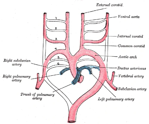
Long description
Scheme of the aortic arches and their destination. (Modified from Kollmann.)

Figure 18: Scheme of the aortic arches and their destination (Modified from Kollmann.).
Figure 19Gray471
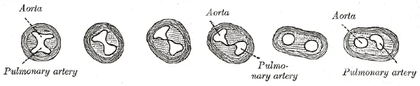
Long description
Transverse sections through the aortic bulb to show the growth of the aortic septum. The lowest section is on the left, the highest on the right of the figure. (After His.)

Figure 19: Transverse sections through the aortic bulb to show the growth of the aortic septum.
Figure 20Gray470
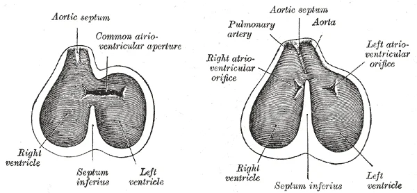
Long description
Diagrams to show the development of the septum of the aortic bulb and of the ventricles. (Born.)

Figure 20: Diagrams to show the development of the septum of the aortic bulb and of the ventricles (Born.).
Circulation patterns are clearly established by the fourth week of embryonic life[1]. The precursor cells for blood vessels are known as hemangioblasts[1]. These differentiate into angioblasts, which give rise to blood vessels, and pluripotent stem cells, which differentiate into the formed elements of blood[1]. Together, these cells form masses known as blood islands scattered throughout the embryonic disc[1]. Spaces appear on the blood islands that develop into vessel lumens[1]. The endothelial lining of the vessels arises from the angioblasts within these islands[1]. Surrounding mesenchymal cells give rise to the smooth muscle and connective tissue layers of the vessels[1]. [2]
DefinitionCardiogenic Area
The region near the head of the embryo where the heart begins to develop from mesoderm, guided by signals from the underlying endoderm.
Clinical pearlPrimitive Heart Tube
The fused endocardial tubes form a single primitive heart tube with five distinct regions, which will undergo looping to establish the final cardiac anatomy.
The development of the cardiovascular system is orchestrated by a complex interplay of molecular signals that guide cell differentiation, migration, and morphogenesis. While the specific genetic pathways are intricate, the fundamental principle is the establishment of polarity and the formation of the primary heart tube. This process begins with the specification of cardiac progenitor cells, which are induced by signals from the underlying endoderm and lateral plate mesoderm. Key signalling molecules, including members of the TGF-beta superfamily such as Nodal and BMPs, play crucial roles in this initial specification. Subsequently, Wnt signalling pathways regulate the anterior-posterior axis of the developing heart, determining where the heart field will form.
As the heart tube forms, it undergoes looping, a critical step that establishes the correct spatial arrangement of the future chambers. This looping is driven by asymmetric gene expression, particularly involving Nodal and Lefty, which break bilateral symmetry. The rightward flow of fluid in the node, known as nodal flow, is essential for establishing left-right asymmetry. Disruption of this flow or the subsequent signalling cascade can lead to laterality defects, such as situs inversus or heterotaxy. The molecular regulation continues as the heart tube partitions into distinct chambers and outflow tracts. Transcription factors such as Nkx2-5, GATA4, and Tbx5 are vital for chamber specification and septation. These factors interact with signalling pathways like Notch and Hedgehog to coordinate the growth and differentiation of cardiac tissues.
The development of the great vessels is also tightly regulated by molecular cues. Endothelial cells respond to shear stress and chemical signals to form the vascular network. VEGF (Vascular Endothelial Growth Factor) is a key mediator of angiogenesis, guiding the formation of new blood vessels from pre-existing ones. The interaction between the endocardium and the myocardium is mediated by signals such as VEGF and FGF (Fibroblast Growth Factor), which promote the formation of the endocardial cushions. These cushions eventually fuse to form the atrioventricular and semilunar valves.
The final stages of cardiovascular development involve the refinement of the septa and the maturation of the valves. Molecular signals continue to play a role in this process, ensuring that the heart functions correctly after birth. The transition from fetal to neonatal circulation involves significant changes in gene expression and signalling pathways, adapting the heart to the new demands of extrauterine life. Understanding these molecular mechanisms is essential for comprehending congenital heart defects and developing potential therapeutic strategies.
The cardiovascular system undergoes profound structural and functional reorganisation at birth, transitioning from a parallel fetal circulation to a series adult circulation. This shift is driven by the cessation of placental blood flow and the initiation of pulmonary respiration. The primary fetal shunts—the foramen ovale, ductus arteriosus, and ductus venosus—close functionally and anatomically to become adult ligaments. Understanding these derivatives is essential for interpreting congenital heart defects and postnatal haemodynamics.
To the right of the heart, the ductus arteriosus connects the pulmonary artery to the aortic arch, diverting blood away from the pulmonary circulation.
Figure 21Gray474
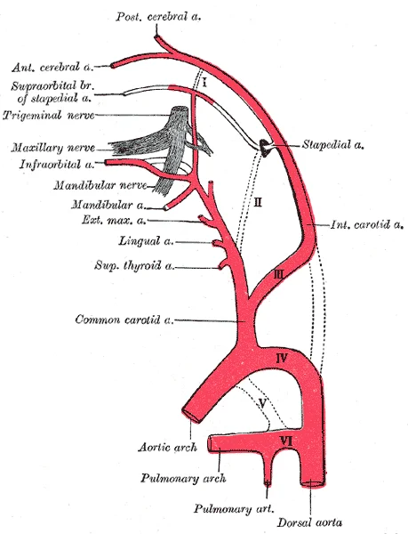
Long description
Diagram showing the origins of the main branches of the carotid arteries. (Founded on Tandler.)

Figure 21: Diagram showing the origins of the main branches of the carotid arteries (Founded on Tandler.).
The closure of fetal shunts results in specific adult ligamentous structures. These are formed by the fibrosis and obliteration of the original vascular channels.
| Fetal Structure | Adult Derivative | Location |
|---|---|---|
| Foramen ovale | Fossa ovalis | Interatrial septum |
| Ductus arteriosus | Ligamentum arteriosum | Between pulmonary artery and aortic arch |
| Ductus venosus | Ligamentum venosum | Within the liver, connecting portal vein to IVC |
| Umbilical vein | Ligamentum teres hepatis | Free edge of the falciform ligament |
| Umbilical arteries | Medial umbilical ligaments | Anterior abdominal wall |
The foramen ovale is a flap-like valve in the right atrium. After birth, increased left atrial pressure pushes the septum primum against the septum secundum, functionally closing the shunt. Over months, the tissues fuse, leaving the fossa ovalis[1]. The ductus arteriosus constricts in response to increased arterial oxygen tension and decreased prostaglandin E2 levels. This fibrosis creates the ligamentum arteriosum, a small ligamentous band[1]. The ductus venosus closes due to the cessation of umbilical blood flow, becoming the ligamentum venosum[1].
The transition is initiated by two main events: clamping of the umbilical cord and the first breath. Clamping the cord removes the low-resistance placental circuit, causing systemic vascular resistance to rise sharply. This increases afterload on the left ventricle. Simultaneously, lung expansion decreases pulmonary vascular resistance dramatically. The combination of increased left atrial pressure and decreased right atrial pressure reverses the flow gradient across the foramen ovale, ensuring it closes[1].
The lungs begin producing surfactant, a substance that reduces surface tension in the lungs and assists proper lung expansion after birth[1]. Inadequate surfactant production in premature newborns may result in respiratory distress syndrome, and as a result, the newborn may require surfactant replacement therapy, supplemental oxygen, or maintenance in a continuous positive airway pressure (CPAP) chamber during their first days or weeks of life[1]. This respiratory support is critical to maintain adequate oxygenation, which further promotes the closure of the ductus arteriosus.
Embryonic organ structures that were primitive and nonfunctional develop to the point that the newborn can survive in the outside world[1]. The heart must now pump the entire cardiac output through the pulmonary circuit. These changes ensure that oxygenated blood from the lungs is efficiently distributed to the body tissues.
Cardiovascular development is a rapid and complex process, beginning with the formation of the heart as the first functional organ. Initially, paired cardiogenic cords form a hollow lumen known as endocardial tubes, which fuse into a single primitive heart tube by day 22. This tube differentiates into five distinct regions: the truncus arteriosus, bulbus cordis, primitive ventricle, primitive atrium, and sinus venosus. Between days 23 and 28, the heart assumes an S-shaped configuration within the pericardium. [3]
Figure 22Gray487
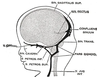
Long description
80 mm. crown-rump length.(After Streeter.)

Figure 22: 80 mm (crown-rump length.(after streeter.)).
Figure 23Gray486
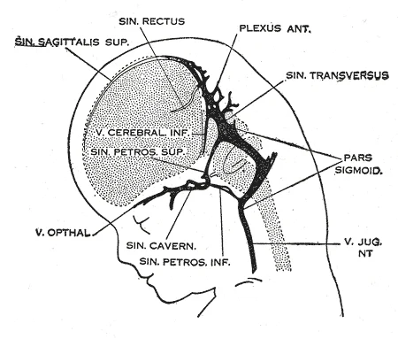
Long description
50 mm. crown-rump length. Labelled illustration from Wikimedia Commons, used under its open licence.

Figure 23: 50 mm (crown-rump length).
DefinitionCardiac Tube Looping
The primitive heart tube undergoes looping to position the future chambers correctly, a critical step whose failure leads to major structural defects.
Finally, the transition to adult vessels and the resulting congenital heart defects are mapped out.
The five regions of the primitive heart tube develop into recognizable structures in a fully developed heart. The truncus arteriosus divides to give rise to the ascending aorta and pulmonary trunk. The bulbus cordis develops into the right ventricle, while the primitive ventricle forms the left ventricle. The primitive atrium becomes the anterior portions of both atria and the auricles. The sinus venosus develops into the posterior right atrium, the SA node, and the coronary sinus. [3]
Figure 24Gray476
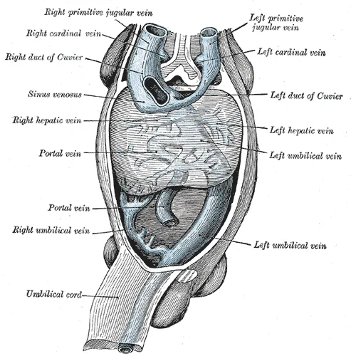
Long description
Human embryo with heart and anterior body-wall removed to show the sinus venosus and its tributaries. (After His.)

Figure 24: Human embryo with heart and anterior body-wall removed to show the sinus venosus and its tributaries (After His.).
Figure 25Gray469
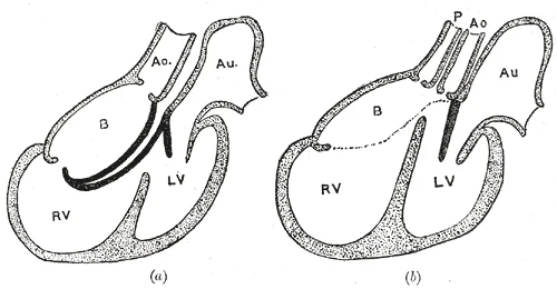
Long description
Diagrams to illustrate the transformation of the bulbus cordis. (Keith.) Ao. Truncus arteriosus. Au. Atrium. B. Bulbus cordis. RV. Right ventricle. LV. Left ventricle. P. Pulmonary artery.

Figure 25: Diagrams to illustrate the transformation of the bulbus cordis (Keith.) Ao.
In the fetus, the lungs are nonfunctional, so circulation bypasses them. Blood shifts through the foramen ovale, a shunt connecting the right and left atria, avoiding the pulmonary trunk. Most blood is pumped to the right ventricle and into the pulmonary trunk. However, the ductus arteriosus diverts a portion of this blood into the aorta. This ensures only a small volume of oxygenated blood passes through the immature pulmonary circuit, which has minor metabolic requirements. Blood vessels in uninflated lungs have high resistance, encouraging flow to the lower-resistance aorta. [1]
Angiogenesis, the creation of new blood vessels from existing ones, continues throughout life. Vascular tubes develop on blood islands and connect to the tubular heart, occurring in many regions simultaneously. [2]
Figure 26Gray461
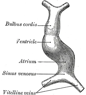
Long description
Diagram to illustrate the simple tubular condition of the heart. (Drawn from Ecker-Ziegler model.)

Figure 26: Diagram to illustrate the simple tubular condition of the heart (Drawn from Ecker-Ziegler model.).
Congenital heart defects arise from errors in these processes. Defects in septation can lead to atrial or ventricular septal defects. Abnormalities in valve formation can cause stenosis or regurgitation. Errors in aortic arch development can result in coarctation of the aorta or vascular rings. Understanding the embryological basis of these structures is essential for diagnosing and managing congenital anomalies.
Congenital heart defects represent the most common class of birth anomalies, occurring in approximately one per cent of live births. The majority of these structural abnormalities involve the septation of the heart chambers or the great vessels. A critical period for cardiac development occurs during the embryonic period, during which the primitive heart tube undergoes looping and septation. Disruption during this window can lead to significant haemodynamic compromise. [1]
DefinitionSeptal Defects
Failures in the formation of the atrial or ventricular septa, leading to left-to-right shunting of blood.
Atrial septal defects (ASD) are often asymptomatic in childhood but may present with right heart volume overload in adulthood. Ventricular septal defects (VSD) are the most frequent congenital heart defect. Small VSDs may close spontaneously, whereas larger defects require surgical intervention to prevent pulmonary hypertension. [1]
| Defect | Pathophysiology | Clinical Feature |
|---|---|---|
| ASD | Left-to-right shunt at atrial level | Fixed split S2 |
| VSD | Left-to-right shunt at ventricular level | Holosystolic murmur |
| PDA | Persistent connection between aorta and pulmonary artery | Continuous machinery murmur |
Patent ductus arteriosus (PDA) results from the failure of the ductus arteriosus to close after birth. This condition is more common in premature infants and those with congenital rubella syndrome. The persistent shunt increases pulmonary blood flow, potentially leading to heart failure if untreated. [1]
Prenatal diagnosis relies heavily on ultrasound technology, which allows for the visualization of cardiac structures in utero. Fetal echocardiography is typically performed during the second trimester. This timing ensures adequate fetal size and amniotic fluid volume for clear imaging. [1]
Clinical pearlScreening Tip
Routine obstetric ultrasound should include a four-chamber view of the fetal heart to screen for major structural anomalies.
Abnormalities detected on screening may warrant further investigation with detailed fetal echocardiography. Specific markers such as increased nuchal translucency may suggest underlying conditions. Early diagnosis allows for planning of delivery at a centre with paediatric cardiac surgery capabilities. [1]
Management depends on the type and severity of the defect. Many simple defects, such as small ASDs or VSDs, may only require observation. Complex defects, such as tetralogy of Fallot, require surgical correction. [1]
NotePrognosis
With modern surgical techniques, the majority of children with congenital heart defects survive into adulthood.
Long-term follow-up is essential for patients with repaired congenital heart disease. Late complications can occur. Multidisciplinary care involving cardiologists, surgeons, and obstetricians ensures optimal outcomes for both mother and child. [1]
Cardiovascular development is a complex, time-sensitive process where precise molecular signalling and mechanical forces converge to form the four-chambered heart. Understanding the embryological timeline is critical for diagnosing congenital heart defects, which are among the most common birth anomalies. The heart begins as a pair of endocardial tubes that fuse to form the primitive heart tube by the end of the third week. This tube undergoes looping, a process driven by differential growth rates, transforming the linear structure into an S-shaped configuration. By the fourth week, the heart is already beating and circulating blood. The septation of the heart chambers occurs between the fourth and seventh weeks, a period vulnerable to teratogenic influences. Defects in this window lead to septal defects, such as atrial septal defects (ASD) or ventricular septal defects (VSD). The great arteries also develop during this period, with the aorticopulmonary septum dividing the truncus arteriosus into the aorta and pulmonary trunk. Neural crest cells play a pivotal role in the formation of the aorticopulmonary septum and the outflow tracts. Migration defects of these cells can result in conotruncal anomalies, such as tetralogy of Fallot or transposition of the great arteries. The fetal circulation is distinct from postnatal circulation, relying on shunts to bypass the non-functional lungs and liver. The foramen ovale allows blood to flow from the right atrium to the left atrium, while the ductus arteriosus connects the pulmonary artery to the aorta. The ductus venosus shunts oxygenated blood from the umbilical vein to the inferior vena cava, bypassing the liver. These shunts close shortly after birth due to changes in pressure and oxygen tension. Failure of closure leads to persistent patent ductus arteriosus (PDA) or patent foramen ovale (PFO). Abnormalities in coronary artery origin can lead to sudden cardiac death in young athletes. Disruptions in lymphatic development can lead to lymphoedema. Vasculogenesis involves the formation of blood vessels from angioblasts, while angiogenesis involves the sprouting of new vessels from existing ones. [1] [2]
Clinical pearlTiming of Septation
The critical period for cardiac septation is weeks 4–7. Defects arising here are often structural and require surgical intervention.
Exam trapFetal vs Postnatal Circulation
Do not confuse fetal shunts with adult anatomy. The foramen ovale is a flap-like valve in the fetus, not a hole in the adult septum unless PFO persists.
DefinitionNeural Crest Cells
These cells migrate to the pharyngeal arches and contribute to the aorticopulmonary septum. Their failure leads to conotruncal defects.
NoteLooping Direction
The heart loops to the right (dextral looping). Leftward looping is a rare anomaly associated with heterotaxy syndromes.
Clinical pearlDuctus Arteriosus Closure
Closure is triggered by increased oxygen tension and decreased prostaglandins. Indomethacin can be used to close a PDA pharmacologically.
Exam trapCoronary Artery Origin
Always check the origin of coronary arteries in cases of unexplained syncope. Anomalous origin from the opposite sinus is a cause of sudden death.
DefinitionAngiogenesis
The formation of new blood vessels from pre-existing ones, driven by VEGF and other growth factors.
NoteLymphatic Development
Lymph sacs appear in the neck, axillae, and abdomen. They connect to form the lymphatic ducts.
Clinical pearlForamen Ovale
This shunt allows right-to-left flow in the fetus. It closes functionally at birth due to increased left atrial pressure.
Exam trapVSD Location
Most VSDs are membranous, located in the upper part of the interventricular septum. Muscular VSDs are less common and often close spontaneously.
DefinitionTruncus Arteriosus
A single vessel arising from the heart that supplies the systemic, pulmonary, and coronary circulations. It results from failure of aorticopulmonary septation.
NoteTetralogy of Fallot
Comprises VSD, overriding aorta, pulmonary stenosis, and right ventricular hypertrophy. It is a common cyanotic heart defect.
Clinical pearlDuctus Venosus
This shunt bypasses the liver, allowing oxygenated blood from the placenta to reach the heart directly.
Exam trapPFO Prevalence
A patent foramen ovale is present in approximately 25% of the adult population. It is usually asymptomatic but can be a source of paradoxical emboli.
DefinitionAorticopulmonary Septum
The structure that divides the truncus arteriosus into the aorta and pulmonary trunk. It is formed by neural crest cells.
NoteHeart Rate
The fetal heart rate is higher than the adult rate, typically 120–160 beats per minute.
Clinical pearlTeratogens
Rubella infection during the first trimester can cause PDA and pulmonary artery stenosis.
Exam trapCoarctation of Aorta
This is a narrowing of the aorta, usually near the ductus arteriosus. It can present with differential blood pressure in the upper and lower limbs.
DefinitionEndocardial Cushions
These structures contribute to the atrial and ventricular septa and the atrioventricular valves. Defects lead to AV septal defects.
NotePlacental Circulation
The umbilical vein carries oxygenated blood to the fetus. The umbilical arteries carry deoxygenated blood away from the fetus.
Clinical pearlTransposition of Great Arteries
The aorta arises from the right ventricle and the pulmonary artery from the left ventricle. It is incompatible with life without shunts.
Exam trapHeterotaxy
This condition involves abnormal arrangement of internal organs. It is often associated with complex congenital heart defects.
DefinitionAngioblasts
These are the precursor cells that form the blood islands and primitive vessels.
NoteVEGF Role
VEGF is a key regulator of angiogenesis. It promotes the proliferation and migration of endothelial cells.
Clinical pearlLymphoedema
This can result from congenital absence or obstruction of lymphatic vessels. It presents as swelling of the limbs.
Exam trapPatent Ductus Arteriosus
This is a persistent connection between the aorta and pulmonary artery. It causes a left-to-right shunt and can lead to heart failure.
DefinitionConotruncal Anomalies
These are defects involving the outflow tracts of the heart. They are often associated with DiGeorge syndrome.
NoteNeural Crest Migration
The migration of neural crest cells is guided by molecular signals. Disruptions lead to various craniofacial and cardiac defects.
Clinical pearlAortic Arch Derivatives
The third aortic arch forms the common carotid artery. The fourth arch forms the aortic arch on the left and the subclavian artery on the right.
Exam trapDouble Outlet Right Ventricle
Both great arteries arise from the right ventricle. It is often associated with a VSD.
DefinitionSinus Venosus
This structure receives blood from the vitelline, umbilical, and common cardinal veins. It becomes incorporated into the right atrium.
NoteRight Atrium Development
The smooth part of the right atrium is derived from the sinus venosus. The rough part is derived from the primitive atrium.
Clinical pearlLeft Atrium Development
The left atrium is primarily derived from the primitive atrium. The smooth part is formed by the absorption of the pulmonary veins.
Exam trapInterventricular Septum
The muscular part forms first. The membranous part forms later from the endocardial cushions.
DefinitionAtrial Septum
The septum primum and septum secundum form the interatrial septum. The foramen ovale is the opening between them.
NoteForamen Primum
This is the initial opening in the septum primum. It closes as the septum primum fuses with the endocardial cushions.
Clinical pearlForamen Secundum
This opening forms in the septum primum before the foramen primum closes. It allows right-to-left shunting.
Exam trapSeptum Secundum
This is a second fold that grows to the right of the septum primum. It overlaps the foramen secundum.
DefinitionEndocardial Cushions
These are swellings in the atrioventricular canal. They fuse to divide the canal into right and left orifices.
NoteAV Valves
The tricuspid and mitral valves develop from the endocardial cushions and the surrounding myocardium.
Clinical pearlSemilunar Valves
The aortic and pulmonary valves develop from the truncal ridges in the truncus arteriosus.
Exam trapTruncus Arteriosus
This is a single vessel that fails to divide. It is associated with DiGeorge syndrome.
DefinitionDiGeorge Syndrome
This is a genetic disorder caused by a deletion on chromosome 22. It is associated with conotruncal anomalies.
NoteNeural Crest Cells
These cells also contribute to the pharyngeal arches. They form the cartilage and bones of the face and neck.
Clinical pearlPharyngeal Arches
There are six pairs of pharyngeal arches. Each arch has an artery, a nerve, and cartilage.
Exam trapAortic Arch Arteries
The first and second arches largely regress. The third, fourth, and sixth arches form the major vessels.
DefinitionThird Aortic Arch
This forms the common carotid artery and the proximal part of the internal carotid artery.
NoteFourth Aortic Arch
On the left, this forms the aortic arch. On the right, it forms the proximal part of the right subclavian artery.
Clinical pearlSixth Aortic Arch
This forms the proximal part of the pulmonary arteries. The left sixth arch forms the ductus arteriosus.
Exam trapDuctus Arteriosus
This connects the left pulmonary artery to the aortic arch. It closes after birth to form the ligamentum arteriosum.
DefinitionLigamentum Arteriosum
This is the fibrous remnant of the ductus arteriosus. It connects the pulmonary artery to the aortic arch.
NoteUmbilical Vein
This carries oxygenated blood from the placenta to the fetus. It becomes the ligamentum teres hepatis after birth.
Clinical pearlUmbilical Arteries
These carry deoxygenated blood from the fetus to the placenta. They become the medial umbilical ligaments.
Exam trapDuctus Venosus
This shunts blood from the umbilical vein to the inferior vena cava. It becomes the ligamentum venosum.
DefinitionLigamentum Venosum
This is the fibrous remnant of the ductus venosus. It connects the left portal vein to the inferior vena cava.
NoteFetal Circulation
The fetus relies on the placenta for gas exchange. The lungs are fluid-filled and have high resistance. [2]
Clinical pearlPostnatal Circulation
After birth, the lungs expand and resistance drops. The shunts close, and the circulation becomes parallel.
Exam trapPatent Foramen Ovale
This is a failure of the foramen ovale to close. It is common and usually asymptomatic.
DefinitionPatent Ductus Arteriosus
This is a failure of the ductus arteriosus to close. It causes a left-to-right shunt.
NoteTreatment of PDA
Indomethacin or ibuprofen can be used to close a PDA. Surgical ligation is reserved for cases where pharmacological treatment fails.
Clinical pearlCongenital Heart Defects
These are the most common type of birth defect. They can be acyanotic or cyanotic.
Exam trapCyanotic Defects
These include tetralogy of Fallot, transposition of the great arteries, and truncus arteriosus. They cause right-to-left shunting.
DefinitionAcyanotic Defects
These include VSD, ASD, and PDA. They cause left-to-right shunting.
NoteDiagnosis
Echocardiography is the primary diagnostic tool for congenital heart defects.
Clinical pearlManagement
Management depends on the type and severity of the defect. It may include medical therapy, catheter-based interventions, or surgery.
Exam trapPrognosis
The prognosis for congenital heart defects has improved significantly. Many patients survive into adulthood.
DefinitionAdult Congenital Heart Disease
This refers to patients with congenital heart defects who have survived into adulthood. They require specialized care.
NotePregnancy
Women with congenital heart disease require careful monitoring during pregnancy. Some defects are contraindications to pregnancy.
Clinical pearlGenetic Counselling
Genetic counselling is important for families with a history of congenital heart defects.
Exam trapTeratogens
Alcohol, rubella, and certain medications can cause congenital heart defects.
DefinitionPrevention
Folic acid supplementation before conception can reduce the risk of neural tube defects, but its role in heart defects is less clear.
NoteResearch
Research into the genetic basis of congenital heart defects is ongoing.
Clinical pearlFuture Directions
Advances in gene therapy may offer new treatments for congenital heart defects.
Exam trapEthical Issues
The management of congenital heart defects raises ethical issues, particularly regarding resource allocation.
DefinitionGlobal Health
Congenital heart defects are a major cause of morbidity and mortality in low-resource settings.
NoteIntervention
Surgical interventions can be life-saving in low-resource settings.
Clinical pearlTraining
Training healthcare workers in the diagnosis and management of congenital heart defects is essential.
Exam trapAwareness
Increasing awareness of congenital heart defects can lead to earlier diagnosis and better outcomes.
DefinitionAdvocacy
Advocacy for patients with congenital heart defects is important.
NoteSupport
Support groups can provide valuable resources for patients and families.
Clinical pearlQuality of Life
Many patients with congenital heart defects lead full and active lives.
Exam trapStigma
Stigma associated with congenital heart defects can affect patients' mental health.
DefinitionPsychosocial Support
Psychosocial support is an important part of the management of congenital heart defects.
NoteEducation
Education about congenital heart defects is important for the general public.
Clinical pearlSchools
Schools should be aware of the needs of students with congenital heart defects.
Exam trapEmployment
Employers should be aware of the capabilities of individuals with congenital heart defects.
DefinitionInsurance
Insurance coverage for congenital heart defects can be a barrier to care.
NotePolicy
Policy changes can improve access to care for patients with congenital heart defects.
Clinical pearlCollaboration
Collaboration between healthcare providers, researchers, and patients is essential.
Exam trapInnovation
Innovation in the diagnosis and treatment of congenital heart defects is ongoing.
DefinitionHope
There is hope for a future where congenital heart defects are preventable and curable.
NoteAction
Action is needed to achieve this goal.
Clinical pearlSummary
Cardiovascular development is a complex process. Understanding it is key to managing congenital heart defects.
Exam trapKey Points
Remember the timeline, the shunts, and the common defects.
DefinitionFinal Thought
The heart is a remarkable organ. Its development is a testament to the complexity of life.
Students frequently confuse the timing of septation events, leading to errors in understanding congenital heart defects. A critical distinction lies between the formation of the atrial septum and the ventricular septum. The septum primum grows downward to meet the endocardial cushions, but before it fuses, the foramen primum closes. Simultaneously, perforations appear in the upper part of the septum primum, forming the foramen secundum. This ensures that blood can still flow from the right atriu
Another frequent mistake involves the aorticopulmonary septum. Students often think this structure forms from a single neural crest cell migration. In fact, the aorticopulmonary septum is formed by the fusion of two ridges: the truncal ridges and the bulbar ridges. These ridges spiral around each other as they grow. This spiral is crucial because it ensures that the aorta connects to the left ventricle and the pulmonary artery to the right ventricle. If the ridges fail to spiral or fuse correctly, transposition of the great arteries or truncus arteriosus may result. The spiral nature means the aorta is posterior and to the right of the pulmonary artery in the adult. Misunderstanding this spatial relationship leads to errors in interpreting echocardiograms.
Exam trapSeptal Defects
Confusing a patent foramen ovale with an atrial septal defect is a common error. A PFO is a failure of fusion, while an ASD is a tissue deficiency.
The development of the ventricular septum is also prone to misunderstanding. The muscular interventricular septum grows upward from the floor of the primitive ventricle. However, it does not reach the endocardial cushions immediately. The gap remaining between the muscular septum and the cushions is the interventricular foramen. This foramen is closed by the membranous part of the interventricular septum, which is derived from endocardial cushion tissue and neural crest cells. A common pitfall i [1]
DefinitionMembranous Septum
The membranous part of the interventricular septum closes the interventricular foramen. [1]
In the arterial system, the pharyngeal arch arteries undergo complex remodeling. Students often memorize the derivatives without understanding the pattern of regression. The third arch gives rise to the common carotid artery and the proximal internal carotid artery. The fourth arch on the left forms the arch of the aorta, while on the right it forms the proximal part of the right subclavian artery. The sixth arch gives rise to the pulmonary arteries and the ductus arteriosus on the left. The rig [1]
NoteArch Derivatives
The left fourth arch forms the arch of the aorta. The right fourth arch forms the proximal right subclavian artery.
Knowledge of cardiovascular development is essential at the bedside for interpreting congenital heart defects. When a newborn presents with cyanosis, understanding the fetal circulation helps determine the cause. For example, in transposition of the great arteries, the aorta arises from the right ventricle and the pulmonary artery from the left ventricle. Survival depends on a shunt, such as a patent foramen ovale or a ventricular septal defect. Recognizing this anatomy guides immediate management, such as maintaining patency of the ductus arteriosus with prostaglandins. Without this knowledge, clinicians might miss the urgency of surgical intervention. [2]
Clinical pearlCyanotic Heart Disease
Transposition of the great arteries results in parallel circulations. Survival depends on a shunt like a PFO or VSD.
In the operating theatre, surgeons rely on embryological landmarks to repair defects. For instance, in the repair of tetralogy of Fallot, the surgeon must understand the anterior displacement of the aortic root. This displacement results from the abnormal spiraling of the aorticopulmonary septum. The defect involves a large ventricular septal defect, right ventricular outflow tract obstruction, overriding aorta, and right ventricular hypertrophy. Understanding the embryological origin of the outflow tract obstruction helps in planning the resection of hypertrophic muscle or the enlargement of the pulmonary valve annulus. Misunderstanding the spiral nature of the septum can lead to incomplete relief of the obstruction.
On imaging, particularly echocardiography and MRI, embryological knowledge aids in identifying structural anomalies. In coarctation of the aorta, the narrowing is typically just distal to the left subclavian artery. This location corresponds to the site where the ductus arteriosus inserts. In preductal coarctation, the narrowing is proximal to the ductus, often associated with other defects. Recognizing these patterns helps in distinguishing between different types of coarctation and planning su [1]
DefinitionCoarctation Location
Coarctation is typically distal to the left subclavian artery, near the insertion of the ductus arteriosus.
In examinations, students are tested on the derivatives of the pharyngeal arches and pouches. Questions often ask for the specific vessel or structure derived from a particular arch. For example, the third pharyngeal pouch gives rise to the inferior parathyroid glands and the thymus. The fourth pouch gives rise to the superior parathyroid glands and the ultimobranchial body. Memorizing these derivatives is crucial for answering multiple-choice questions correctly. Additionally, questions may ask [1]
Fetal Circulation Shunts
- Oxygenated blood from placentaEnters via umbilical vein
- Question: Passes through liver?
Yes
- Some enters hepatic sinusoids
No
- Most passes via ductus venosus
- Blood enters right atriumMixes with deoxygenated blood from SVC
- Question: Flow direction?
Through foramen ovale
- Enters left atrium
To right ventricle
- Pumps to pulmonary artery
- Blood from pulmonary arteryMost bypasses lungs via ductus arteriosus
- Enters descending aortaSupplies lower body and placenta
| Arch | Artery Derivative | Other Structures |
|---|---|---|
| 1st | Maxillary artery | Muscles of mastication |
| 2nd | Stapedial artery | Muscles of facial expression |
| 3rd | Common carotid, proximal ICA | Inferior parathyroids, thymus |
| 4th (Left) | Arch of aorta | Superior parathyroids |
| 4th (Right) | Proximal right subclavian | Ultimobranchial body |
| 6th | Pulmonary arteries, ductus arteriosus |
The foramen secundum forms before the foramen primum closes to maintain right-to-left shunting. The aorticopulmonary septum is formed by the fusion of truncal and bulbar ridges. The membranous part of the interventricular septum closes the interventricular foramen. The left fourth arch forms the arch of the aorta. The right fourth arch forms the proximal right subclavian artery. Transposition of the great arteries results from failure of the aorticopulmonary septum to spiral. [1] Coarctation of the aorta is typically located distal to the left subclavian artery. The third pharyngeal pouch gives rise to the inferior parathyroid glands and the thymus.
Answer each one in your head first, then open the answer.
Which structure closes the interventricular foramen?
Show answerHide answer
Membranous part of the interventricular septumWhat artery does the left fourth pharyngeal arch form?
Show answerHide answer
Arch of the aortaWhat is the opening in the septum secundum called?
Show answerHide answer
Foramen ovaleWhich arch gives rise to the common carotid artery?
Show answerHide answer
Third pharyngeal archWhat is the result of failure of the aorticopulmonary septum to spiral?
Show answerHide answer
Transposition of the great arteries
- Betts JG, Young KA, Wise JA, et al. Fetal Development. OpenStax, Anatomy and Physiology 2e (CC BY-NC-SA 4.0). Accessed .
- Betts JG, Young KA, Wise JA, et al. Development of Blood Vessels and Fetal Circulation. OpenStax, Anatomy and Physiology 2e (CC BY-NC-SA 4.0). Accessed .
- Betts JG, Young KA, Wise JA, et al. Development of the Heart. OpenStax, Anatomy and Physiology 2e (CC BY-NC-SA 4.0). Accessed .
Figure credits
- Figure 1: Henry Vandyke Carter, via Wikimedia Commons, Public domain, Public domain.
- Figure 2: Henry Vandyke Carter, via Wikimedia Commons, Public domain, Public domain.
- Figure 3: Henry Vandyke Carter, via Wikimedia Commons, Public domain, Public domain.
- Figure 4: Henry Vandyke Carter, via Wikimedia Commons, Public domain, Public domain.
- Figure 5: Henry Vandyke Carter, via Wikimedia Commons, Public domain, Public domain.
- Figure 6: Henry Vandyke Carter, via Wikimedia Commons, Public domain, Public domain.
- Figure 7: Henry Vandyke Carter, via Wikimedia Commons, Public domain, Public domain.
- Figure 8: Henry Vandyke Carter, via Wikimedia Commons, Public domain, Public domain.
- Figure 9: Henry Vandyke Carter, via Wikimedia Commons, Public domain, Public domain.
- Figure 10: Henry Vandyke Carter, via Wikimedia Commons, Public domain, Public domain.
- Figure 11: Henry Vandyke Carter, via Wikimedia Commons, Public domain, Public domain.
- Figure 12: Henry Vandyke Carter, via Wikimedia Commons, Public domain, Public domain.
- Figure 13: Henry Vandyke Carter, via Wikimedia Commons, Public domain, Public domain.
- Figure 14: Henry Vandyke Carter, via Wikimedia Commons, Public domain, Public domain.
- Figure 15: Henry Vandyke Carter, via Wikimedia Commons, Public domain, Public domain.
- Figure 16: Henry Vandyke Carter, via Wikimedia Commons, Public domain, Public domain.
- Figure 17: Henry Vandyke Carter, via Wikimedia Commons, Public domain, Public domain.
- Figure 18: Henry Vandyke Carter, via Wikimedia Commons, Public domain, Public domain.
- Figure 19: Henry Vandyke Carter, via Wikimedia Commons, Public domain, Public domain.
- Figure 20: Henry Vandyke Carter, via Wikimedia Commons, Public domain, Public domain.
- Figure 21: Henry Vandyke Carter, via Wikimedia Commons, Public domain, Public domain.
- Figure 22: Henry Vandyke Carter, via Wikimedia Commons, Public domain, Public domain.
- Figure 23: Henry Vandyke Carter, via Wikimedia Commons, Public domain, Public domain.
- Figure 24: Henry Vandyke Carter, via Wikimedia Commons, Public domain, Public domain.
- Figure 25: Henry Vandyke Carter, via Wikimedia Commons, Public domain, Public domain.
- Figure 26: Henry Vandyke Carter, via Wikimedia Commons, Public domain, Public domain.
Educational content for medical students and doctors. It is not a substitute for clinical judgement or local guidelines.