Respiratory development
Embryological timeline of human respiratory system development from laryngotracheal bud to functional alveoli.
The development of the respiratory system begins at about week 4 of gestation[1]. This process involves the transformation of simple epithelial structures into a complex gas-exchange organ. The respiratory system, however, is not fully developed until early childhood, when a full complement of mature alveoli is present[1].
Respiratory development in the embryo begins around week 4[1]. An olfactory pit is one of a pair of structures that will enlarge to become the nasal cavity[1]. At about this same time, the lung bud forms[1]. The lung bud is a dome-shaped structure composed of tissue that bulges from the foregut[1]. The foregut is endoderm just inferior to the pharyngeal pouches[1]. The laryngotracheal bud is a structure that forms from the longitudinal extension of the lung bud as development progresses[1]. The portion of this structure nearest the pharynx becomes the trachea, whereas the distal end becomes more bulbous, forming bronchial buds[1]. [2]
Bronchial buds continue to branch as development progresses until all of the segmental bronchi have been formed[1]. Beginning around week 13, the lumens of the bronchi begin to expand in diameter[1]. The fetus now has all major lung structures involved in the airway[1]. These buds then branch repeatedly, so that at about week 16, all major airway structures are present[1]. [2]
Once the respiratory bronchioles form, further development includes extensive vascularization, or the development of the blood vessels, as well as the formation of alveolar ducts and alveolar precursors[1]. In addition, cells lining the respiratory structures begin to differentiate to form type I and type II pneumocytes[1]. Once type II cells have differentiated, they begin to secrete small amounts of pulmonary surfactant[1]. Around week 20, fetal breathing movement
Major growth and maturation of the respiratory system occurs from week 24 until term[1]. More alveolar precursors develop, and larger amounts of pulmonary surfactant are produced[1]. Surfactant levels are not generally adequate to create effective lung compliance until about the eighth month of pregnancy[1]. The major milestone of respiratory development occurs at around week 28, when sufficient alveolar precursors have matured so that a baby born prematurely at this time can usually breathe on its own[1]. However, alveoli continue to develop and mature into childhood[1]. A full complement of functional alveoli does not appear until around 8 years of age[1]. [3]
Prior to birth, the lungs are filled with amniotic fluid, mucus, and surfactant[1]. As the fetus is squeezed through the birth canal, the fetal thoracic cavity is compressed, expelling much of this fluid[1]. Some fluid remains, however, but is rapidly absorbed by the body shortly after birth[1]. The first inhalation occurs within 10 seconds after birth and not only serves as the first inspiration, but also acts to inflate the lungs[1]. Pulmonary surfactant
Structural errors during the branching morphogenesis can lead to significant pathology. Choanal atresia results from the failure of the nasal passages to open, obstructing airflow. Lung malformations, such as bronchogenic cysts, arise from abnormal budding of the tracheobronchial tree. Pulmonary hypoplasia represents incomplete development of lung tissue, often secondary to restricted thoracic space or oligohydramnios. These conditions highlight the precision required during the embryonic weeks 4–16 when the conducting zone is established.
DefinitionConducting vs Respiratory Zone
Functionally, the respiratory system can be divided into a conducting zone and a respiratory zone. The conducting zone includes structures not directly involved in gas exchange, while gas exchange occurs in the respiratory zone. [3]
Figure 1Development of the respiratory system in mice.
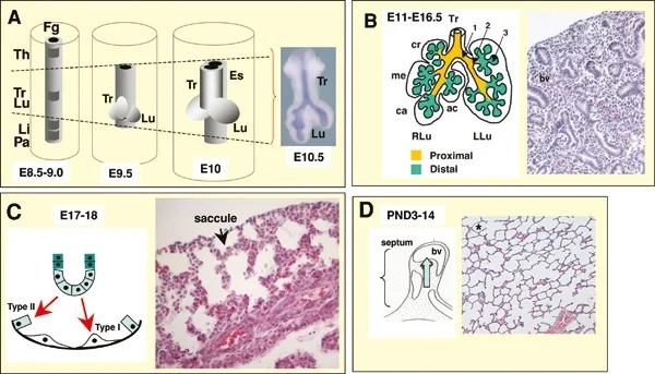
Long description
A. Initial events. From around embryonic day E8.5-9.5 the foregut endoderm (represented in grey) is specified into organ-specific domains, such as the thyroid (Th), trachea (Tr), lung (Lu), liver (Li) and pancreas (Pa).

Figure 1: A (initial events).
The respiratory system originates from the ventral wall of the foregut. A median outpouching, the laryngotracheal diverticulum, appears during the fourth week of gestation. This structure separates from the oesophagus by the tracheoesophageal septum. The cranial end of the diverticulum forms the larynx, while the caudal end elongates to become the trachea. At the distal end, a pair of bronchial buds emerges, which will develop into the primary bronchi and all subsequent lower respiratory structu [1]
During the embryonic stage, the trachea bifurcates into the right and left primary bronchi at the carina[1]. The primary bronchi enter the lungs at the hilum, a concave area where vessels and nerves also pass[1]. The bronchi continue to branch, forming the bronchial tree[1]. This branching continues through the pseudoglandular stage, where the conducting airways are established but no gas exchange occurs. The epithelium lining these structures is pseudostratified ciliated columnar with mucus-producing goblet cells[1]. [3]
As development progresses into the canalicular stage, the respiratory zone begins to form. This zone starts where terminal bronchioles join respiratory bronchioles, the smallest bronchioles[1]. These lead to alveolar ducts, which are tubes of smooth muscle and connective tissue opening into clusters of alveoli[1]. The saccular stage follows, characterized by the formation of terminal sacs. Type II pulmonary cells begin to produce small amounts of surfactant during this phase. Surfactant is composed of phospholipids and proteins that reduce alveolar surface tension[1].
The alveolar stage begins around week 36 and extends into childhood. Alveolar precursors mature into fully functional alveoli. Each alveolus is approximately 200 μm in diameter with elastic walls[1]. The alveolar wall comprises type I cells, type II cells, and alveolar macrophages[1]. Type I cells are squamous epithelial cells, about 25 nm thick, covering up to 97 percent of the surface area[1]. Type II cells secrete surfactant[1]. The respiratory membrane, formed by the alveolar and capillary membranes, is approximately 0.5 μm thick[1]. [3]
Inadequate surfactant production in premature newborns can cause respiratory distress syndrome. This condition may require surfactant replacement therapy, supplemental oxygen, or continuous positive airway pressure. Fetal breathing movements, involving the inhalation and exhalation of amniotic fluid, begin around week 20 or 21. These movements tone respiratory muscles and indicate fetal health. At birth, thoracic compression expels lung fluid, and the first inhalation inflates the lungs. [4] [1]
The respiratory system is fundamentally defined by its dual role in gas exchange and non-vital functions such as speech and olfaction. To understand its development, one must first distinguish between the conducting zone and the respiratory zone. The conducting zone comprises all structures that provide passageways for air, including the nasal cavity, pharynx, trachea, bronchi, and most bronchioles. This zone is lined primarily by pseudostratified ciliated columnar epithelium with goblet cells. [3]
The larynx itself is lined with stratified squamous epithelium superiorly, transitioning to pseudostratified ciliated columnar epithelium inferiorly. This specialized epithelium produces mucus to trap debris as air enters the trachea. The trachea bifurcates into the bronchi, which further divide into bronchioles.
The molecular regulation of these structures begins early. While folate deficiency is critical for neural tube development, causing defects like spina bifida or anencephaly, the respiratory system's structural integrity relies on precise epithelial differentiation. The respiratory epithelium, found in t [2]
The respiratory system is fundamentally defined by its dual role in gas exchange and non-vital functions such as speech and olfaction. To understand its development, one must first distinguish between the conducting zone and the respiratory zone. The conducting zone comprises all structures that provide passageways for air, including the nasal cavity, pharynx, trachea, bronchi, and most bronchioles. This zone is lined primarily by pseudostratified ciliated columnar epithelium with goblet cells.
The respiratory system originates from the foregut, but its embryological foundation is established during the earliest stages of conceptus development. Understanding the derivatives requires tracing the lineage from the fertilised ovum through the formation of the germ layers and extra-embryonic membranes.
The molecular regulation of these structures begins early. During the second week of development, cells within the blastocyst organize into layers, forming extra-embryonic membranes like the amnion, yolk sac, allantois, and chorion to support the embryo. While folate deficiency is critical for neural tube development, causing defects like spina bifida or anencephaly, the respiratory system's structural integrity relies on precise epithelial differentiation. The respiratory epithelium, found in t [2]
| Embryonic Structure | Adult Structure |
|---|---|
| Inner cell mass | Embryo proper |
| Trophoblast | Placenta and fetal membranes |
| Epiblast | Amnion |
| Laryngeal cartilages | Larynx framework |
| Allantois | Urinary bladder and umbilical vessels |
The inner cell mass is totipotent initially, meaning each cell can differentiate into any cell type. This totipotency lasts only a few days before cell fates are determined. The epiblast cells extend around the amniotic cavity to form the amnion. The yolk sac provides primitive blood circulation during the second and third weeks. By week 4, the placenta takes over nourishment. The stalks of the yolk sac and allantois [2]
The larynx regulates air volume entering and leaving the lungs. It is inferior to the laryngopharynx. The thyroid cartilage consists of the laryngeal prominence, or Adam’s apple. This prominence is more prominent in males. The cricoid cartilage is thick and ring-shaped. [3]
Embryonic males and females are morphologically identical until this stage. Bipotential gonads connect to the cloaca via Müllerian and Wolffian ducts. The cloaca is an extension of the primitive gut. By week 12, the fetus measures approximately 9 cm from crown to rump. The fetal period lasts until birth. The fetus grows from approximately 3.3 cm and 7 g to 51 cm and 3.4 kg. Embryonic organ structures develop to allow newborn survival. [4]
Respiratory development begins with the formation of the respiratory diverticulum, or lung bud, from the ventral wall of the foregut during the fourth week of gestation. This early event sets the stage for the complex branching morphogenesis that defines the lower respiratory tract. Disruptions in this timeline or in the separation of the foregut from the trachea lead to significant congenital anomalies.
Surfactant production is critical for reducing surface tension in the alveoli, preventing collapse during expiration. Although fetal breathing movements involving the inhalation of amniotic fluid can be observed starting at 20–21 weeks of development, these movements do not guarantee adequate surfactant levels. Pulmonary hypoplasia, or underdevelopment of the lungs, often results from conditions that restrict lung expansion in utero, such as oligohydramnios or congenital diaphragmatic hernia. [1]
Choanal atresia is a failure of the nasal passages to canalize, resulting in a bony or membranous obstruction of the posterior nasal aperture. This condition is clinically significant because newborns are obligate nasal breathers for the first few months of life. An infant with choanal atresia will present with cyclic cyanosis that improves when crying, as crying forces mouth breathing.
Bronchogenic cysts are congenital malformations arising from abnormal budding of the tracheobronchial tree. These cysts are lined by respiratory epithelium and contain cartilage, smooth muscle, and mucous glands. They are typically located in the mediastinum near the carina or within the lung parenchyma. The embryological basis lies in the aberrant development of the ventral foregut, where accessory lung buds form and detach from the main tracheobronchial tree. These cysts may remain asymptomatic until they become infected or compress adjacent structures.
Tracheo-oesophageal anomalies result from the incomplete separation of the foregut into the trachea and oesophagus. The most common form is oesophageal atresia with a distal tracheo-oesophageal fistula. During the fourth week, the tracheoesophageal septum divides the foregut; failure of this septum to form correctly leads to these anomalies. Infants present with excessive drooling, choking, and cyanosis during feeding. The embryological error involves the abnormal migration of the laryngotracheal groove, leading to a connection between the oesophagus and the trachea.
Neonatal respiratory distress syndrome (NRDS) is a condition primarily affecting premature infants, resulting from a deficiency of pulmonary surfactant. Surfactant is a lipoprotein complex produced by type II pneumocytes that reduces surface tension within the alveoli, preventing their collapse at the end of expiration. Without sufficient surfactant, the alveoli tend to collapse, leading to decreased lung compliance and significant work of breathing.
The production of surfactant begins around week 24 of gestation but does not reach adequate levels for independent respiration until approximately week 35 to 36. Consequently, infants born before this period are at high risk. The clinical presentation typically includes tachypnoea, grunting, nasal flaring, and intercostal retractions shortly after birth. Chest radiography often reveals a characteristic 'ground-glass' appearance with air bronchograms, reflecting diffuse atelectasis.
Management focuses on supporting respiration and replacing the missing surfactant. Exogenous surfactant administration is a cornerstone of therapy, usually delivered directly into the trachea via an endotracheal tube. This intervention significantly improves oxygenation and reduces the need for high ventilatory pressures, thereby lowering the risk of bronchopulmonary dysplasia. Concurrently, respiratory support is provided through continuous positive airway pressure (CPAP) or mechanical ventilation to maintain alveolar patency.
Prevention strategies include the administration of antenatal corticosteroids to mothers at risk of preterm delivery. Corticosteroids accelerate the maturation of type II pneumocytes and stimulate surfactant production, thereby reducing the incidence and severity of NRDS. This intervention is most effective when given between 24 and 34 weeks of gestation, ideally at least 24 hours before delivery.
Understanding the embryological basis of lung development is crucial for managing NRDS. The lung develops from the respiratory diverticulum, which emerges from the foregut around week 4. The branching morphogenesis continues through the pseudoglandular, canalicular, and saccular stages, with alveolar maturation extending into childhood. The transition from fetal to neonatal circulation involves the closure of fetal shunts, including the ductus arteriosus and foramen ovale, and the onset of pulmonary ventilation.
In the fetus, the respiratory system is immature and cannot yet oxygenate blood on its own. The placenta provides the fetus with necessary oxygen and nutrients via the umbilical vein. The liver receives just a trickle of blood, which is all that it needs in its immature, semifunctional state. Blood flows from the inferior vena cava to the right atrium, mixing with fetal venous blood along the way. [4]
After birth, the first breaths expand the fluid-filled alveoli. The drop in pulmonary vascular resistance and the rise in systemic vascular resistance facilitate the closure of fetal shunts. Surfactant plays a critical role in this transition by stabilising the alveoli and preventing atelectasis. Without it, the lungs become stiff and difficult to inflate, leading to the clinical syndrome of NRDS.
Early recognition and aggressive management are essential to improve outcomes. Supportive care, including thermoregulation and nutritional support, complements respiratory interventions. Long-term follow-up is often required for survivors of severe NRDS to monitor for potential complications such as chronic lung disease or neurodevelopmental delays.
The respiratory system begins its structural organisation during the fourth week of embryonic development, a critical period for understanding congenital anomalies. The nasal cavity and pharynx form early as part of the foregut, but their specific skeletal and cartilaginous components develop at distinct times.
The external nose is a composite structure. The root and bridge are supported by bone, specifically the paired nasal bones. These articulate superiorly with the frontal bone and laterally with the maxillary bones. In contrast, the protruding portion of the nose is composed of cartilage, which is why the nose is missing from a dry skull specimen. The septal cartilage is flexible hyaline cartilage that connects to the nasal bone to form the dorsum nasi. The alar cartilage forms the apex of the nose and surrounds the naris. [3]
The pharynx is divided into three regions, each with distinct epithelial linings and functions. The nasopharynx serves only as an airway and is flanked by the conchae of the nasal cavity. At the top of the nasopharynx lie the pharyngeal tonsils, also known as adenoids. These are aggregates of lymphoid reticular tissue covered with ciliated epithelium that traps and destroys invading pathogens. Unlike other tonsils, pharyngeal tonsils are large in children but tend to regress with age and may disappear. [3]
The oropharynx is a passageway for both air and food, bordered superiorly by the nasopharynx and anteriorly by the oral cavity. The fauces is the opening connecting the oral cavity to the oropharynx. A key histological transition occurs here: as the nasopharynx becomes the oropharynx, the epithelium changes from pseudostratified ciliated columnar epithelium to stratified squamous epithelium. This change protects against the mechanical stress of food passage. The oropharynx contains the palatine [3]
The larynx acts as a gateway to the lower respiratory tract. The epiglottis is a flexible piece of elastic cartilage attached to the thyroid cartilage. It covers the opening of the trachea to prevent aspiration. When in the “closed” position, the unattached end of the epiglottis rests on the glottis. The glottis is composed of the vestibular folds, the true vocal cords, and the space between these folds. The true vocal cords are white, membranous folds attached by muscle to the thyroid and arytenoid cartilages. Their inner edges are free, allowing oscillation to produce sound. The size of these folds differs between individuals; folds in males tend to be larger than those in females, creating a deeper voice. [3]
The conducting zone provides a route for incoming and outgoing air. Its major functions include removing debris and pathogens from the incoming air, and warming and humidifying it. The mucous membrane traps debris and pathogens in the bronchi. Additionally, the bronchial epithelium can metabolize some airborne carcinogens. [3]
While not part of the respiratory tract itself, understanding fetal circulation is essential for respiratory physiology at birth. The descending aorta carries partially oxygenated and partially deoxygenated blood into the lower regions of the body. It eventually passes into the umbilical arteries through branches of the internal iliac arteries. The two umbilical arteries carry blood low in oxygen and high in carbon dioxide and fetal wastes. This blood is filtered through the placenta, where wastes diffuse into the maternal circulation. [4]
Students frequently conflate the timing of structural formation with functional maturation, leading to errors in clinical reasoning. A primary error is assuming that the presence of a structure implies it is fully functional or ready for extrauterine life. For instance, the trachea is formed by stacked, C-shaped pieces of hyaline cartilage that provide structural support and prevent collapse. However, the presence of this cartilaginous framework does not equate to respiratory competence. The cri
Another frequent mistake involves the timeline of fetal movement and its relation to maternal perception. Students may assume that fetal movement is constant and obvious from early in the second trimester. In reality, while limb movements become more powerful during weeks 16–20, the pregnant person may only begin to feel quickening, or fetal movements, during this window. Before this period, movements are too subtle to be detected. Furthermore, space restrictions limit these movements, typically forcing the growing fetus into the “fetal position,” with the arms crossed and the legs bent at the knees. Misinterpreting the absence of felt movement in earlier weeks as pathological is a common clinical error. [4]
Confusion also arises regarding the composition of the nasal septum. The nasal septum is formed anteriorly by a portion of the septal cartilage, which is the flexible portion you can touch with your fingers. Posteriorly, it is formed by the perpendicular plate of the ethmoid bone and the thin vomer bones. Students often mistake the entire septum for cartilage, failing to recognize the bony posterior support. This distinction is vital during nasal examinations or procedures, as the cartilaginous portion is pliable while the bony portion is rigid. [3]
The role of the conchae is another area of confusion. The conchae serve to increase the surface area of the nasal cavity and to disrupt the flow of air as it enters the nose. This disruption causes air to bounce along the epithelium, where it is cleaned and warmed. Students may underestimate the importance of this turbulent flow, assuming laminar flow is sufficient. Additionally, the conchae and meatuses conserve water and prevent dehydration of the nasal epithelium by trapping water during exhalation. Failure to appreciate this humidification function can lead to underestimating the consequences of nasal obstruction.
Finally, students often misinterpret the significance of lanugo and vernix caseosa. A silky hair called lanugo covers the skin during weeks 17–20, but it is shed as the fetus continues to grow. Extremely premature infants sometimes exhibit residual lanugo. Similarly, sebaceous glands coat the skin with a waxy, protective substance called vernix caseosa that protects and moisturizes the skin and may provide lubrication during childbirth. Mistaking residual lanugo in a term infant for a developmental anomaly, or ignoring the protective role of vernix, are common errors. The fetus continues to lay down subcutaneous fat from week 31 until birth, filling out the hypodermis. This fat is crucial for thermoregulation, as developmental weeks 21–30 are characterized by rapid weight gain, which is important for maintaining a stable body temperature after birth. [4]
This knowledge is applied at the bedside when assessing preterm infants. The production of surfactant begins during weeks 21–30, assisting proper lung expansion after birth. Clinicians use this timeline to predict respiratory distress syndrome in infants born before this period. The trachea is formed by 16 to 20 stacked, C-shaped pieces of hyaline cartilage. Understanding this structure helps in managing airway obstruction, as the cartilage prevents collapse during inspiration. The fibroelastic membrane allows the trachea to stretch and expand slightly during inhalation and exhalation. This flexibility is critical during positive pressure ventilation. [3]
In the operating theatre, knowledge of the nasal septum is essential. The nasal septum is formed anteriorly by a portion of the septal cartilage. Surgeons must distinguish this from the posterior bony components, the perpendicular plate of the ethmoid bone and the vomer bones. This distinction guides septoplasty and prevents injury to the cranial base. The external nose consists of the surface and skeletal structures that result in the outward appearance of the nose. The root is the region of the nose located between the eyebrows. The bridge is the part of the nose that connects the root to the rest of the nose. The dorsum nasi is the length of the nose. The apex is the tip of the nose. On either side of the apex, the nostrils are formed by the alae. An ala is a cartilaginous structure that forms the lateral side of each naris. The philtrum is the concave surface that connects the apex of the nose to the upper lip. These landmarks are used in facial trauma assessment and reconstructive surgery.
On imaging, the conchae are visible as bony projections. Each lateral wall of the nasal cavity has three bony projections, called the superior, middle, and inferior nasal conchae. The inferior conchae are separate bones, whereas the superior and middle conchae are portions of the ethmoid bone. Radiologists use this anatomy to identify sinusitis or structural abnormalities. The floor of the nasal cavity is composed of the palate. The hard palate at the anterior region of the nasal cavity is composed of bone. The soft palate at the posterior portion of the nasal cavity consists of muscle tissue. This distinction is important in evaluating cleft palate or swallowing disorders.
In examinations, students must recall the timeline of fetal development. During approximately weeks 16–20, the pregnant person may begin to feel quickening. The fetus grows eyelashes during weeks 21–30. The eyelids are no longer fused and can be opened and closed. The lungs begin producing surfactant. The fetus continues to lay down subcutaneous fat from week 31 until birth. Immediately before birth, the average crown-to-rump length is 35.5–40.5 cm. The fetus weighs approximat [4]
Respiratory Readiness at Birth
- Assess Gestational AgeDetermine weeks of pregnancy
- Question: Is gestational age ≥ 21 weeks?
Yes
- Check Surfactant StatusLungs begin producing surfactant
- Question: Is gestational age ≥ 30 weeks?
Yes
- High Respiratory ReadinessSurfactant production established
No
- Moderate Respiratory RiskSurfactant production initiated but limited
No
- High Respiratory RiskNo surfactant production yet
Implantation is complete by the middle of the second week. [2] The embryo folds laterally and again at either end, forming a C-shape. [2] The nasal septum is formed anteriorly by septal cartilage and posteriorly by bone. The trachea is formed by 16 to 20 stacked, C-shaped pieces of hyaline cartilage. Quickening, or fetal movements, is typically felt during weeks 16–20. Lanugo covers the skin during weeks 17–20 but is shed as the fetus grows. [4] The lungs begin producing surfactant during weeks 21–30. The fetus continues to lay down subcutaneous fat from week 31 until birth.
Answer each one in your head first, then open the answer.
What hormone directs the corpus luteum to survive and continue producing progesterone?
Show answerHide answer
hCGWhat structure forms the anterior part of the nasal septum?
Show answerHide answer
septal cartilageWhat substance reduces surface tension in the lungs and assists proper lung expansion?
Show answerHide answer
surfactantWhat is the waxy, protective substance that coats the fetal skin?
Show answerHide answer
vernix caseosaWhat is the average crown-to-rump length immediately before birth?
Show answerHide answer
35.5–40.5 cm
- Betts JG, Young KA, Wise JA, et al. Embryonic Development of the Respiratory System. OpenStax, Anatomy and Physiology 2e (CC BY-NC-SA 4.0). Accessed .
- Betts JG, Young KA, Wise JA, et al. Embryonic Development. OpenStax, Anatomy and Physiology 2e (CC BY-NC-SA 4.0). Accessed .
- Betts JG, Young KA, Wise JA, et al. Organs and Structures of the Respiratory System. OpenStax, Anatomy and Physiology 2e (CC BY-NC-SA 4.0). Accessed .
- Betts JG, Young KA, Wise JA, et al. Fetal Development. OpenStax, Anatomy and Physiology 2e (CC BY-NC-SA 4.0). Accessed .
Figure credits
Educational content for medical students and doctors. It is not a substitute for clinical judgement or local guidelines.