Gastrointestinal development
Embryonic origins, rotations, and anomalies of the foregut, midgut, and hindgut.
Gastrointestinal development begins in the fourth week of gestation when the primitive gut tube differentiates into three distinct segments: the foregut, midgut, and hindgut. The foregut gives rise to the esophagus, stomach, liver, and pancreas, while the midgut forms the small intestine and proximal colon. The hindgut develops into the distal colon, rectum, and anal canal. A critical event in midgut development is its rotation and temporary herniation into the umbilical region. During the sixth week, the rapidly growing midgut herniates into the umbilical cord to accommodate the expanding liver. It remains there until the tenth week, when it returns to the abdominal cavity. If this rotation fails or is incomplete, the condition is known as malrotation, which predisposes patients to midgut volvulus.
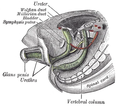
Figure 1Gray993
Long description
Tail end of human embryo, from eight and a half to nine weeks old. (From model by Keibel.)

Figure 1: Tail end of human embryo, from eight and a half to nine weeks old (From model by Keibel.).
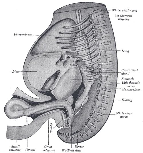
Figure 2Gray986
Long description
Reconstruction of a human embryo of 17 mm. (After Mall.)

Figure 2: Reconstruction of a human embryo of 17 mm (After Mall.).
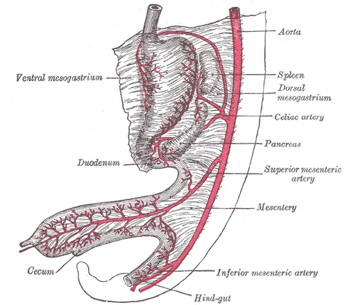
Figure 3Gray985
Long description
Abdominal part of digestive tube and its attachment to the primitive or common mesentery. Human embryo of six weeks. (After Toldt.)

Figure 3: Abdominal part of digestive tube and its attachment to the primitive or common mesentery (human embryo of six weeks).
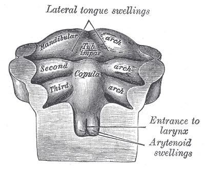
Figure 4Gray980
Long description
Floor of pharynx of human embryo of about the end of the fourth week. (From model by Peters.)

Figure 4: Floor of pharynx of human embryo of about the end of the fourth week (From model by Peters.).
The foregut initially forms a single tube that must divide into the trachea and esophagus. This separation is mediated by the tracheoesophageal septum. Failure of this septum to complete the division results in tracheoesophageal fistula or esophageal atresia. These anomalies are frequently associated with other congenital defects, including annular pancreas and intestinal atresia.
Hypertrophic pyloric stenosis is a postnatal condition characterized by the thickening of the pyloric sphincter muscle. While not a direct embryological defect, it represents a failure of normal muscular maturation. It typically presents in the first few weeks of life with projectile vomiting. The pylorus is the junction between the stomach and the duodenum, derived from the foregut.
Intestinal atresia refers to a complete obstruction of the intestinal lumen. It can occur in the stomach, duodenum, jejunum, ileum, or colon. Jejunal and ileal atresia often result from vascular accidents in utero, where a thrombus occludes the mesenteric vessels, leading to necrosis and subsequent resorption of the affected segment. This leaves a blind end on either side of the atretic segment.
The pancreas develops from two buds: the dorsal and ventral pancreatic buds, both arising from the foregut endoderm. The dorsal bud forms the majority of the gland, while the ventral bud rotates to join it. The spleen develops from mesenchymal tissue in the dorsal mesogastrium. Pancreas divisum is a common anatomical variant where the dorsal and ventral ducts fail to fuse, leaving two separate drainage pathways.
Annular pancreas is a rare congenital anomaly affecting approximately 1 in 20,000 newborns. It occurs when the ventral pancreatic bud fails to rotate properly, resulting in a ring of pancreatic tissue surrounding the second portion of the duodenum. This ring can cause duodenal obstruction. Annular pancreas is associated with other congenital anomalies, including Down’s syndrome, tracheoesophageal fistula, and intestinal atresia. [3]
The retroperitoneal position of the duodenum, pancreas, and ascending and descending colon is established during embryonic development. The midgut initially lies in the ventral mesentery but becomes secondarily retroperitoneal as the mesentery fuses with the posterior abdominal wall. This fusion is crucial for the stable positioning of these organs.
Exam trapMeckel Diverticulum
It is located in the ileum and is a common cause of lower gastrointestinal bleeding in children.
| Anomaly | Embryological Origin | Clinical Significance |
|---|---|---|
| Annular Pancreas | Ventral bud rotation failure | Duodenal obstruction |
| Meckel Diverticulum | Vitelline duct persistence | Bleeding, obstruction |
| Malrotation | Incomplete midgut rotation | Volvulus, ischemia |
| Pyloric Stenosis | Muscular hypertrophy | Projectile vomiting |
The spleen is not yet morphologically distinct but is indicated by a cluster of mesenchymal cells in the dorsal mesogastrium, located posterior to the stomach and lateral to the pancreatic tail. The liver bud lies anterior to the stomach, while the dorsal pancreatic bud rests against the posterior abdominal wall. The ventral pancreatic bud is tucked beneath the liver bud, ready to migrate. This spatial arrangement is critical for understanding the final anatomical positions of these organs.
The liver originates from the hepatic diverticulum, an outpouching of the foregut endoderm. This diverticulum grows into the septum transversum, a mass of mesoderm that forms the primitive diaphragm. The hepatic cells proliferate and form cords that invade the surrounding mesenchyme. These cords eventually differentiate into hepatocytes and biliary epithelial cells. The vascular system of the liver develops concurrently, with the vitelline and umbilical veins contributing to the portal venous system and hepatic sinusoids. The liver begins its functional role in hematopoiesis during the fetal period. By week 12, the liver is a major site of red blood cell production. The structural organization of the liver lobules is established through the branching of the bile ducts and the invasion of blood vessels. The gallbladder and cystic duct develop from a distal outpouching of the hepatic diverticulum.
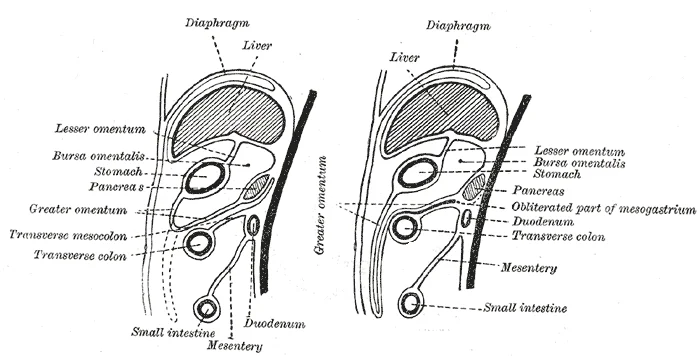
Figure 5Gray990
Long description
Diagrams to illustrate the development of the greater omentum and transverse mesocolon.

Figure 5: Diagrams to illustrate the development of the greater omentum and transverse mesocolon.
The pancreas develops from two separate endodermal buds: the dorsal and ventral pancreatic buds. The dorsal bud arises from the dorsal wall of the duodenum, while the ventral bud arises from the base of the hepatic diverticulum. As the duodenum rotates, the ventral bud migrates posteriorly and fuses with the dorsal bud. The main pancreatic duct forms from the union of the ducts of the two buds. The head of the pancreas is derived primarily from the ventral bud, while the body and tail arise from the dorsal bud. The acinar cells, which produce digestive enzymes, and the islets of Langerhans, which produce hormones, differentiate from the endodermal epithelium. The exocrine and endocrine functions of the pancreas begin to develop in the second trimester. The fusion of the two buds is a critical step; failure of proper fusion can lead to annular pancreas, a condition where pancreatic tissue encircles the duodenum, potentially causing obstruction.
The spleen develops from mesenchymal cells within the dorsal mesogastrium. Unlike the liver and pancreas, the spleen is of mesodermal origin. These mesenchymal cells aggregate to form a solid mass that later becomes vascularized. The spleen does not have an endodermal component. It begins to function as a site of hematopoiesis during the fetal period, producing lymphocytes and red blood cells. The white pulp, which contains lymphoid tissue, and the red pulp, which filters blood, differentiate during the later stages of fetal development. The spleen is located in the left upper quadrant of the abdomen, posterior to the stomach. Its development is independent of the gut tube endoderm, distinguishing it from the liver and pancreas. The splenic vessels grow into the mesenchymal mass, establishing its blood supply.
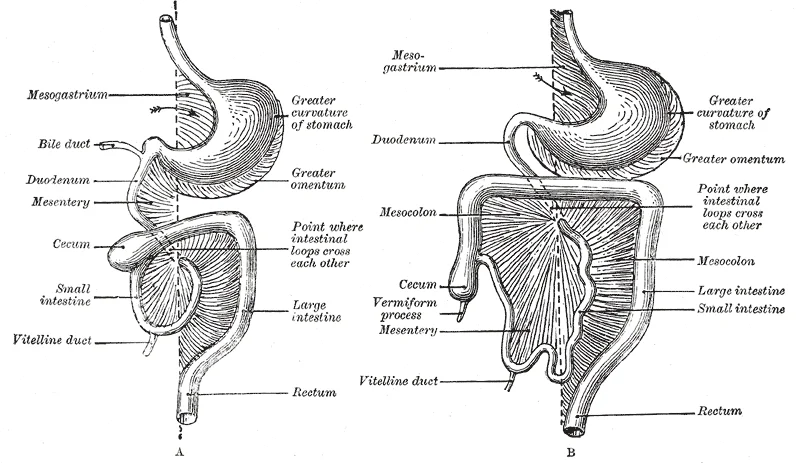
Figure 6Gray987
Long description
Diagrams to illustrate two stages in the development of the digestive tube and its mesentery. The arrow indicates the entrance to the bursa omentalis.

Figure 6: Diagrams to illustrate two stages in the development of the digestive tube and its mesentery.
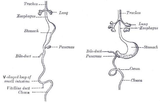
Figure 7Gray983
Long description
Front view of two successive stages in the development of the digestive tube. (His.)

Figure 7: Front view of two successive stages in the development of the digestive tube (His.).
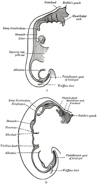
Figure 8Gray982
Long description
Sketches in profile of two stages in the development of the human digestive tube. (His.) A X 30. B X 20.

Figure 8: Sketches in profile of two stages in the development of the human digestive tube (His.) A X 30.
The molecular regulation of gastrointestinal development involves complex signaling pathways that coordinate the formation and function of the digestive system. This timing coincides with increases in gene expression of various ion channels known to modulate enteric action potentials. The ENS consists of three main components: enteric neurons, enteric glial cells (EGCs), and interstitial cells of Cajal (ICCs), which form two major ganglionated structures—the submucosal plexus (Meissner's plexus) and the myenteric plexus (Auerbach's plexus). [2]
The gut-brain axis (GBA) represents a crucial signaling network that develops during fetal life and continues to mature postnatally. This connection involves both top-down and bottom-up mechanisms, with signals from the brain modulating gastrointestinal functions while GI-derived molecules influence brain processes through different pathways. The exact mechanisms through which the gut and brain communicate in young infants remain limited in our current understanding. Humoral components of the gut-brain axis include the hypothalamic-pituitary-adrenal (HPA) axis, the enteroendocrine system, and the immune system. Enteroendocrine cells (EECs) in the gut produce gastrointestinal hormones such as ghrelin, glucagon-like peptide (GLP-1), cholecystokinin, and peptide YY (PYY), which regulate food intake and satiety while also being involved in the regulation of emotions and mood. [2]
The maturation of the gastrointestinal system in full-term and preterm infants is significantly influenced by molecular signaling mechanisms. In preterm infants less than 28 gestational weeks, the relatively underdeveloped GI system limits their ability to utilize enteral nutrition, contributing to a nutritional emergency. Disorders of gastrointestinal ontogenesis, combined with other factors such as early postnatal stress and microbiota alterations, result in impaired activation of intestinal peristalsis and the gut-brain axis. The temporal development of neural subtypes and enteric glia occurs between 12 and 16 GW, establishing the foundation for proper gastrointestinal function. [2]
Figure 9Gray989
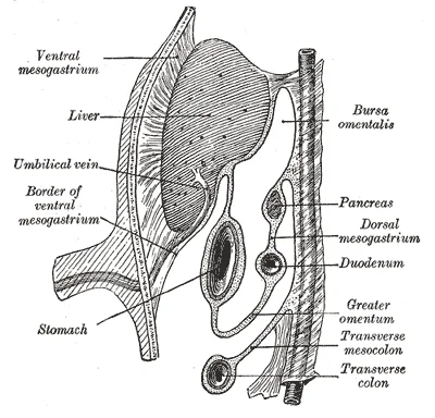
Long description
Schematic figure of the bursa omentalis, etc. Human embryo of eight weeks. (Kollmann.)

Figure 9: Schematic figure of the bursa omentalis, etc (human embryo of eight weeks).
The gastrointestinal tract develops from the primitive gut tube, which is divided into three main parts: the foregut, midgut, and hindgut. Each of these sections gives rise to specific structures in the adult gastrointestinal system through complex embryological processes involving cell migration, differentiation, and organogenesis.
Figure 10Gray984
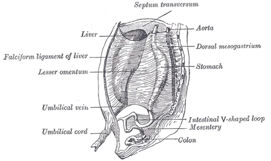
Long description
The primitive mesentery of a six weeks’ human embryo, half schematic. (Kollmann.)

Figure 10: The primitive mesentery of a six weeks’ human embryo, half schematic (Kollmann.).
Figure 11Gray977
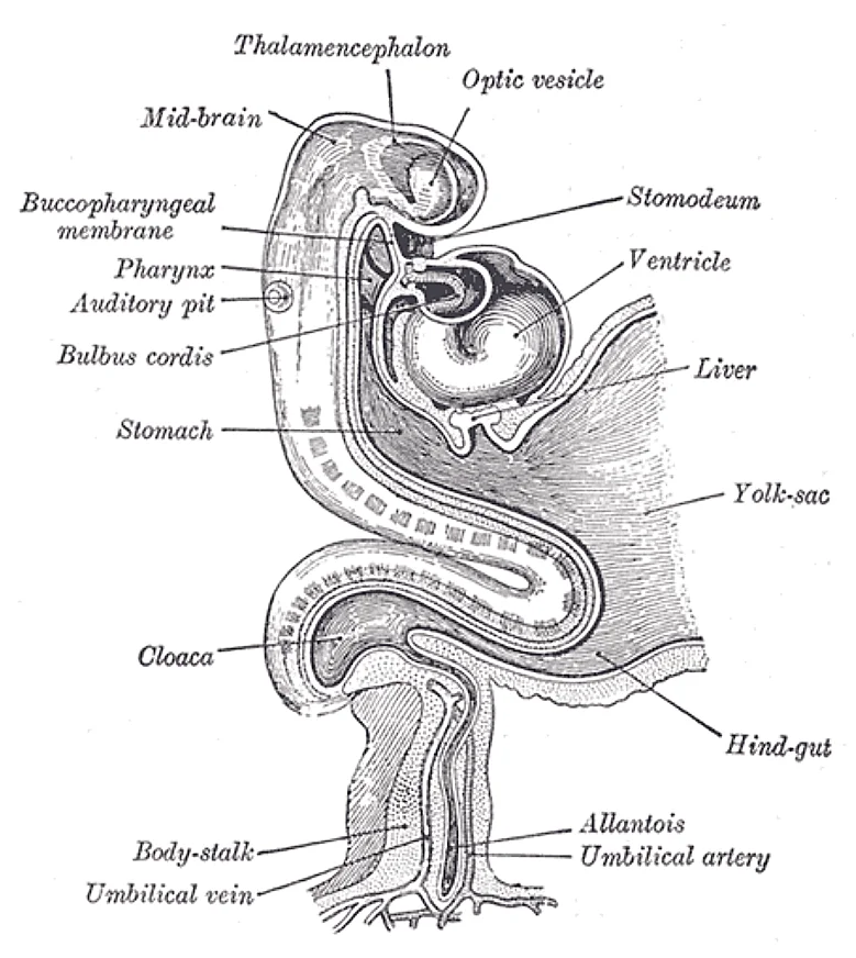
Long description
Human embryo about fifteen days old. Brain and heart represented from right side. Digestive tube and yolk sac in median section. (After His.)

Figure 11: Human embryo about fifteen days old.
| Embryonic Structure | Adult Derivatives |
|---|---|
| Foregut | Pharynx, esophagus, stomach, proximal duodenum (up to the entrance of the bile duct), liver, gallbladder, pancreas, and lower respiratory tract |
| Midgut | Distal duodenum (after bile duct entrance), jejunum, ileum, cecum, appendix, ascending colon, and proximal two-thirds of the transverse colon |
| Hindgut | Distal one-third of the transverse colon, descending colon, sigmoid colon, rectum, and upper anal canal (above the pectinate line) |
The intestinal length increases significantly during development. Autopsy data show that the intestine measures 125 cm by 20 weeks gestation, 200 cm by 30 weeks gestation, and 275 cm at term. This growth continues postnatally, reaching approximately 575 cm by 20 years of life. The large intestine is approximately 60 cm in a full-term infant, which increases to about 150 cm by adulthood. When including villus and microvillus structures, the surface area in the adult is estimated to be 200 m², whi [2]
The enteric nervous system (ENS) originates from neural crest precursor cells that enter the foregut at 9 embryonic days and migrate rostrocaudally to colonize the entire intestine by 14 embryonic days. The ENS is a complex network of neurons and glial cells that controls gastrointestinal functions independently of the central nervous system. Diverse populations of fibroblast-like interstitial cells are present in the adult gut, including various subpopulations of Kit-positive ICCs and fibroblas [2]
Figure 12Gray992
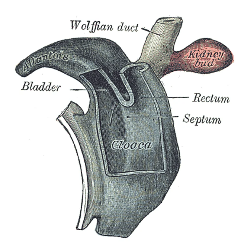
Long description
Cloaca of human embryo from twenty-five to twenty-seven days old. (From model by Keibel.)

Figure 12: Cloaca of human embryo from twenty-five to twenty-seven days old (From model by Keibel.).
Figure 13Gray991
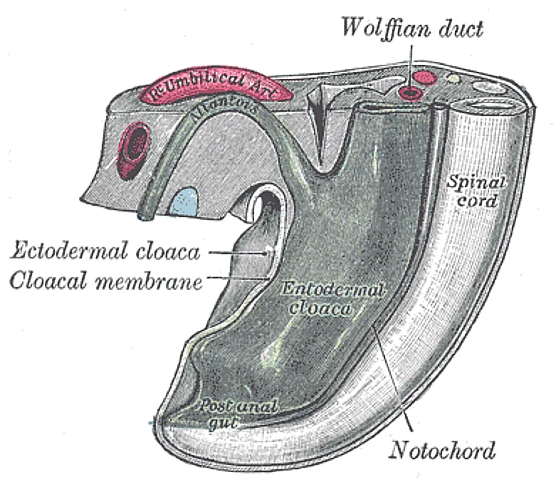
Long description
Tail end of human embryo from fifteen to eighteen days old. (From model by Keibel.)

Figure 13: Tail end of human embryo from fifteen to eighteen days old (From model by Keibel.).
Figure 14Gray981
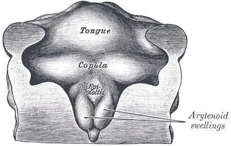
Long description
Floor of pharynx of human embryo about thirty days old. (From model by Peter.)

Figure 14: Floor of pharynx of human embryo about thirty days old (From model by Peter.).
Figure 15Gray979
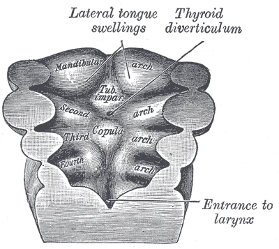
Long description
Floor of pharynx of human embryo about twenty-six days old. (From model by Peters.)

Figure 15: Floor of pharynx of human embryo about twenty-six days old (From model by Peters.).
Figure 16Gray978
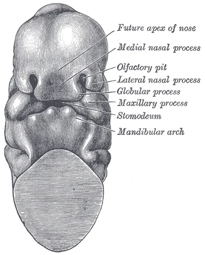
Long description
Head end of human embryo of about thirty to thirty-one days. (From model by Peters.)

Figure 16: Head end of human embryo of about thirty to thirty-one days (From model by Peters.).
The development of neural connections involves the central nervous system (CNS), the enteric nervous system (ENS), and the autonomic nervous system (ANS). The ANS comprises sympathetic and parasympathetic nerves: the sympathetic system exerts mainly an inhibitory influence on the gut, while the vagus nerve (VN) appears to be able to sense hormones, cytokines, and metabolites from the GI tract, leading to afferent signals to the brain.
The foregut gives rise to the esophagus, stomach, and proximal duodenum, as well as the liver, gallbladder, and pancreas. The midgut develops into the jejunum, ileum, cecum, appendix, and ascending colon. The hindgut forms the descending colon, sigmoid colon, rectum, and upper anal canal.
Congenital anomalies of the gastrointestinal tract arise from errors in the complex processes of gut tube formation, rotation, and differentiation. Understanding these defects requires tracing the embryological journey from the primitive gut tube to the mature organ system. The gut tube is divided into the foregut, midgut, and hindgut, each giving rise to specific structures with distinct vascular supplies and developmental timelines.
The midgut undergoes a physiological herniation into the umbilical cord around week 6, followed by a return to the abdominal cavity and a 270-degree counter-clockwise rotation around the superior mesenteric artery. Failure of this rotation or retraction leads to malrotation. Malrotation can result in volvulus, where the bowel twists around its mesentery, compromising blood supply. This is a surgical emergency. The liver and pancreas develop from outpouchings of the foregut. Abnormal fusion or mi [1]
Anomalies of the liver and pancreas often involve structural variations rather than complete absence. Pancreatic anomalies include pancreas divisum, where the dorsal and ventral ducts fail to fuse, which may be asymptomatic or cause recurrent pancreatitis. These structures are closely related; the common bile duct and pancreatic duct usually join before entering the duodenum.
Atresia refers to the complete closure of a lumen, while stenosis is a narrowing. Oesophageal atresia with tracheo-oesophageal fistula is the most common foregut anomaly. In this condition, the oesophagus ends in a blind pouch, and a fistula connects the trachea to the lower oesophageal segment. This prevents swallowing of amniotic fluid and allows air to enter the stomach. The classic "double bubble" sign on abdominal X-ray reflects air in the stomach and proximal duodenum with no distal gas.
The cloaca is divided by the urorectal septum into the urogenital sinus and the anorectal canal. Failure of this division or improper descent of the hindgut leads to imperforate anus or rectal atresia. These anomalies are classified as high or low based on the relationship of the rectal pouch to the levator ani muscle. Early surgical correction is essential to preserve continence and bowel function.
The development of the gastrointestinal and respiratory systems is closely linked in terms of functional maturation, particularly regarding the transition to extrauterine life. While the structural formation of the gut occurs early in embryogenesis, its functional capacity to handle nutrition matures later. Newborns require the structural and functional maturation of the GI tract for digestion and absorption of the nutrients from colostrum and breast milk. This process relies on a complete development of intestinal motor function which includes suck–swallow coordination, continence of the gastroesophageal sphincter tone, adequate gastric emptying, and intestinal peristalsis. [2]
DefinitionEnteral Feeding Readiness
The ability to tolerate full enteral feeding depends on coordinated motor functions and adequate gastric emptying, which are often immature in preterm infants.
Full-term babies can acquire adequate amounts of nutrients to promote the rapid growth that occurs shortly after birth. However, preterm infants often face significant challenges. Half of preterm infants are delayed in reaching full enteral feeding volumes and have gastroesophageal reflux, gastric residuals, and constipation due to delayed gastric emptying, prolonged bowel transit, abdominal distension, and delayed passage of meconium, all of which are GI functions describing immaturity. These symptoms reflect the underlying immaturity of the gastrointestinal system, which must coordinate with respiratory maturity to ensure survival. [2]
Exam trapRespiratory vs GI Maturity
While respiratory maturity is often assessed by surfactant levels and alveolar development, GI maturity is assessed by the ability to tolerate enteral feeds without residual or reflux. These milestones do not always align perfectly.
The respiratory system continues to expand, and the surfaces that will form the respiratory membrane develop further. At this point, pulmonary capillaries have formed and continue to expand, creating a large surface area for gas exchange. The major milestone of respiratory development occurs at around week 28, when sufficient alveolar precursors have matured so that a baby born prematurely at this time can usually breathe on its own. However, alveoli continue to develop and mature into childhood. A full complement of functional alveoli does not appear until around 8 years of age. [1]
| System | Key Milestone | Approximate Timeframe | Clinical Significance |
|---|---|---|---|
| Respiratory | Sufficient alveolar precursors for independent breathing | Week 28 | Premature infants may breathe spontaneously |
| Respiratory | Full complement of functional alveoli | Around 8 years of age | Continued lung development into childhood |
| Gastrointestinal | Adequate nutrient acquisition for rapid growth | Full-term birth | Depends on coordinated motor functions |
| Gastrointestinal | Delayed enteral feeding volumes | Preterm infants | Associated with reflux, residuals, and constipation |
Congenital anomalies of the gastrointestinal tract, such as annular pancreas, can present with obstruction. Useful diagnostic modalities for the diagnosis of AP include ultrasonography or plain abdominal radiographs, which usually show the classic “double bubble” sign when a duodenal obstruction is present. Surgical management is usually required to relieve the obstruction when the diagnosis of AP is confirmed operatively. Although surgery has been considered the “gold standard” for the diagnosis of annular pancreas, non operative modalities (CT scanning, MRI, MRCP, ERCP and EUS) may all suggest the presence of pancreatic tissue encircling the duodenum. MRI and CT scanning have the advantage of being non invasive, although the correct diagnosis may be overlooked by both scan techniques if the pancreatic tissue is present as a thin band incorporated in the duodenal wall. [3]
NoteDiagnostic Imaging
The “double bubble” sign on plain abdominal radiographs is a classic indicator of duodenal obstruction, such as that caused by annular pancreas. Non-invasive imaging like MRI and CT can also suggest the diagnosis by visualizing pancreatic tissue encircling the duodenum.
In adults, annular pancreas may remain asymptomatic or present with non-specific symptoms. These findings highlight the variable presentation of gastrointestinal anomalies across different age groups. [3]
While primarily a digestive organ, it also exerts neural, endocrine, exocrine, and immunologic functions, a multifunctional role that is a frequent exam trap if students focus solely on digestion. The bronchial bud is a paired embryonic structure that gives rise to the bronchi and all lower respiratory structures, distinct from the GI tract but developing in close proximity. Understanding this separation is crucial for identifying foregut derivatives. The enteric nervous system (ENS) matures in stages; by 12 gestational weeks, the fetal colon possesses a dense neuronal network in the myenteric plexus with excitatory neurotransmitter markers. However, inhibitory neurotransmitter markers do not appear until 14 gestational weeks, marking a key developmental milestone. Electrical stimulation of internodal strands fails to evoke activity in the ENS at 12 or 14 gestational weeks, indicating functional immaturity despite structural presence. The gut–brain axis represents a bidirectional communication system between the gut and the brain, mediated by interrelated components. This axis is now commonly referred to as the microbiota-gut–brain axis, highlighting the extensive role of microbiota in this dialogue. Alterations in gut microbiota are directly linked to physical and mental health, though the specific mechanisms are not yet fully elucidated. Lactose is the primary carbohydrate source in milk, and lactose intolerance is rare in the preterm population. Unabsorbed lactose is converted to short-chain fatty acids, providing an efficient salvage pathway for energy. Lactase activity in preterm infants can be induced through enteral feedings of human milk, a clinical pearl for nutritional management. Triglycerides constitute half of the non-protein energy content in both human milk and formula. Their digestion begins with bile acid micellar emulsification, which creates smaller droplets and increases surface area for lipase interaction. Pancreatic lipase is the most important enzyme hydrolyzing long-chain triglycerides into monoglycerides and free fatty acids. These products are then absorbed into the small intestinal epithelium, completing the lipid digestion process. [2] [1]
Students often confuse the timing of excitatory versus inhibitory neurotransmitter expression in the ENS, incorrectly assuming they develop simultaneously. Another trap is overlooking the non-digestive functions of the GI tract, such as its immunologic and endocrine roles. In lipid digestion, candidates may forget that emulsification by bile acids is the prerequisite step before pancreatic lipase can effectively hydrolyze triglycerides. Finally, regarding the gut–brain axis, it is a common error to view it as unidirectional; it is strictly a bidirectional communication system.
Exam trapIncomplete understanding of enzyme development
Students often assume that all digestive enzymes are fully functional at birth. In reality, lactase activity is low in preterm infants, while sucrase, maltase, and isomaltase are fully active. [2]
Exam trapOverlooking gastric motility differences
Many students mistakenly believe that gastric motility functions similarly in preterm and full-term infants. Gastric motility is poorly organized in preterm infants, particularly in those of 28-32 weeks gestational age, while preterm infants of 32-36 weeks show patterns much closer to full-term newborns. [2]
Exam trapIgnoring essential fatty acid requirements
Students may not recognize that preterm infants lack the enzymatic capability to convert linoleic and linolenic acids into long-chain polyunsaturated fatty acids (LCPUFAs), which are required for eicosanoid formation and central nervous system structures.
Exam trapConfusing enteric nervous system components
The distinction between Meissner's plexus (located in the submucosa and innervating muscularis mucosae, intestinal neuroendocrine cells, glandular epithelium and submucosal blood vessels) and Auerbach's plexus (located between circular and longitudinal muscle layers and associated with their contractility) is often misunderstood. [2]
Exam trapMisunderstanding gut-brain axis mediators
Students may overlook the central role of metabolic mediators in the microbiome-gut-brain axis, including serotonin (5-HT), melatonin, and short-chain fatty acids (SCFAs), which influence gut-brain communication through various pathways.
Exam trapIncorrect treatment approach for annular pancreatitis
A common misconception is that resection of annular pancreatic tissue is the preferred treatment. In fact, bypass operations like gastro-jejunostomy or duodeno-jejunostomy are preferred, as resection has been associated with complications including pancreatitis, pancreatic fistula formation, and incomplete relief of obstruction. [3]
NoteClinical assessment of fetal wellbeing
Fetal breathing movements are considered a sign of robust health and may help the alveoli to form and mature. This knowledge is used in obstetrics to assess fetal wellbeing.
NoteNeonatal nutrition management
Understanding developmental differences in intestinal digestion and absorption functionality is crucial for managing nutrition in term and preterm infants. This knowledge guides dietary interventions, particularly regarding enzyme supplementation and fatty acid requirements.
Assessment of gastric motility in preterm infants
- Assess gestational ageDetermine if infant is preterm or term
- Question: Is infant preterm (28-32 weeks GA)?
Yes
- Expect impaired gastric motilityPoorly organized gastric electrical activity and delayed gastric emptying
- Monitor feeding toleranceSmall, frequent feeds may be necessary
No
- Expect mature motility patternsSimilar to full-term newborns
- Proceed with standard feeding protocolsLarger volumes less frequently
- Consider motilin involvementMotor migratory complexes occur every 2-4 hours and are initiated by motilin release
NoteSurgical decision making
Knowledge of annular pancreatitis treatment is essential in surgical practice. Up to 40% of cases require surgery, with duodeno-duodenostomy or duodeno-jejunostomy performed to bypass the obstructed duodenal segment. Division of the pancreatic ring is not recommended due to high post-operative complication rates. [3]
NoteImaging interpretation
Understanding the appropriate imaging techniques for gastrointestinal conditions is vital in clinical practice. For annular pancreatitis, computed tomography (CT), endoscopic retrograde cholangiopancreatography (ERCP), and magnetic resonance cholangiopancreatography (MRCP) are the imaging techniques of choice.
NoteNeurogastroenterology applications
Knowledge of the microbiome-gut-brain axis is increasingly applied in clinical practice, particularly in conditions involving altered gut-brain communication. Understanding the role of serotonin and short-chain fatty acids in this axis helps inform treatment approaches for functional gastrointestinal disorders.
- Fetal breathing movements are a sign of robust health and help alveoli form and mature
- Lactase activity is low in preterm infants while sucrase, maltase, and isomaltase are fully active
- Preterm infants lack enzymatic capability to convert linoleic and linolenic acids into LCPUFAs
- Gastric motility is poorly organized in preterm infants of 28-32 weeks GA
- Meissner's plexus is in the submucosa while Auerbach's plexus is between muscle layers
- Serotonin and SCFAs play central roles in microbiome-gut-brain axis communication
- Bypass operations are preferred over resection for treating symptomatic annular pancreatitis
Answer each one in your head first, then open the answer.
Which enzyme activity is low in preterm infants?
Show answerHide answer
Lactase activityWhat is the preferred surgical treatment for symptomatic annular pancreatitis with duodenal obstruction?
Show answerHide answer
Bypass operations such as gastro-jejunostomy or duodeno-jejunostomyWhere is Meissner's plexus located?
Show answerHide answer
In the connective tissue of submucosaWhich metabolic mediator is involved in most branches of the microbiome-gut-brain axis?
Show answerHide answer
Serotonin (5-HT)What percentage of annular pancreatitis cases requires surgery?
Show answerHide answer
Up to 40%
- Betts JG, Young KA, Wise JA, et al. Embryonic Development of the Respiratory System. OpenStax, Anatomy and Physiology 2e (CC BY-NC-SA 4.0). Accessed .
- Indrio F, Neu J, Pettoello-Mantovani M, et al. Development of the Gastrointestinal Tract in Newborns as a Challenge for an Appropriate Nutrition: A Narrative Review. Nutrients 2022;14(7):1405. doi:10.3390/nu14071405. Accessed .
- Rondelli F, Bugiantella W, Stella P, et al. Symptomatic annular pancreas in adult: Report of two different presentations and treatments and review of the literature. International journal of surgery case reports 2016;20S:21-24. doi:10.1016/j.ijscr.2016.02.001. Accessed .
Figure credits
- Figure 1: Henry Vandyke Carter, via Wikimedia Commons, Public domain, Public domain.
- Figure 2: Henry Vandyke Carter, via Wikimedia Commons, Public domain, Public domain.
- Figure 3: Henry Vandyke Carter, via Wikimedia Commons, Public domain, Public domain.
- Figure 4: Henry Vandyke Carter, via Wikimedia Commons, Public domain, Public domain.
- Figure 5: Henry Vandyke Carter, via Wikimedia Commons, Public domain, Public domain.
- Figure 6: Henry Vandyke Carter, via Wikimedia Commons, Public domain, Public domain.
- Figure 7: Henry Vandyke Carter, via Wikimedia Commons, Public domain, Public domain.
- Figure 8: Henry Vandyke Carter, via Wikimedia Commons, Public domain, Public domain.
- Figure 9: Henry Vandyke Carter, via Wikimedia Commons, Public domain, Public domain.
- Figure 10: Henry Vandyke Carter, via Wikimedia Commons, Public domain, Public domain.
- Figure 11: Henry Vandyke Carter, via Wikimedia Commons, Public domain, Public domain.
- Figure 12: Henry Vandyke Carter, via Wikimedia Commons, Public domain, Public domain.
- Figure 13: Henry Vandyke Carter, via Wikimedia Commons, Public domain, Public domain.
- Figure 14: Henry Vandyke Carter, via Wikimedia Commons, Public domain, Public domain.
- Figure 15: Henry Vandyke Carter, via Wikimedia Commons, Public domain, Public domain.
- Figure 16: Henry Vandyke Carter, via Wikimedia Commons, Public domain, Public domain.
Educational content for medical students and doctors. It is not a substitute for clinical judgement or local guidelines.