Urogenital development
Embryonic origins of kidneys, ureters, bladder and urethra with key anomalies.
Urogenital development begins in the early embryo with the sequential formation of three nephrogenic structures: the pronephros, mesonephros, and metanephros. The pronephros appears around week 4 but degenerates in humans. The mesonephros forms next and contributes to the male reproductive system, while the metanephros, which forms from the metanephric mesoderm and the ureteric bud, becomes the permanent kidney. The ureteric bud is an outgrowth of the mesonephric (Wolffian) duct that invades the metanephric mesoderm, inducing it to form nephrons while the bud itself branches to form the collecting system.
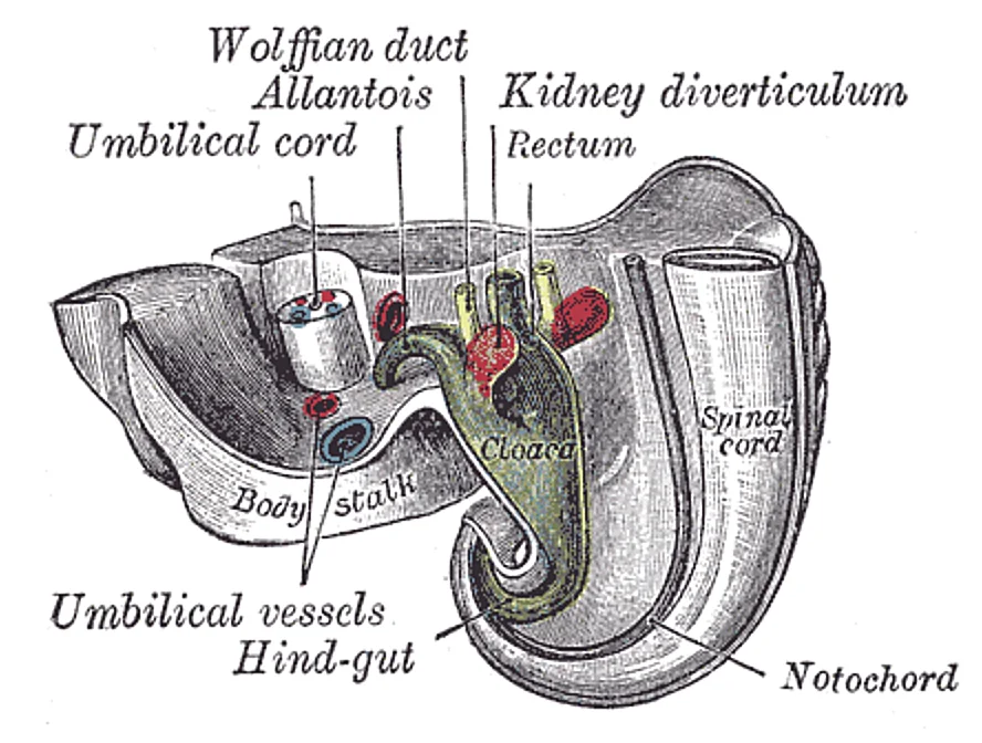
Figure 1Gray1115
Long description
Tail end of human embryo twenty-five to twenty-nine days old. (From model by Keibel.)

Figure 1: Tail end of human embryo twenty-five to twenty-nine days old (From model by Keibel.).
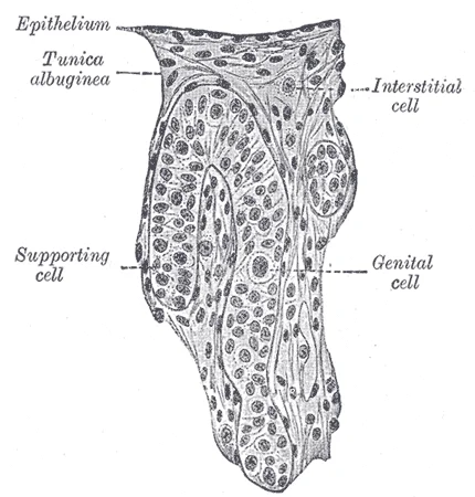
Figure 2Gray1114
Long description
Section of a genital cord of the testis of a human embryo 3.5 cm. long. (Felix and Bühler.)

Figure 2: Section of a genital cord of the testis of a human embryo 3.5 cm (long).
The kidneys initially develop in the pelvis and ascend to the upper abdomen between weeks 6 and 10. Failure of ascent results in a pelvic kidney. The bladder and urethra develop from the urogenital sinus, which is the ventral part of the cloaca. The cloaca is divided by the urorectal septum into the urogenital sinus and the anorectal canal. The upper part of the urogenital sinus forms the bladder, while the lower part forms the urethra.
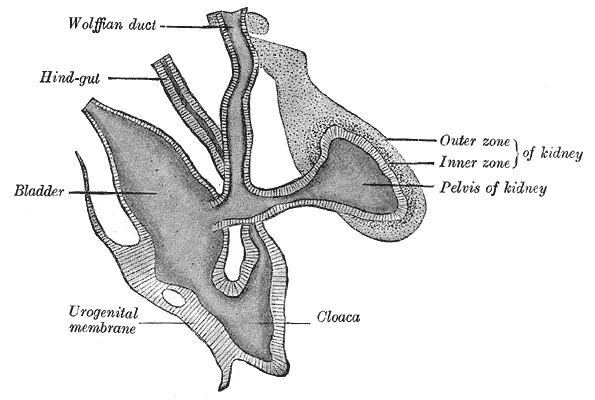
Figure 3Gray1118
Long description
Primitive kidney and bladder, from a reconstruction. (After Schreiner.)

Figure 3: Primitive kidney and bladder, from a reconstruction (After Schreiner.).
Several congenital anomalies arise from disruptions in this process. Potter sequence is a constellation of findings including pulmonary hypoplasia, facial dysmorphism, and limb deformities, resulting from severe oligohydramnios due to bilateral renal agenesis or non-functioning kidneys. Unilateral renal agenesis is the absence of one kidney, often asymptomatic if the contralateral [1]
Sexual differentiation of the urogenital system occurs during weeks 9–12 of fetal development. In males, the SRY gene on the Y chromosome triggers testis development, and testosterone stimulates the Wolffian ducts to form the vas deferens, while the Müllerian ducts degenerate. In females, the absence of SRY leads to ovarian development, Müllerian ducts form the uterus and uterine tubes, and Wolffian ducts degenerate. [1][2]
Urogenital development begins with chromosomal determination. Mammalian sex is generally determined by X and Y chromosomes[1]. Individuals homozygous for X (XX) are female in sex and heterozygous individuals (XY) are male[1]. The presence of a Y chromosome triggers the development of a certain set of male characteristics and its absence results in female characteristics[1]. Nondisjunction during meiosis can produce other combinations such as XXY, XYY, and XO which are called chromosomal intersex[1].
In both male and female embryos, the same group of cells has the potential to develop into either the male or female gonads; this tissue is considered bipotential[1]. The SRY gene actively recruits other genes that begin to develop the testes, and suppresses genes that are important in female development[1]. As part of this SRY-prompted cascade, germ cells in the bipotential gonads differentiate into spermatogonia[1]. Without SRY, different genes are expressed, oogonia form, and primordial follicles develop in the primitive ovary[1].
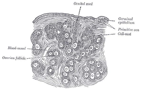
Figure 4Gray1113
Long description
Section of the ovary of a newly born child. (Waldeyer.)

Figure 4: Section of the ovary of a newly born child (Waldeyer.).
Figure 5Gray1111
Long description
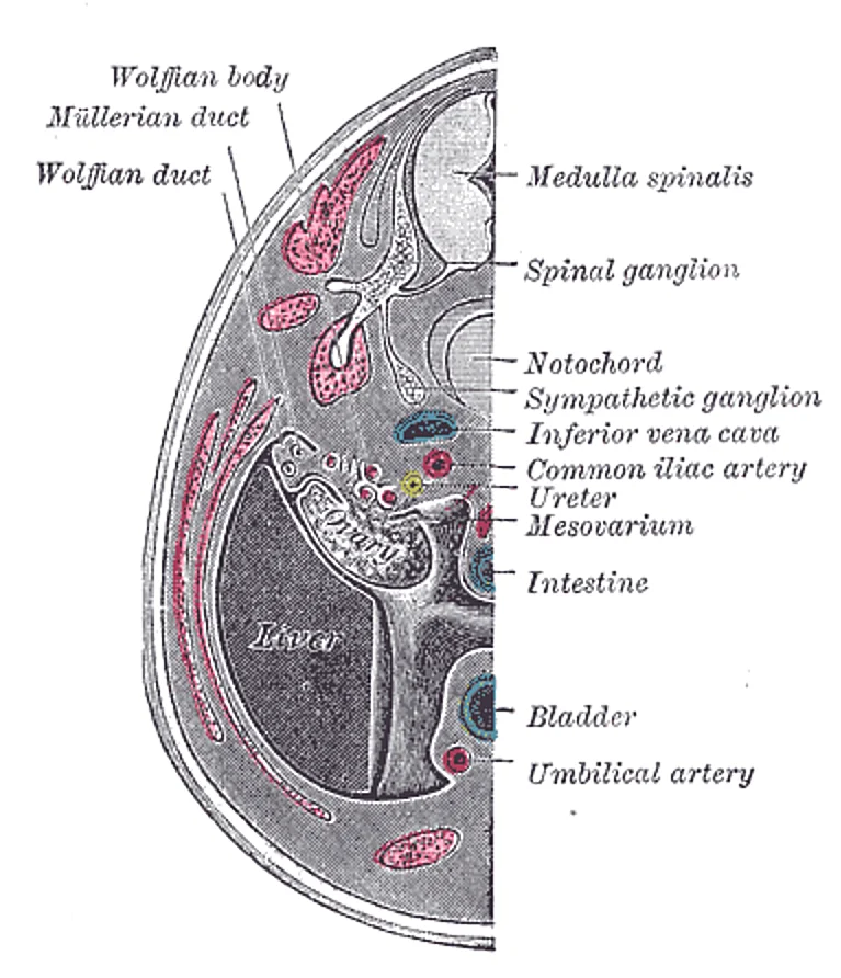
Transverse section of human embryo eight and a half to nine weeks old. (From model by Keibel.)

Figure 5: Transverse section of human embryo eight and a half to nine weeks old (From model by Keibel.).
The fetal period lasts from the ninth week of development until birth. During this period, male and female gonads differentiate. [2]
Further development of the reproductive systems occurs at puberty[1]. The initiation of the changes that occur in puberty is the result of a decrease in sensitivity to negative feedback in the hypothalamus and pituitary gland, and an increase in sensitivity of the gonads to FSH and LH stimulation[1]. These changes lead to increases in either estrogen or testosterone[1]. The increase in sex steroid hormones leads to maturation of the gonads and other reproductive organs[1]. The initiation of spermatogenesis begins, as well as ovulation and menstruation[1]. Increases in sex steroid hormones also lead to the development of secondary sex characteristics[1].
In genetic males (46,XY), defects in androgen synthesis or action can lead to ambiguous genitalia. For instance, 5-alpha-reductase deficiency prevents the conversion of testosterone to dihydrotestosterone (DHT). Since DHT is crucial for the development of the external male genitalia, these individuals may be born with ambiguous or female-appearing external genitalia. However, their internal Wolffian structures develop normally due to the presence of testosterone, which can influence tissues to become male reproductive structures. [1]
As a female reaches puberty, typically the first change that is visible is the development of the breast tissue[1]. This is followed by the growth of axillary and pubic hair[1]. A growth spurt normally starts at approximately age 9 to 11, and may last two years or more[1]. During this time, height can increase 3 inches a year[1]. The next step in puberty is menarche, the start of menstruation[1].
Figure 6Gray1110
Long description
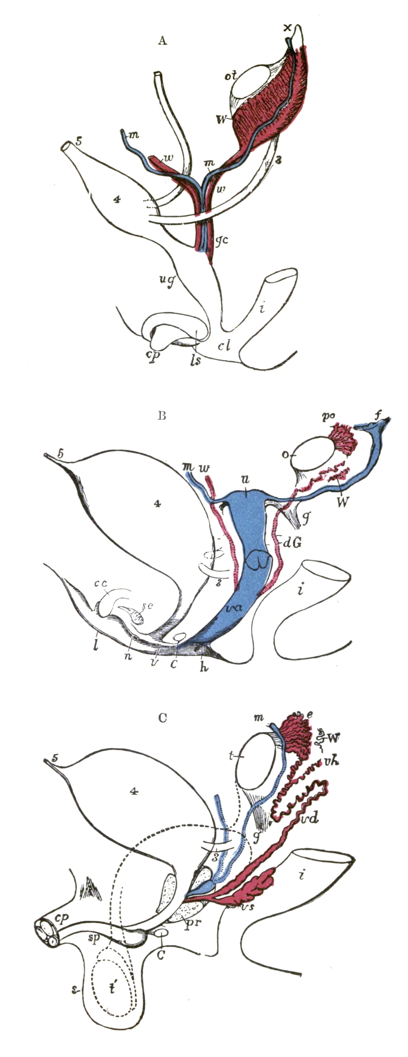
Diagrams to show the development of male and female generative organs from a common type. (Allen Thomson.) A.—Diagram of the primitive urogenital organs in the embryo previous to sexual distinction. 3. Ureter. 4. Urinary bladder. 5. Urachus. cl. Cloaca. cp. Elevation which becomes clitoris or penis

Figure 6: Diagrams to show the development of male and female generative organs from a common type.
Figure 7Gray1107
Long description
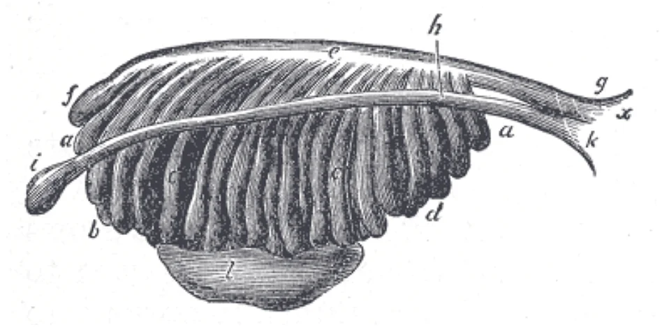
Enlarged view from the front of the left Wolffian body before the establishment of the distinction of sex. (From Farre, after Kobelt.) a, a, b, c, d. Tubular structure of the Wolffian body. e. Wolffian duct. f. Its upper extremity. g. Its termination in x, the urogenital sinus. h. The duct of Müller

Figure 7: Enlarged view from the front of the left Wolffian body before the establishment of the distinction of sex.
The development of the urogenital system is a complex process orchestrated by precise molecular signals and anatomical migrations. Understanding these signals requires looking at the interaction between the intermediate mesoderm and surrounding tissues.
Adjacent to the nephrogenic cord is the genital ridge, which will form the gonads. The relationship between the ureteric bud and the metanephric mesenchyme is critical for kidney development. The ureteric bud emerges from the mesonephric duct and invades the metanephric mesenchyme. This interaction is driven by signals such as GDNF from the mesenchyme and RET receptors on the bud. Without this signalling, the kidney fails to develop.
DefinitionNephrogenic Cord
A longitudinal ridge of intermediate mesoderm that gives rise to the urinary and genital systems.
The molecular regulation involves a cascade of gene expressions. The PAX2 gene is expressed in the ureteric bud and metanephric mesenchyme. It is essential for the survival and branching of the ureteric bud. This pathway determines sexual differentiation. In the absence of SRY on the Y chromosome, the default pathway leads to ovarian development.
Exam trapCommon Error
Do not confuse the mesonephric duct with the paramesonephric duct. The mesonephric duct gives rise to male structures, while the paramesonephric duct gives rise to female structures.
| Signal | Source | Target | Function |
|---|---|---|---|
| GDNF | Metanephric Mesenchyme | Ureteric Bud | Induces branching morphogenesis |
| RET | Ureteric Bud | GDNF | Receptor for GDNF |
| PAX2 | Intermediate Mesoderm | Nephrogenic Cord | Regulates kidney development |
| WNT9B | Metanephric Mesenchyme | Gonadal Ridge | Promotes ovarian differentiation |
The integration of these molecular signals with the anatomical structures ensures the proper formation of the urogenital system. Disruptions in these pathways can lead to congenital anomalies such as renal agenesis or ambiguous genitalia.
Figure 8Gray1109
Long description
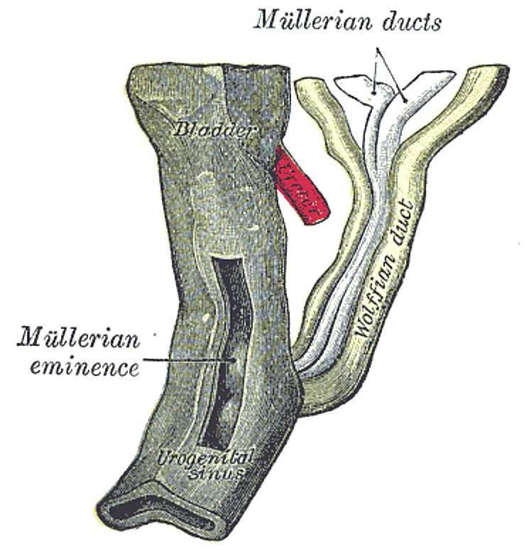
Urogenital sinus of female human embryo of eight and a half to nine weeks old. (From model by Keibel)

Figure 8: Urogenital sinus of female human embryo of eight and a half to nine weeks old (From model by Keibel).
The internal reproductive structures of the adult do not arise from a single uniform tissue but are derived from two distinct, rudimentary duct systems present in the early embryo. These systems are the Wolffian duct and the Müllerian duct. It is crucial to understand that while the gonads themselves are bipotential, the tissues forming the internal reproductive tracts are committed to developing into either male or female structures, but not both. For typical reproductive function in the adult, one set of these ducts must develop properly, and the other must degrade. This selective development ensures that the newborn possesses the necessary anatomical structures to survive and function in the outside world. [1]
Figure 9Gray1108
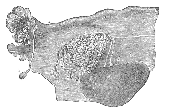
Long description
Broad ligament of adult, showing epoöphoron. (From Farre, after Kobelt.) a, a. Epoöphoron formed from the upper part of the Wolffian body. b. Remains of the uppermost tubes sometimes forming appendices. c. Middle set of tubes. d. Some lower atrophied tubes. e. Atrophied remains of the Wolffian duct.

Figure 9: Broad ligament of adult, showing epoöphoron.
In the male pathway, the Wolffian duct persists and differentiates into the epididymis, ductus deferens, and seminal vesicles. Simultaneously, the Müllerian duct regresses. In the female pathway, the reverse occurs: the Müllerian duct persists to form the uterus, uterine tubes, and part of the vagina, while the Wolffian duct degrades.
In genetic males (46,XY), defects in androgen synthesis or action can lead to ambiguous genitalia. For instance, 5-alpha-reductase deficiency prevents the conversion of testosterone to dihydrotestosterone (DHT). Since DHT is crucial for the development of the external male genitalia, these individuals may be born with ambiguous or female-appearing external genitalia. However, their internal Wolffian structures develop normally due to the presence of testosterone.
The following table summarises the embryonic origins of the adult internal reproductive structures.
| Embryonic Structure | Adult Male Derivatives | Adult Female Derivatives |
|---|---|---|
| Wolffian Duct | Epididymis, Ductus Deferens, Seminal Vesicles | Degenerates |
| Müllerian Duct | Degenerates | Uterus, Uterine Tubes, Part of Vagina |
This table demonstrates the mutual exclusivity of the developmental pathways. The Wolffian duct gives rise to the epididymis, ductus deferens, and seminal vesicles in males. In females, this duct system does not persist and undergoes regression. Conversely, the Müllerian duct forms the uterus, uterine tubes, and part of the vagina in females. In males, this duct system is actively degraded. This clear dichotomy ensures that the internal reproductive anatomy is consistent with the external genitalia and gonadal sex. [1]
Understanding these derivatives is fundamental for diagnosing congenital anomalies. Any disruption in the hormonal signals or the ductal response can lead to intersex conditions or reproductive tract abnormalities. The key principle is that one system must develop properly, and the other must degrade. This process is not merely structural but is essential for the physiological capacity to reproduce as an adult. The embryonic organ structures, initially primitive and nonfunctional, develop through these precise mechanisms to reach a state where the newborn can survive and eventually reproduce. The specificity of the Wolffian and Müllerian duct derivatives underscores the importance of hormonal regulation in human sexual differentiation. [1]
Figure 10Gray1117
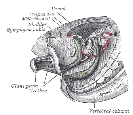
Long description
Tail end of human embryo; from eight and a half to nine weeks old. (From model by Keibel.)

Figure 10: Tail end of human embryo; from eight and a half to nine weeks old (From model by Keibel.).
Figure 11Gray1116
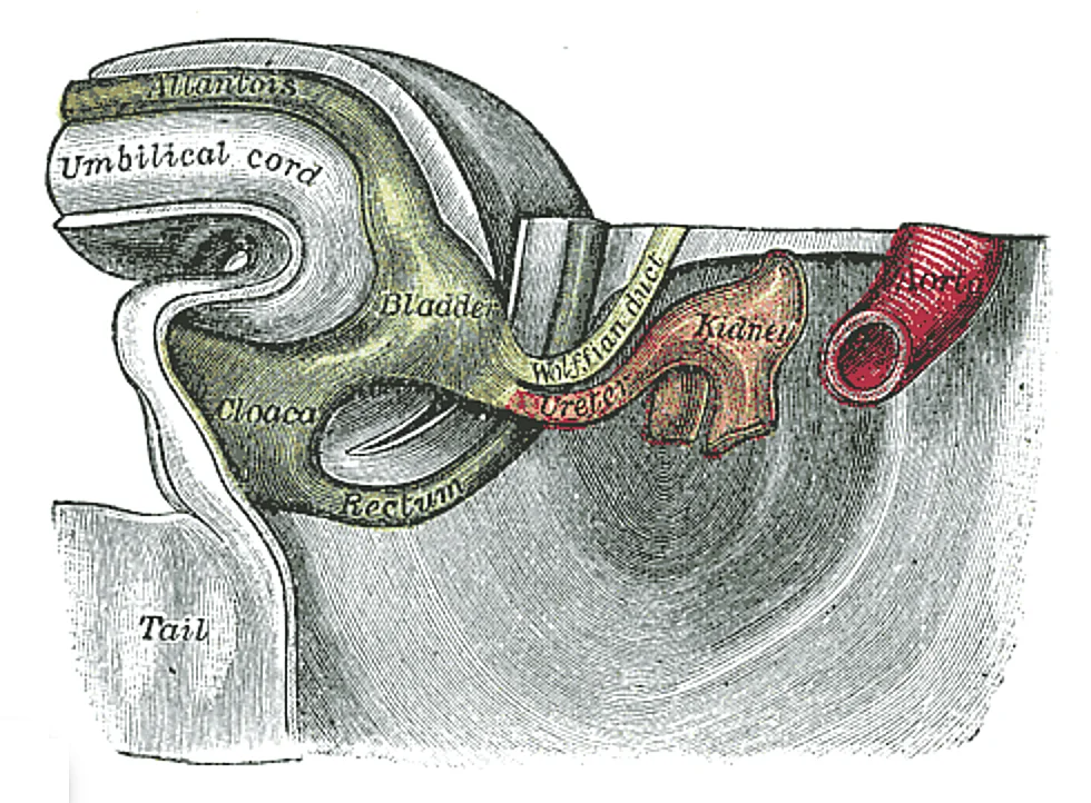
Long description
Tail end of human embryo thirty-two to thirty-three days old. (From model by Keibel.)

Figure 11: Tail end of human embryo thirty-two to thirty-three days old (From model by Keibel.).
Figure 12Gray1110-1
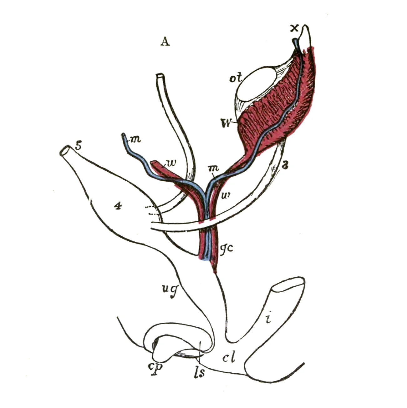
Long description
Diagrams to show the development of male and female generative organs from a common type. (Allen Thomson.) :A.—Diagram of the primitive urogenital organs in the embryo previous to sexual distinction. The common genital cord is labeled with gc. 3. Ureter 4. Urinary bladder 5. Urachus cl. Cloaca cp. E

Figure 12: Diagrams to show the development of male and female generative organs from a common type.
Adjacent structures include the developing bladder at the caudal end and the genital ducts (mesonephric and paramesonephric) running parallel to the kidneys.
The kidney develops from the intermediate mesoderm through three successive stages. The pronephros is the earliest stage but is rudimentary and non-functional in humans, regressing quickly. The mesonephros follows, functioning temporarily as a kidney during early embryonic life before regressing. The metanephros is the final stage, becoming the permanent kidney. It begins development around the fifth week. The ureteric bud, an outgrowth of the mesonephric duct, induces the metanephric mesenchyme to form the nephrons. Failure of the ureteric bud to connect with the metanephric mesenchyme results in renal agenesis.
Disorders of sex development (DSD) encompass a wide range of conditions where chromosomal, gonadal, or anatomical sex is atypical. One classic example is congenital adrenal hyperplasia (CAH), often due to 21-hydroxylase deficiency. In genetic females (46,XX), excess androgen production during fetal life can cause virilisation of the external genitalia. The internal structures, such as the uterus and fallopian tubes, remain female because the Müllerian duct develops in the absence of sustentacular cell secretion. [1]
Sexual differentiation is determined by the presence or absence of the SRY gene on the Y chromosome. In males, the testes produce testosterone and anti-Müllerian hormone, leading to male internal and external genitalia. In females, the absence of these hormones leads to the development of female internal and external genitalia. The gonads initially develop in the abdominal cavity. In males, the testes descend into the scrotum through the inguinal canal, guided by the gubernaculum. This process is influenced by testosterone and insulin-like factor 3. Cryptorchidism, the failure of the testes to descend, is a common congenital anomaly.
Anomalies can arise at any stage of development. Renal agenesis results from the failure of the ureteric bud to induce the metanephros. Polycystic kidney disease involves the formation of multiple cysts in the kidneys. Hypospadias is a condition where the urethral opening is on the ventral side of the penis, resulting from incomplete fusion of the urethral folds. Epispadias is a rare anomaly where the urethral opening is on the dorsal side. Congenital solitary kidney refers to the presence of only one kidney, which can be due to agenesis or fusion anomalies. These anomalies highlight the critical importance of precise embryological development.
One of the most common is multicystic dysplastic kidney, which results from the obstruction of the ureteric bud early in development. This condition is typically unilateral and non-hereditary. The affected kidney appears as a cluster of cysts with no functional renal parenchyma. Prenatal ultrasound often reveals this mass, which may regress spontaneously over time. [1]
Another significant anomaly is posterior urethral valves, a condition affecting males exclusively. These are obstructive membranes located in the posterior urethra that impede urine flow from the bladder. This obstruction leads to bladder distension, hydronephrosis, and potentially renal dysplasia if untreated. Postnatal confirmation is achieved through a voiding cystourethrogram, which v [1]
Disorders of sex development (DSD) encompass a wide range of conditions where chromosomal, gonadal, or anatomical sex is atypical. One classic example is congenital adrenal hyperplasia (CAH), often due to 21-hydroxylase deficiency.
In genetic males (46,XY), defects in androgen synthesis or action can lead to ambiguous genitalia. For instance, 5-alpha-reductase deficiency prevents the conversion of testosterone to dihydrotestosterone (DHT).
Prenatal ultrasound is the primary tool for monitoring urogenital development. Key markers include the presence of the fetal bladder, which indicates patency of the lower urinary tract. Absence of the bladder may suggest renal agenesis or severe obstruction. Amniotic fluid volume is also critical; oligohydramnios can result from bilateral renal agenesis or posterior urethral valves, leading to Potter sequence. This sequence includes pulmonary hypoplasia, limb deformities, and characteristic faci [1]
Clinical pearlKey Diagnostic Insight
Prenatal ultrasound assessment of amniotic fluid volume is a vital indicator of fetal renal function and urinary tract patency.
| Anomaly | Key Feature | Diagnostic Tool |
|---|---|---|
| Multicystic Dysplastic Kidney | Unilateral cystic mass | Ultrasound |
| Posterior Urethral Valves | Bladder distension in males | Cystourethrogram |
| 21-Hydroxylase Deficiency | Virilised female genitalia | Hormone assays |
| 5-Alpha-Reductase Deficiency | Ambiguous genitalia in males | Genetic testing |
Prenatal Urogenital Assessment
- Prenatal UltrasoundAssess kidney and bladder
- Question: Bladder visible?
No
- Check amniotic fluid
- Question: Oligohydramnios?
Yes
- Suspect renal agenesis
No
- Consider transient anuria
Yes
- Assess bladder wall thickness
- Question: Thickened?
Yes
- Suspect obstruction
No
- Normal development
Puberty onset is influenced by genetics, environment, and psychological stress. Nutrition is a significant factor, with better nutrition lowering the age of menarche. Historical data show menarche age decreased from approximately 17 years in 1860 to approximately 12.75 years in 1960. This age remains approximately 12.75 years today. Body fat correlates with puberty onset, linked to leptin secretion by adipose cells. This effect is more pronounced in females but documented in both sexes. The mechanism may reflect high metabolic costs of gestation and lactation. [1]
Exam trapNutritional Impact
Do not assume puberty timing is fixed; it shifts with nutritional status and body fat percentage.
Leptin, secreted by adipose cells, plays a strong role in determining menarche. This hormonal link explains the correlation between stored fat and puberty onset. The biological rationale involves the energy demands of future reproduction. [1]
DefinitionLeptin Role
Leptin signals sufficient energy reserves to initiate the reproductive axis.
Clinicians must consider body composition when assessing delayed or early puberty. Historical trends indicate secular changes in pubertal timing due to environmental factors. Genetic predisposition sets the baseline, but environment modulates the expression.
| Factor | Influence | Mechanism/Note |
|---|---|---|
| Nutrition | Decreases age of menarche | Historical shift from 17 to 12.75 years |
| Body Fat | Promotes onset | Linked to leptin secretion |
| Genetics | Sets baseline | Interacts with environmental factors |
| Stress | Modulates onset | Psychological component |
It is incorrect to attribute puberty timing solely to genetics. Environmental factors, particularly nutrition, have demonstrable historical impacts. The decrease in menarche age is not linear but stabilised.
NoteHistorical Context
The drop from 17 to 12.75 years highlights environmental plasticity.
The average age of menarche was approximately 17 years in 1860. By 1960, this decreased to approximately 12.75 years. Current data confirms this level remains stable.
Clinical pearlMenarche Timeline
Remember the 12.75-year benchmark for modern populations.
The effect of body fat on puberty is more pronounced in females. However, it is documented in both sexes. This suggests a universal metabolic requirement for reproductive maturity.
DefinitionSexual Dimorphism
Females show stronger correlation between adiposity and pubertal timing.
Gestation and lactation impose high metabolic costs. Leptin levels may ensure these costs can be met. This evolutionary perspective explains the fat-puberty link.
Exam trapEvolutionary Logic
Puberty initiates only when energy reserves support potential pregnancy.
Multiple factors interact to determine puberty onset. Age is just one variable among many. Nutrition and body fat are critical modifiable factors.
NoteHolistic View
Assess nutrition and body composition in pubertal disorders.
A frequent error in embryology is assuming that the default developmental pathway for the human embryo is male. This is incorrect. Without much chemical prompting, all fertilized eggs would develop into females. To become a male, an individual must be exposed to the cascade of factors initiated by a single gene on the male Y chromosome. This is called the SRY (Sex-determining Region of the Y chromosome). Because females do not have a Y chromosome, they do not have the SRY gene. Without a functional SRY gene, an individual will be female. Students often overlook that the presence of the Y chromosome and specifically the SRY gene is the active trigger for male differentiation, rather than the absence of female signals. [1]
Another common mistake involves the origin of external genitalia. Students frequently confuse the homologous structures without understanding the hormonal dependency. Soon after the formation of the testis, the Leydig cells begin to secrete testosterone. Testosterone can influence tissues that are bipotential to become male reproductive structures. For example, with exposure to testosterone, cells that could become either the glans penis or the glans clitoris form the glans penis. Without testosterone, these same cells differentiate into the clitoris. It is crucial to remember that the underlying tissue is identical until hormonal exposure directs the divergence. [1]
Exam trapHormonal Dependency
External genital development is not autonomous; it relies entirely on the presence or absence of testosterone from Leydig cells during specific windows of gestation.
A third pitfall relates to the timing of fetal movements and physical changes. Students may misremember the timeline for quickening and lanugo. During approximately weeks 16–20, as the fetus grows and limb movements become more powerful, the pregnant person may begin to feel quickening, or fetal movements[1]. A silky hair called lanugo also covers the skin during weeks 17–20, but it is shed as the fetus continues to grow[1]. Extremely premature infants sometimes exhibit residual lanugo[1]. Confusing these weeks with earlier or later developmental milestones can lead to errors in clinical dating or assessment of prematurity. [2]
DefinitionQuickening
The perception of fetal movements by the pregnant person, typically occurring during weeks 16–20[1].
A fourth error concerns the composition of fetal circulation, specifically the oxygenation status of blood in the umbilical vein. Students often assume that veins carry deoxygenated blood. The placenta provides the fetus with necessary oxygen and nutrients via the umbilical vein[1]. (Remember that veins carry blood toward the heart. In this case, the blood flowing to the fetal heart is oxygenated because it comes from the placenta. The respiratory system is immature and cannot yet oxyg [2]
NoteUmbilical Vein Oxygenation
Unlike systemic veins, the umbilical vein carries oxygenated blood from the placenta to the fetus[1].
A fifth pitfall involves the function of the fetal lungs and the rationale for shunts. Students may question why blood bypasses the lungs if the heart is pumping. Although the fetal liver is semifunctional, the fetal lungs are nonfunctional[1]. The fetal circulation therefore bypasses the lungs by shifting some of the blood through the foramen ovale, a shunt that directly connects the right and left atria and avoids the pulmonary trunk altogether[1]. Most of the rest of the blood is pumped to the right ventricle, and from there, into the pulmonary trunk, which splits into pulmonary arteries[1]. However, a shunt within the pulmonary artery, the ductus arteriosus, diverts a portion of this blood into the aorta[1]. This ensures that only a small volume of oxygenated blood passes through the immature pulmonary circuit, which has only minor metabolic requirements[1]. Blood vessels of uninflated lungs have high resistance to flow, a condition that encourages blood to flow to the aorta, which presents much lower resistance[1]. Understanding that high pulmonary vascular resistance drives this shunting is key. [2]
Clinical pearlPulmonary Resistance
Uninflated fetal lungs have high resistance to flow, encouraging blood to bypass them via the ductus arteriosus[1].
A sixth mistake is regarding the physical characteristics of the term fetus. Students might assume lanugo persists until birth. The fetus continues to lay down subcutaneous fat from week 31 until birth[1]. The added fat fills out the hypodermis, and the skin transitions from red and wrinkled to soft and pink[1]. Lanugo is shed, and the nails grow to the tips of the fingers and toes[1]. Immediately before birth, the average crown-to-rump length is 35.5–40.5 cm (14–16 in), and the fetus weighs approximately 2.5–4 kg (5.5–8.8 lbs)[1]. Once born, the newborn is no longer confined to the fetal position, so subsequent measurements are made from head-to-toe instead of from crown-to-rump[1]. At birth, the average length is approximately 51 cm (20 in)[1]. Confusing crown-to-rump with head-to-toe measurements can lead to significant errors in assessing growth. [2]
DefinitionCrown-to-Rump
A measurement from the top of the head to the bottom of the buttocks, used for fetuses and newborns before they can stand[1].
A final common error is linking maternal lifestyle factors incorrectly to developmental timing. In females who are extremely lean and highly active, such as gymnasts, there is often a delay in the onset of puberty. This is a post-natal/pubertal phenomenon, not a direct embryological defect of urogenital formation, but students often conflate pubertal timing with embryonic sexual differentiation. The embryonic process is driven by SRY and testosterone, not maternal leanness. [1]
This knowledge is used at the bedside to interpret ambiguous genitalia in newborns. If a newborn presents with ambiguous genitalia, clinicians must consider whether testosterone was present during the critical window of weeks 8–12. The fact that without testosterone, cells differentiate into the clitoris helps explain female external anatomy. Conversely, the presence of SRY indicates testicular development, which should lead to testosterone secretion. If testosterone is absent or ineffective, the external genitalia will resemble female structures despite the presence of testes.
In the operating theatre, understanding fetal circulation is vital for neonatal surgery. If a newborn has a patent ductus arteriosus (PDA) that fails to close, blood may shunt from the aorta to the pulmonary artery. This is the reverse of the fetal flow. The fetal shunt, the ductus arteriosus, diverts a portion of this blood into the aorta[1]. Recognizing the normal fetal pattern helps surgeons understand the hemodynamic consequences of surgical ligation. Similarly, a patent foramen ovale is common, but the foramen ovale, a shunt that directly connects the right and left atria and avoids the pulmonary trunk altogether[1], is designed to bypass the lungs.
On imaging, ultrasound is used to assess fetal development. Quickening, or fetal movements, is felt by the mother during approximately weeks 16–20[1]. Ultrasound can visualize these movements and confirm gestational age. The presence of lanugo during weeks 17–20[1] can be seen on high-resolution scans. Lanugo is shed as the fetus continues to grow[1]. Extremely premature infants sometimes exhibit residual lanugo[1]. This helps radiologists estimate gestati
In examinations, students must accurately describe the fetal circulation. The umbilical vein carries oxygenated blood[1]. From the umbilical vein, the oxygenated blood flows toward the inferior vena cava, all but bypassing the immature liver, via the ductus venosus shunt[1]. The liver receives just a trickle of blood, which is all that it needs in its immature, semifunctional state[1]. Blood flows from the inferior vena cava to the right atrium, mixing with fetal venous blood along the way[1]. Although the fetal liver is semifunctional, the fetal lungs are nonfunctional[1]. The fetal circulation therefore bypasses the lungs by shifting some of the blood through the foramen ovale, a shunt that directly connects the right and left atria and avoids the pulmonary trunk altogether[1]. Most of the rest of the blood is pumped to the right ventricle, and from there, into the pulmonary trunk, which splits into pulmonary arteries[1]. However, a shunt within the pulmonary artery, the ductus arteriosus, diverts a portion of this blood into the aorta[1]. This ensures that only a small volume of oxygenated blood passes through the immature pulmonary circuit, which has only minor metabolic requirements[1]. Blood vessels of uninflated lungs have high resistance to flow, a condition that encourages blood to flow to the aorta, which presents much lower resistance[1]. [2]
Fetal Circulation Shunts
- Oxygenated Blood from PlacentaVia Umbilical Vein
- Question: Bypass Liver?
Yes
- Ductus Venosus Shunt
- Inferior Vena Cava
No
- Liver Sinusoids
- Question: Bypass Lungs?
Yes (Right Atrium)
- Foramen Ovale
- Left Atrium
No (Right Ventricle)
- Pulmonary Trunk
- Question: Bypass Lungs?
Yes
- Ductus Arteriosus
- Aorta
No
- Pulmonary Arteries
- Lungs (High Resistance)
At birth, the average length is approximately 51 cm (20 in)[1]. Immediately before birth, the average crown-to-rump length is 35.5–40.5 cm (14–16 in), and the fetus weighs approximately 2.5–4 kg (5.5–8.8 lbs)[1]. Once born, the newborn is no longer confined to the fetal position, so subsequent measurements are made from head-to-toe instead of from crown-to-rump[1]. This transition is critical for pediatricians to note when assessing growth charts.
NoteBirth Measurements
Post-birth measurements are head-to-toe, whereas fetal measurements are crown-to-rump[1].
In genetic males (46,XY), defects in androgen synthesis or action can lead to ambiguous genitalia. For instance, 5-alpha-reductase deficiency prevents the conversion of testosterone to dihydrotestosterone (DHT). Since DHT is crucial for the development of the external male genitalia, these individuals may be born with ambiguous or female-appearing external genitalia. However, their internal Wolffian structures develop normally due to the presence of testosterone. At puberty, the surge in testosterone leads to the development of secondary sex characteristics.
DefinitionSRY Gene
The Sex-determining Region of the Y chromosome, essential for male development.
Understanding these principles allows clinicians to diagnose disorders of sexual development, interpret fetal ultrasound findings, and manage neonatal care effectively. The detailed knowledge of shunts, hormonal influences, and physical milestones provides a comprehensive framework for clinical practice.
- Default development is female; male development requires SRY gene.
- SRY initiates testis formation and testosterone secretion.
- Testosterone directs bipotential tissues to form male structures.
- Without testosterone, tissues form female structures like the clitoris.
- Umbilical vein carries oxygenated blood from placenta[1].
- Ductus venosus bypasses the immature liver[1].
- Foramen ovale and ductus arteriosus bypass nonfunctional lungs[1].
- Quickening occurs weeks 16–20; lanugo sheds by term[1].
Answer each one in your head first, then open the answer.
What gene is responsible for initiating male sexual differentiation?
Show answerHide answer
SRYWhich hormone is secreted by Leydig cells to influence male reproductive structures?
Show answerHide answer
TestosteroneWhat structure bypasses the immature liver in fetal circulation?
Show answerHide answer
Ductus venosusDuring which weeks do pregnant persons typically feel quickening?
Show answerHide answer
16–20What is the average birth length of a newborn?
Show answerHide answer
51 cm
- Betts JG, Young KA, Wise JA, et al. Development of the Male and Female Reproductive Systems. OpenStax, Anatomy and Physiology 2e (CC BY-NC-SA 4.0). Accessed .
- Betts JG, Young KA, Wise JA, et al. Fetal Development. OpenStax, Anatomy and Physiology 2e (CC BY-NC-SA 4.0). Accessed .
Figure credits
- Figure 1: Henry Vandyke Carter, via Wikimedia Commons, Public domain, Public domain.
- Figure 2: Henry Vandyke Carter, via Wikimedia Commons, Public domain, Public domain.
- Figure 3: Henry Vandyke Carter, via Wikimedia Commons, Public domain, Public domain.
- Figure 4: Henry Vandyke Carter, via Wikimedia Commons, Public domain, Public domain.
- Figure 5: Henry Vandyke Carter, via Wikimedia Commons, Public domain, Public domain.
- Figure 6: Henry Vandyke Carter, via Wikimedia Commons, Public domain, Public domain.
- Figure 7: Henry Vandyke Carter, via Wikimedia Commons, Public domain, Public domain.
- Figure 8: Henry Vandyke Carter, via Wikimedia Commons, Public domain, Public domain.
- Figure 9: Henry Vandyke Carter, via Wikimedia Commons, Public domain, Public domain.
- Figure 10: Henry Vandyke Carter, via Wikimedia Commons, Public domain, Public domain.
- Figure 11: Henry Vandyke Carter, via Wikimedia Commons, Public domain, Public domain.
- Figure 12: Henry Vandyke Carter, via Wikimedia Commons, Public domain, Public domain.
Educational content for medical students and doctors. It is not a substitute for clinical judgement or local guidelines.