Musculoskeletal development
Embryological development of the appendicular skeleton from limb buds to ossified bones.
The appendicular skeleton originates from mesenchyme, an embryonic tissue capable of differentiating into bone or muscle[1]. The upper limb bud appears near the end of the fourth week of development, with the lower limb bud appearing shortly after[1]. Each limb starts as a small bulge on the lateral side of the embryo, consisting of a mesenchymal core covered by ectoderm[1]. The ectoderm at the distal end thickens to form the apical ectodermal ridge, wh [4]
By the sixth week, the distal ends of the limb buds expand into paddle shapes, representing the future hands or feet[1]. Constrictions appear at the future wrist/ankle and elbow/knee levels[1]. Within the paddle, selective cell death separates the developing digits[1]. Simultaneously, mesenchyme differentiates into hyaline cartilage models for the future bones[1]. The clavicle is unique among appendicular bones as it develops via intramembranous ossification, where mesenchyme converts directly to bone without a cartilage intermediate[1]. Ossification of the clavicle begins in the fifth week and continues until age 25[1]. [4]
In contrast, most limb bones form via endochondral ossification, where hyaline cartilage serves as a template for bone replacement[1]. This process begins in the limbs by the twelfth week[1]. During the seventh week, limb rotation occurs: the upper limb rotates 90 degrees laterally, positioning the palm anteriorly and thumb laterally, while the lower limb rotates 90 degrees medially, bringing the big toe to the medial side[1]. [3]
DefinitionApical Ectodermal Ridge
A thickened ectodermal crest at the limb bud tip that stimulates mesenchymal proliferation and limb elongation.
DefinitionIntramembranous Ossification
The direct differentiation of mesenchyme into bone tissue, bypassing a cartilage model phase.
DefinitionEndochondral Ossification
The process where bone develops by completely replacing a hyaline cartilage template.
| Week | Event |
|---|---|
| 4 | Limb buds appear; upper limbs precede lower limbs. |
| 5 | Clavicle ossification begins via intramembranous ossification. |
| 6 | Paddle shape forms; digit separation via cell death; cartilage models form. |
| 7 | Limb rotation: upper lateral, lower medial. |
| 12 | Endochondral ossification of limb bones begins. |
Polydactyly, clubfoot, and limb reduction defects represent common congenital anomalies arising from disruptions in these precise developmental windows. Clubfoot involves abnormal positioning, while polydactyly results from failed separation of digits during the sixth week[1]. Limb reduction defects may stem from early disruptions in mesenchymal proliferation or vascular supply[1]. Understanding the timeline is critical, as the sequence of ossification and rotation dictates the final anatomical configuration of the adult skeleton.
Muscle masses are indicated as dense regions surrounding the skeletal elements.
The musculoskeletal system originates from mesenchyme, which differentiates into various tissues. Somites form along the neural tube and give rise to the vertebrae and associated muscles. Bone formation occurs via two primary mechanisms: intramembranous and endochondral ossification. All bone formation is a replacement process. Embryos initially develop a cartilaginous skeleton and various membranes, which are subsequently replaced by bone during ossification. [3] [1]
The bones of the skull arise from mesenchyme through two distinct pathways. The bones of the brain case, including the flat bones of the face and most cranial bones, develop via intramembranous ossification. In this process, compact and spongy bone develops directly from sheets of mesenchymal connective tissue. At birth, these bones are separated by fontanelles, which are wide areas of fibrous connective tissue. These fontanelles allow the skull to change shape during birth and permit continued growth as the brain enlarges. As the bones grow, the fontanelles reduce to sutures, which allow for continued growth throughout childhood. The last bones to ossify via this method are the flat bones of the face, reaching adult size at the end of the adolescent growth spurt. [1] [2][3]
Mesenchyme initially produces a hyaline cartilage model of the future bone. This cartilage model allows for growth and is gradually converted into bone over many years. This conversion is a slow process, and the cartilage is not completely converted to bone until the skull achieves its full adult size. [2] [4]
The girdle and limb bones, with the exception of the clavicle, develop through endochondral ossification. This process begins as mesenchyme within the limb bud differentiates into hyaline cartilage, forming cartilage models for future bones. Endochondral ossification begins in most appendicular bones by the twelfth fetal week. A primary ossification center appears in the diaphysis (shaft) region of the long bones, initiating the conversion of the cartilage model into bone. By the second or third month of fetal life, bone cell development ramps up, creating this primary ossification center deep in the periosteal collar. [4] [1][3]
For example, ossification of the femur begins at the end of the seventh week with the appearance of the primary ossification center in the diaphysis. [1]
Muscle development accompanies skeletal formation. Mesenchyme in the head region produces skull bones, while somites contribute to axial muscles. Limb muscles develop from the myotomes of the somites, migrating into the limb buds to surround the developing cartilage models.
Figure 1Gray373
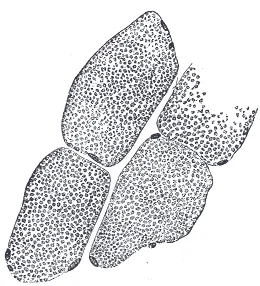
Long description
Transverse section of human striped muscle fibers. x 255.

Figure 1: Transverse section of human striped muscle fibers (x 255).
Clinical pearlKey Distinction
Intramembranous ossification forms flat bones directly from mesenchyme, while endochondral ossification replaces a hyaline cartilage model with bone.
The axial skeleton begins to form during early embryonic development. This process is fundamental for understanding skeletal abnormalities. During the third week of embryonic development, a rod-like structure called the notochord develops dorsally along the length of the embryo. The tissue overlying the notochord enlarges and forms the neural tube, which will give rise to the brain and spinal cord. By the fourth week, mesoderm tissue located on either side of the notochord thickens and separates into a repeating series of block-like tissue structures, each of which is called a somite. As the somites enlarge, each one will split into several parts. The most medial of these parts is called a sclerotome. The sclerotomes consist of an embryonic tissue called mesenchyme, which will give rise to the fibrous connective tissues, cartilages, and bones of the body. [2]
Figure 2Gray20
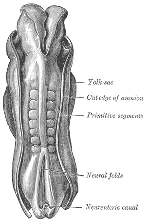
Long description
Dorsum of human embryo, 2.11 mm. in length. (After Eternod.)

Figure 2: Dorsum of human embryo, 2.11 mm (in length).
Figure 3Gray375
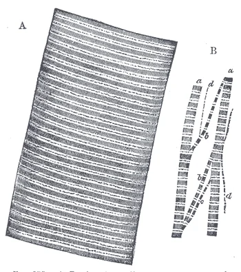
Long description
A. Portion of a medium-sized human muscular fiber. Magnified nearly 800 diameters. B. Separated bundles of fibrils, equally magnified. a, a. Larger, and b, b, smaller collections. c. Still smaller. d, d. The smallest which could be detached.

Figure 3: A.
Flanking the notochord are the somites, appearing as segmented blocks. Medially, the sclerotome is indicated, adjacent to the neural tube.
Development of the vertebrae begins with the accumulation of mesenchyme cells from each sclerotome around the notochord. These cells differentiate into a hyaline cartilage model for each vertebra, which then grow and eventually ossify into bone through the process of endochondral ossification. As the developing vertebrae grow, the notochord largely disappears. However, small areas of notochord tissue persist between the adjacent vertebrae and this contributes to the formation of each intervertebral disc. [2]
Bone is a replacement tissue; that is, it uses a model tissue on which to lay down its mineral matrix. For skeletal development, the most common template is cartilage. During fetal development, a framework is laid down that determines where bones will form. This framework is a flexible, semi-solid matrix produced by chondroblasts and consists of hyaluronic acid, chondroitin sulfate, collagen fibers, and water. As the matrix surrounds and isolates chondroblasts, they are called chondrocytes. Unlike most connective tissues, cartilage is avascular, meaning that it has no blood vessels supplying nutrients and removing metabolic wastes. All of these functions are carried on by diffusion through the matrix. This is why damaged cartilage does not repair itself as readily as most tissues do. [3]
The epiphyseal plate is composed of four zones of cells and activity. [3]
The process in which matrix is resorbed on one surface of a bone and deposited on another is known as bone modeling. Modeling primarily takes place during a bone’s growth. However, in adult life, bone undergoes remodeling, in which resorption of old or damaged bone takes place on the same surface where osteoblasts lay new bone to replace that which is resorbed. Injury, exercise, and other activities lead to remodeling. Those influences are discussed later in the chapter, but even without injury or exercise, about 5 to 10 percent of the skeleton is remodeled annually just by destroying old bone and renewing it with fresh bone. [3]
Joints form during embryonic development in conjunction with the formation and growth of the associated bones. The embryonic tissue that gives rise to all bones, cartilages, and connective tissues of the body is called mesenchyme. In the head, mesenchyme will accumulate at those areas that will become the bones that form the top and sides of the skull. The mesenchyme in these areas will develop directly into bone through the process of intramembranous ossification, in which mesenchymal cells differentiate into bone-producing cells that then generate bone tissue. The mesenchyme between the areas of bone production will become the fibrous connective tissue that fills the spaces between the developing bones. Initially, the connective tissue-filled gaps between the bones are wide, and are called fontanelles. [1] [4]
The musculoskeletal system arises primarily from the mesoderm, specifically through the segmentation of the paraxial mesoderm into somites. These somites are transient, block-like structures that form along the neural tube and provide the foundational blueprint for the axial skeleton and associated musculature. Understanding the precise fate of these embryonic structures is critical for correlating congenital anomalies with their developmental origins.
Within each somite, three distinct regions are identifiable. The sclerotome, situated medially and ventrally, surrounds the notochord and neural tube. The dermomyotome, located dorsally and laterally, gives rise to the skin and muscle components. The relationship between the sclerotome and the notochord is crucial, as the sclerotome cells resegment to form the vertebral bodies, while the intervertebral discs are derived from the remaining notochordal tissue. The dermomyotome’s position relative to the somite’s dorsal midline determines the innervation pattern of the resulting muscles, with epaxial muscles receiving dorsal rami and hypaxial muscles receiving ventral rami.
The somite itself differentiates into three primary components: the sclerotome, the dermomyotome, and the syndetome. The sclerotome gives rise to the vertebrae and ribs. The syndetome, a recently recognized component, contributes to the tendons.
| Embryonic Structure | Adult Derivative |
|---|---|
| Sclerotome | Vertebrae, Ribs |
| Dermomyotome (Dermatome) | Dermis of the back |
| Dermomyotome (Myotome) | Skeletal muscles of the trunk and limbs |
| Syndetome | Tendons |
Beyond the axial skeleton, limb muscles are derived from myoblasts that migrate from the ventral medial lip of the dermomyotome into the limb buds. These myoblasts proliferate and differentiate into myotubes, which eventually fuse to form multinucleated muscle fibres. The innervation of these muscles follows the pathway of the migrating myoblasts, with nerves growing in ahead of the muscle mass.
In the context of bone development, the process begins when mesenchymal cells in the embryonic skeleton gather together and begin to differentiate into specialized cells. Some of these cells will differentiate into capillaries, while others will become osteogenic cells and then osteoblasts. Although they will ultimately be spread out by the formation of bone tissue, early osteoblasts appear in a cluster called an ossification center. This is particularly evident in endochondral ossification, where hyaline cartilage models are replaced by bone. The epiphyseal plate is retained for many years, until the bone reaches its final, adult size, at which time the epiphyseal plate disappears and the epiphysis fuses to the diaphysis. [1] [3]
Joint formation involves the cavitation of the interzone. Mesenchyme cells at the margins of the interzone will give rise to the articular capsule, while cell death at the center forms the space that will become the joint cavity of the future synovial joint. The hyaline cartilage model of each limb bone will eventually be converted into bone via the process of endochondral ossification. However, hyaline cartilage will remain, covering the ends of the adult bone as the articular cartilage. This ensures smooth movement and shock absorption in the mature skeleton. [4]
Congenital anomalies of the musculoskeletal system arise from disruptions in the complex choreography of limb bud formation, somite segmentation, and chondrogenesis. These defects provide a window into the underlying embryological principles that govern normal development. Understanding the timing and nature of the insult allows us to predict the resulting phenotype.
The humerus appears shortened and internally rotated. The anomaly demonstrates the failure of the scapula to descend properly from its initial cervical position to its normal thoracic location. This descent normally occurs during the seventh week of gestation. The relationship between the scapula and the ribs is disrupted, leading to restricted shoulder abduction.
DefinitionSprengel Deformity
A congenital condition where the scapula fails to descend to its normal position, remaining elevated and often fused to the cervical spine. [1]
Another common anomaly is clubfoot, or talipes equinovarus. This condition involves multiple deformities of the foot and ankle. The embryological basis lies in abnormal positioning of the foot in utero or intrinsic muscle imbalance. The talus bone is often displaced medially and plantarflexed. The calcaneus is inverted and adducted. The forefoot is adducted relative to the hindfoot. The condition may be isolated or associated with other syndromes such as arthrogryposis.
Exam trapClubfoot Misconception
Clubfoot is not merely a soft tissue contracture; it involves bony malalignment and joint subluxation that must be corrected structurally.
Limb reduction defects result from vascular accidents or teratogenic exposure during critical periods of limb bud outgrowth. If the insult occurs early, before the sixth week, major segments of the limb may be absent. For example, phocomelia involves the absence of long bones with hands or feet attached directly to the trunk. This was historically associated with thalidomide exposure. The severity depends on the timing and extent of the vascular disruption.
| Anomaly | Embryological Basis | Key Feature |
|---|---|---|
| Sprengel Deformity | Failure of scapular descent | Elevated, hypoplastic scapula |
| Clubfoot | Abnormal foot positioning | Equinovarus deformity |
| Phocomelia | Vascular disruption | Absent long bones |
| Polydactyly | Abnormal apical ectodermal ridge signaling | Extra digits |
Polydactyly is another frequent anomaly, resulting from errors in the regulation of the zone of polarising activity (ZPA). The ZPA secretes Sonic Hedgehog (SHH), which determines anterior-posterior patterning. Overexpression or ectopic expression of SHH can lead to the formation of additional digits. Postaxial polydactyly, involving the fifth digit, is more common than preaxial polydactyly. The extra digit may be fully formed or a small nodule of tissue. Genetic factors play a significant role, with many autosomal dominant patterns described.
NoteSHH Role
Sonic Hedgehog is crucial for digit number and identity; its gradient determines the polarity of the limb.
These anomalies underscore the precision required in embryonic development. Each structure has a specific origin, course, and termination that must be followed correctly. Deviations lead to functional impairment and cosmetic deformity. Early diagnosis allows for timely intervention, whether surgical or orthotic, to improve outcomes. The study of these anomalies reinforces our understanding of normal musculoskeletal anatomy and physiology.
The neural tube forms during the fourth week of embryonic development, closing at the cranial and caudal neuropores. Failure of this closure results in neural tube defects, which are among the most common congenital anomalies. The most severe form is anencephaly, where the cranial neuropore fails to close, leading to the absence of major portions of the brain, skull, and scalp. This condition is incompatible with life, and affected fetuses are often stillborn or die shortly after birth. Another significant defect is spina bifida, which involves the failure of the caudal neuropore to close. Spina bifida occulta is the mildest form, characterized by a defect in the vertebral arches without protrusion of the meninges or spinal cord. It is often asymptomatic and may present only with a dimple, tuft of hair, or lipoma over the lower spine. In contrast, spina bifida cystica involves the protrusion of meninges (meningocele) or meninges and spinal cord (myelomeningocele) through the defect. Myelomeningocele is the most serious variant, often resulting in neurological deficits such as paralysis, loss of sensation, and bladder or bowel dysfunction below the level of the lesion.
Figure 4Gray376
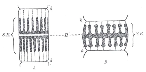
Long description
Diagram of a sarcomere. (After Schäfer.) A. In moderately extended condition. B. In a contracted condition. k, k. Membranes of Krause. H. Line or plane of Hensen. S.E. Poriferous sarcous element.

Figure 4: Diagram of a sarcomere (After Schäfer.) A.
Prenatal diagnosis of neural tube defects relies on maternal serum screening and ultrasound imaging. Maternal serum alpha-fetoprotein (MSAFP) levels are significantly elevated in pregnancies affected by open neural tube defects. Alpha-fetoprotein is a protein produced by the fetal liver and yolk sac, which leaks into the amniotic fluid and subsequently into the maternal circulation when the neural tube is open. Elevated MSAFP levels typically prompt further investigation, including detailed ultrasound examination. Ultrasound can directly visualize structural abnormalities such as the absence of the skull vault in anencephaly or the spinal defect in spina bifida. In cases where ultrasound findings are inconclusive, amniocentesis may be performed to measure amniotic fluid alpha-fetoprotein and acetylcholinesterase levels. Elevated acetylcholinesterase in the amniotic fluid is highly specific for open neural tube defects, as this enzyme is released from damaged neural tissue.
Prevention of neural tube defects is a major public health initiative. Folic acid supplementation before conception and during early pregnancy has been shown to significantly reduce the risk of neural tube defects. The recommended dose for women at average risk is 400 micrograms daily, starting at least one month before conception and continuing through the first trimester. Women with a history of neural tube defects in previous pregnancies or those taking certain anticonvulsant medications are advised to take a higher dose, typically 4 milligrams daily. This preventive strategy underscores the importance of early prenatal care and awareness of risk factors.
Clinical pearlKey Diagnostic Marker
Elevated maternal serum alpha-fetoprotein is a primary screening marker for open neural tube defects, prompting further diagnostic imaging or amniocentesis. [1]
| Defect | Description | Clinical Features |
|---|---|---|
| Anencephaly | Failure of cranial neuropore closure | Absence of brain/skull, incompatible with life |
| Spina Bifida Occulta | Defect in vertebral arches only | Asymptomatic, skin markers over spine |
| Myelomeningocele | Protrusion of meninges and cord | Neurological deficits, paralysis, incontinence |
At birth, the brain case and orbits of the skull are disproportionally large compared to the bones of the jaws and lower face. This disproportion reflects the relative underdevelopment of the maxilla and mandible, which lack teeth, and the small sizes of the paranasal sinuses and nasal cavity. During early childhood, the mastoid process enlarges, the two halves of the mandible and frontal bone fuse together to form single bones, and the paranasal sinuses enlarge. The jaws also expand as the teeth begin to appear. These changes all contribute to the rapid growth and enlargement of the face during childhood. [2]
Clinical pearlNeonatal Skull Proportions
The neonatal skull is dominated by the neurocranium, with the viscerocranium being relatively small due to the absence of teeth and underdeveloped sinuses.
Exam trapMandibular Fusion
Do not assume the mandible is a single bone at birth; it consists of two halves that fuse during early childhood.
While bones are increasing in length, they are also increasing in diameter; growth in diameter can continue even after longitudinal growth ceases. This is called appositional growth. Osteoclasts resorb old bone that lines the medullary cavity, while osteoblasts, via intramembranous ossification, produce new bone tissue beneath the periosteum. The erosion of old bone along the medullary cavity and the deposition of new bone beneath the periosteum not only increase the diameter of the diaphysis but also increase the diameter of the medullary cavity. This process is called modeling. [3]
DefinitionAppositional Growth
The process by which bones increase in diameter through the deposition of new bone tissue beneath the periosteum and resorption within the medullary cavity.
NoteLongitudinal vs. Appositional Growth
Longitudinal growth ceases earlier than appositional growth, which can continue throughout life to adapt bone strength to mechanical stress.
The cellular coordination in bone modeling involves two primary cell types with opposing actions. Osteoclasts are responsible for the resorption of old bone lining the medullary cavity. Simultaneously, osteoblasts produce new bone tissue beneath the periosteum via intramembranous ossification. This dual action ensures that as the bone thickens externally, the internal cavity expands proportionally to maintain appropriate marrow space and reduce excessive weight.
Clinical pearlIntramembranous Ossification in Modeling
New bone formation beneath the periosteum during appositional growth occurs via intramembranous ossification, distinct from the endochondral ossification seen in longitudinal growth.
Exam trapMedullary Cavity Size
The medullary cavity increases in diameter during appositional growth; it does not remain static while the outer diameter expands.
Understanding these developmental milestones is crucial for interpreting radiological findings in paediatric patients. The unfused mandibular halves and frontal bone are normal variants in infants and should not be mistaken for fractures or synostosis disorders. Similarly, the small size of paranasal sinuses at birth explains the susceptibility of infants to certain types of facial trauma and the different presentation of sinusitis compared to adults.
DefinitionViscerocranial Development
The viscerocranium undergoes significant expansion during childhood, driven by dental eruption and sinus enlargement, altering facial proportions dramatically.
NoteBone Remodelling vs. Growth
While remodelling maintains bone health throughout life, appositional growth specifically refers to the increase in bone diameter during developmental stages.
Clinical pearlMastoid Process Enlargement
The enlargement of the mastoid process during early childhood is a key landmark for assessing normal skeletal maturation.
Exam trapDiaphysis Diameter
The diameter of the diaphysis increases due to both new bone deposition and medullary cavity erosion, not just external deposition.
DefinitionFacial Growth Spurt
Rapid facial growth in childhood is characterised by the expansion of jaws concurrent with tooth eruption and sinus development.
A frequent error in interpreting paediatric radiographs is mistaking a normal epiphyseal plate for a fracture. Students often overlook the radiolucency of cartilage, leading to unnecessary anxiety or intervention. The epiphyseal plate consists of cartilage, which is less dense than bone and therefore appears dark on an X-ray. It is crucial to remember that this dark line represents a normal growth zone, not a break in the cortex. [3]
Another common misconception involves the timing of epiphyseal fusion. Many assume all growth plates close simultaneously or in a random order. In reality, the fusion follows a specific reversed sequence relative to ossification. The lesser trochanter is the first to fuse, doing so at the onset of puberty (around 11 years of age). This is followed by the greater trochanter approximately 1 year later. The femoral head fuses between the ages of 14–17 years. The distal condyles of the femur are the last to fuse, between the ages of 16–19 years. Knowing this sequence helps in assessing skeletal maturity and potential growth disorders. [1]
Students also frequently confuse the mechanisms of bone growth in length versus width. Longitudinal growth is strictly dependent on the activity of the epiphyseal plate. The proliferative zone contains stacks of slightly larger chondrocytes that make new chondrocytes via mitosis. These replace those that die at the diaphyseal end. The zone of maturation and hypertrophy contains older, larger cells closer to the diaphysis. Longitudinal growth results from cellular division in the proliferative zone and maturation in the hypertrophy zone. Once chondrocytes cease proliferation and bone replaces the cartilage, longitudinal growth stops. [3]
A third pitfall is misunderstanding the fate of the epiphyseal plate after growth ceases. It does not vanish completely but becomes a fibrous remnant. All that remains of the epiphyseal plate is the epiphyseal line. This line marks the end of longitudinal bone growth in early adulthood. [3]
Finally, students often confuse intramembranous and endochondral ossification in the skull. The base and facial regions develop via endochondral ossification, where hyaline cartilage is gradually displaced by bone. In contrast, the flat bones of the calvaria develop via intramembranous ossification. The fontanelles are the fibrous gaps between these flat bones. The largest fontanelle is located on the anterior head, at the junction of the frontal and parietal bones. These fontanelles decrease in size and disappear by age 2. However, the skull bones remain separated at the sutures, which contain dense fibrous connective tissue. This tissue allows for continued growth of the skull bones as the brain enlarges. [2]
In the operating theatre, understanding ossification centres is vital for surgical planning and fracture management. Surgeons must avoid damaging the epiphyseal plate to prevent growth arrest. The epiphyseal plate is responsible for the longitudinal growth of bones. Injury to this zone can lead to significant limb length discrepancies. Knowledge of the age at which different epiphyseal plates disappear is important when interpreting radiographs taken of children. This knowledge guides decisions on whether to fix a fracture conservatively or surgically. [1] [3]
On imaging, the distinction between cartilage and bone is critical. Since the cartilage of an epiphyseal plate is less dense than bone, the plate will appear dark in a radiograph image. A normal epiphyseal plate may be mistaken for a bone fracture. Radiologists must differentiate this physiological lucency from pathological fractures. Similarly, the fontanelles appear as soft tissue gaps on imaging. The connective tissue of the sutures allows for continued growth of the skull bones as the brain enlarges during childhood growth. Recognising these normal variants prevents misdiagnosis of craniosynostosis or other anomalies. [1] [2]
At the bedside, clinicians use these principles to assess growth disorders. Bones continue to grow in length until early adulthood. The rate of growth is controlled by hormones. Conditions like growth hormone deficiency or precocious puberty alter the timing of epiphyseal fusion. For instance, precocious puberty may cause early fusion of the epiphyseal plates, resulting in short adult stature. Conversely, delayed puberty may prolong growth. The osteoblasts secrete osteoid, uncalcified matrix, which calcifies within a few days as mineral salts are deposited on it. Once entrapped, the osteoblasts become osteocytes. This process is fundamental to bone repair and remodelling. [3]
In examinations, students are often tested on the histological zones of the epiphyseal plate. The zone of calcified matrix is closest to the diaphysis. Most chondrocytes in this zone are dead because the matrix around them has calcified. Capillaries and osteoblasts from the diaphysis penetrate this zone. The osteoblasts secrete bone tissue on the remaining calcified cartilage. Thus, the zone of calcified matrix connects the epiphyseal plate to the diaphysis. A bone grows in length when osseous tissue is added to the diaphysis. [3]
Epiphyseal Plate Histology
- Proliferative ZoneChondrocytes divide via mitosis
- Question: Cells mature?
Yes
- Zone of Maturation/HypertrophyCells enlarge and age
No
- Remain in Proliferative Zone
- Zone of Calcified MatrixMatrix calcifies, chondrocytes die
- Question: Osteoblasts invade?
Yes
- Bone deposition on calcified cartilageLongitudinal growth occurs
No
- Growth arrest
Clinical pearlRadiographic Pearl
A dark line in a paediatric long bone is likely an epiphyseal plate, not a fracture, unless there is clinical evidence of trauma.
DefinitionFontanelle
A fibrous membrane connecting the cranial bones in an infant, allowing for brain growth and moulding during birth.
Exam trapCommon Trap
Do not confuse the epiphyseal line (remnant of the plate) with a fracture line. The line is smooth and parallel to the joint surface.
NoteClinical Note
Hyaline cartilage is retained as articular cartilage at synovial joints and as the epiphyseal plate during growth. [4]
- Epiphyseal plates appear radiolucent (dark) on X-rays due to cartilage density.
- Fusion sequence: lesser trochanter (11y) > greater trochanter (12y) > femoral head (14-17y) > distal femoral condyles (16-19y).
- Longitudinal growth occurs via the epiphyseal plate until early adulthood.
- The fontanelles close by age 2, but sutures remain for brain growth.
- Osteoblasts secrete osteoid which calcifies to form bone.
- The epiphyseal plate consists of proliferative, hypertrophic, and calcified zones.
- Endochondral ossification forms the base of the skull.
- Articular cartilage and epiphyseal plates are hyaline cartilage.
Answer each one in your head first, then open the answer.
What is the first epiphyseal plate to fuse?
Show answerHide answer
The lesser trochanterAt what age do fontanelles typically disappear?
Show answerHide answer
By age 2What cells secrete osteoid?
Show answerHide answer
OsteoblastsWhy do chondrocytes in the zone of calcified matrix die?
Show answerHide answer
The matrix calcifies, preventing nutrient reachWhat remains of the epiphyseal plate after growth stops?
Show answerHide answer
The epiphyseal line
- Betts JG, Young KA, Wise JA, et al. Development of the Appendicular Skeleton. OpenStax, Anatomy and Physiology 2e (CC BY-NC-SA 4.0). Accessed .
- Betts JG, Young KA, Wise JA, et al. Embryonic Development of the Axial Skeleton. OpenStax, Anatomy and Physiology 2e (CC BY-NC-SA 4.0). Accessed .
- Betts JG, Young KA, Wise JA, et al. Bone Formation and Development. OpenStax, Anatomy and Physiology 2e (CC BY-NC-SA 4.0). Accessed .
- Betts JG, Young KA, Wise JA, et al. Development of Joints. OpenStax, Anatomy and Physiology 2e (CC BY-NC-SA 4.0). Accessed .
Figure credits
- Figure 1: Henry Vandyke Carter, via Wikimedia Commons, Public domain, Public domain.
- Figure 2: Henry Vandyke Carter, via Wikimedia Commons, Public domain, Public domain.
- Figure 3: Henry Vandyke Carter, via Wikimedia Commons, Public domain, Public domain.
- Figure 4: Henry Vandyke Carter, via Wikimedia Commons, Public domain, Public domain.
Educational content for medical students and doctors. It is not a substitute for clinical judgement or local guidelines.