Nervous system development
Embryology of the nervous system from fertilisation to organogenesis.
Prenatal development is divided into distinct periods based on weeks from fertilisation, commonly called conception. The first two weeks constitute the pre-embryonic stage, followed by the embryonic period from weeks three to eight. During this critical window, the developing human is termed an embryo, and by the end of the eighth week, all organ systems are structured in rudimentary form. The subsequent fetal period begins at the ninth week and continues until birth. [2]
Figure 1Gray660
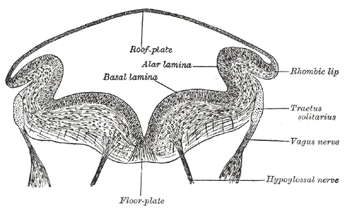
Long description
Transverse section of medulla oblongata of human embryo. (After His.)

Figure 1: Transverse section of medulla oblongata of human embryo (After His.).
Figure 2Gray659
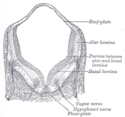
Long description
Transverse section of medulla oblongata of human embryo. X 32. (Kollmann)

Figure 2: Transverse section of medulla oblongata of human embryo (x 32).
Figure 3Gray658
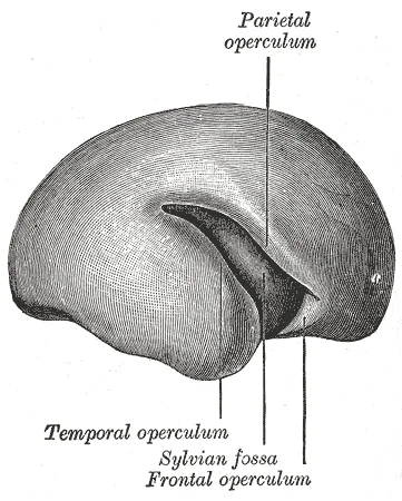
Long description
Outer surface of cerebral hemisphere of human embryo of about five months.

Figure 3: Outer surface of cerebral hemisphere of human embryo of about five months.
Figure 4Gray654
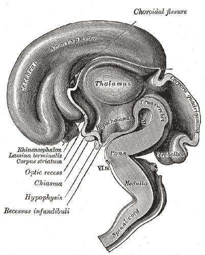
Long description
Median sagittal section of brain of human embryo of three months. (From model by His.)

Figure 4: Median sagittal section of brain of human embryo of three months (From model by His.).
Figure 5Gray653
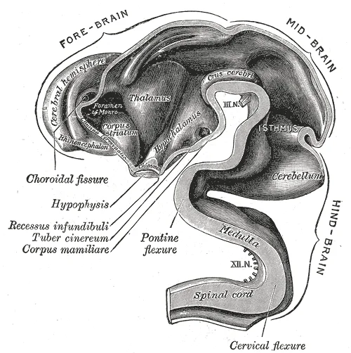
Long description
Interior of brain of human embryo of five weeks. (From model by His.)

Figure 5: Interior of brain of human embryo of five weeks (From model by His.).
Figure 6Gray652
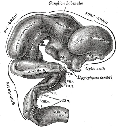
Long description
Exterior of brain of human embryo of five weeks. (From model by His.)

Figure 6: Exterior of brain of human embryo of five weeks (From model by His.).
Figure 7Gray650
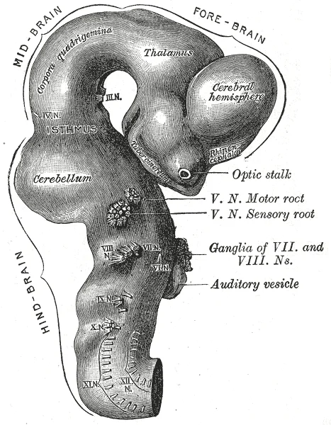
Long description
Exterior of brain of human embryo of four and a half weeks. (From model by His.)

Figure 7: Exterior of brain of human embryo of four and a half weeks (From model by His.).
Figure 8Gray649
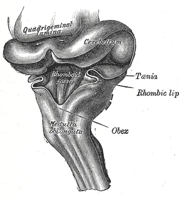
Long description
Hind-brain of a human embryo of three months—viewed from behind and partly from left side. (From model by His.)

Figure 8: Hind-brain of a human embryo of three months—viewed from behind and partly from left side (From model by His.).
Figure 9Gray643
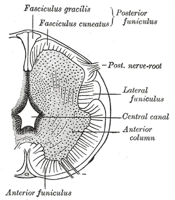
Long description
aged about three months. Labelled illustration from Wikimedia Commons, used under its open licence.

Figure 9: Aged about three months.
Figure 10Gray642
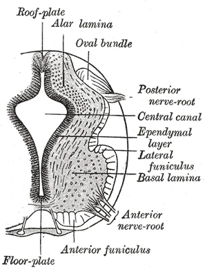
Long description
aged about four and a half weeks. Labelled illustration from Wikimedia Commons, used under its open licence.

Figure 10: Aged about four and a half weeks.
DefinitionOrganogenesis
The process by which the three primary germ layers establish the rudimentary structures of all organs and tissues within the first eight weeks of gestation. [2]
Following implantation, embryonic cells undergo gastrulation to differentiate into the endoderm, mesoderm, and ectoderm. Neurulation specifically initiates the development of central nervous system structures. The central nervous system comprises the brain and spinal cord. The adult brain is described in terms of four major regions: the cerebrum, diencephalon, brain stem, and cerebellum. The diencephalon is the only adult brain region that retains its embryological name. It serves as the connection between the cerebrum and the rest of the nervous system, with the exception of the olfactory system. [1]
Figure 11Gray657
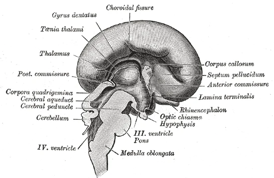
Long description
Median sagittal section of brain of human embryo of four months. (Marchand.)

Figure 11: Median sagittal section of brain of human embryo of four months (Marchand.).
Figure 12Gray655
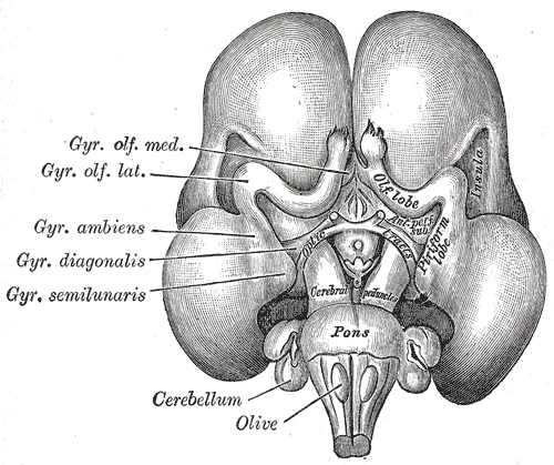
Long description
Inferior surface of brain of embryo at beginning of fourth month. (From Kollmann.)

Figure 12: Inferior surface of brain of embryo at beginning of fourth month (From Kollmann.).
Figure 13Gray651
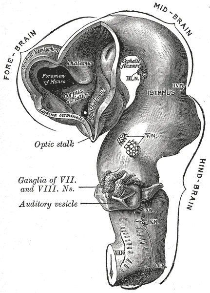
Long description
Brain of human embryo of four and a half weeks, showing interior of fore-brain. (From model by His.)

Figure 13: Brain of human embryo of four and a half weeks, showing interior of fore-brain (From model by His.).
Figure 14Gray641
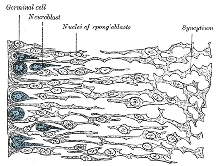
Long description
Transverse section of the medulla spinalis of a human embryo at the beginning of the fourth week. The left edge of the figure corresponds to the lining of the central canal. (His.)

Figure 14: Transverse section of the medulla spinalis of a human embryo at the beginning of the fourth week.
Figure 15Gray640
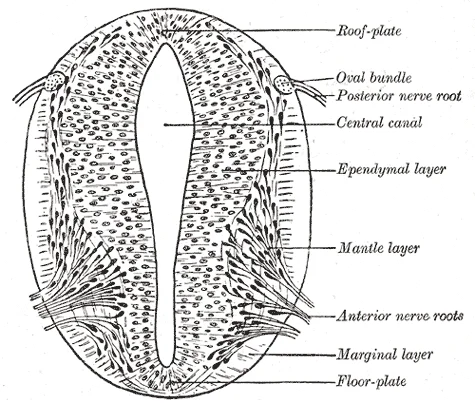
Long description
Section of medulla spinalis of a four weeks’ embryo. (His.)

Figure 15: Section of medulla spinalis of a four weeks’ embryo (His.).
NoteOlfactory Exception
The system associated with olfaction connects directly with the cerebrum, bypassing the diencephalon. [1]
The diencephalon includes the thalamus and the hypothalamus. The thalamus acts as a relay between the cerebrum and the rest of the nervous system. The hypothalamus is located inferior and slightly anterior to the thalamus. It is a collection of nuclei largely involved in regulating homeostasis. The hypothalamus coordinates homeostatic functions through the autonomic and endocrine systems. It regulates the anterior pituitary gland and is involved in memory and emotion as part of the limbic system. [1]
Clinical pearlHomeostasis Hub
The hypothalamus is the executive region in charge of the autonomic nervous system and the endocrine system. [1]
Blood vessels nourishing the central nervous tissue are located between the pia mater and the nervous tissue. Cerebrospinal fluid is produced within the ventricles by the choroid plexus. The fluid is essentially water, small molecules, and electrolytes. The ventricular system opens to the subarachnoid space from the fourth ventricle via the median and lateral apertures. [3]
| Structure | Primary Function |
|---|---|
| Thalamus | Relay between cerebrum and rest of nervous system |
| Hypothalamus | Regulation of homeostasis via autonomic and endocrine systems |
| Olfactory System | Direct connection to cerebrum, bypassing diencephalon |
The central nervous system originates from the ectoderm during neurulation. Specialized neuroectodermal tissues thicken into the neural plate, which folds into neural folds that converge to form the neural tube. This tube lies atop the notochord and subdivides into vesicles during the fourth and fifth weeks.
DefinitionNeural Tube Defects
A deficiency of maternal folate in the first weeks of pregnancy can result in neural tube defects, including spina bifida.
Spina bifida is a birth defect in which spinal tissue protrudes through the newborn’s vertebral column. A more severe neural tube defect is anencephaly, a partial or complete absence of brain tissue.
Clinical pearlBrain Vesicles
The midbrain and hindbrain are collectively referred to as the brain stem. The hindbrain is composed of the pons and the medulla. [1]
The brain stem emerges from the ventral surface of the forebrain as a tapering cone. The midbrain coordinates sensory representations of the visual, auditory, and somatosensory perceptual spaces. The pons is the main connection with the cerebellum. The pons and the medulla regulate several crucial functions, including the cardiovascular and respiratory systems and rates. [1]
DefinitionCerebellum
The cerebellum accounts for approximately 10 percent of the mass of the brain. [1]
The cerebellum is largely responsible for comparing information from the cerebrum with sensory feedback from the periphery. It is covered in gyri and sulci like the cerebrum. [1]
NoteSpinal Cord Regions
The length of the spinal cord is divided into regions that correspond to the regions of the vertebral column. [1]
Immediately adjacent to the brain stem is the cervical region, followed by the thoracic, then the lumbar, and finally the sacral region. The sacral spinal cord is at the level of the upper lumbar vertebral bones. The spinal cord is not the full length of the vertebral column because the spinal cord does not grow significantly longer after the first or second year. The nerves that emerge from the spinal cord pass through the intervertebral foramina at the respective levels. As the vertebral column grows, these nerves grow with it and result in a long bundle of nerves that resembles a horse’s tail and is named the cauda equina. [1]
Figure 16Gray646
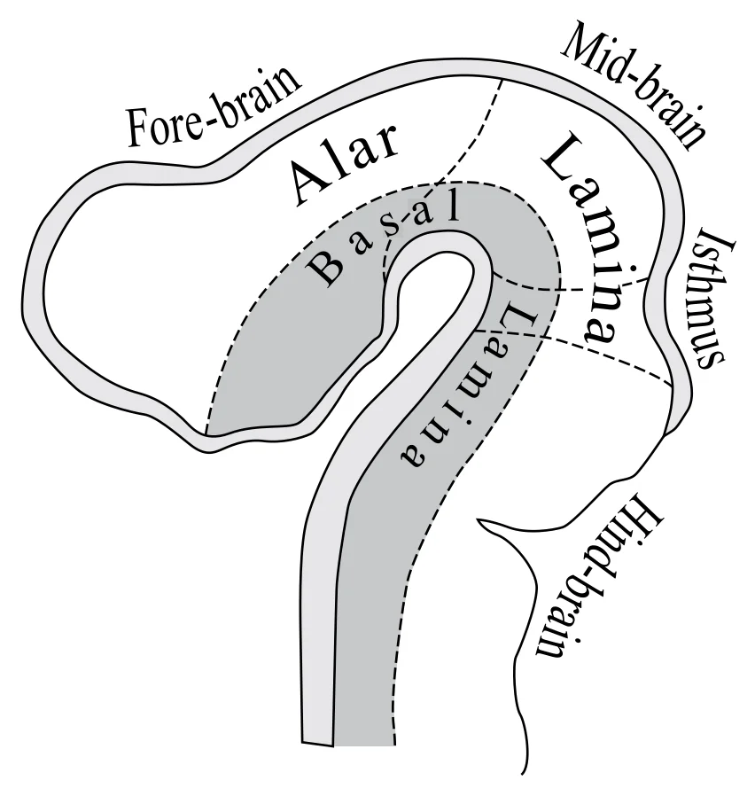
Long description
Diagram to illustrate the alar and basal laminæ of brain vesicles. (His.)

Figure 16: Diagram to illustrate the alar and basal laminæ of brain vesicles (His.).
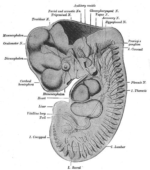
Figure 17Gray645
Long description
Reconstruction of periphera nerves of a human embryo of 10.2 mm. (After His.) The abducent nerve is not labelled, but is seen passing forward to the eye under the mandibular and maxillary nerves.

Figure 17: Reconstruction of periphera nerves of a human embryo of 10.2 mm.
DefinitionAlar and Basal Plates
The basal plate is closest to the ventral midline of the neural tube, which will become the anterior face of the spinal cord and gives rise to motor neurons. [1]
The alar plate is on the dorsal side of the neural tube and gives rise to neurons that will receive sensory input from the periphery. The anterior midline is marked by the anterior median fissure, and the posterior midline is marked by the posterior median sulcus. Axons enter the posterior side through the dorsal (posterior) nerve root, which marks the posterolateral sulcus on either side. The axons emerging from the anterior side do so through the ventral (anterior) nerve root. [1]
Exam trapWhite Matter Columns
The posterior columns are composed of axons of ascending tracts. [1]
Ascending tracts of nervous system fibers in these columns carry sensory information up to the brain, whereas descending tracts carry motor commands from the brain. Between the two posterior horns of gray matter are the posterior columns. Between the two anterior horns, and bounded by the axons of motor neurons emerging from that gray matter area, are the anterior columns. The white matter on either side of the spinal cord, between the posterior horn and the axons of the anterior horn neurons, are the lateral columns. [1]
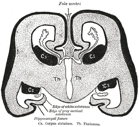
Figure 18Gray656
Long description
Diagrammatic coronal section of brain to show relations of neopallium. (After His.) Cs. Corpus striatum. Th. Thalamus.

Figure 18: Diagrammatic coronal section of brain to show relations of neopallium (After His.) Cs.
The posterior end maintains the tube structure. The notochord lies beneath the neural tube. The somites are block-like structures on either side. The vertebral column is developing around the neural tube. The brain stem connects the forebrain to the spinal cord. The cerebellum is attached to the brain stem. The pons and medulla are part of the brain stem. The midbrain is the most superior part of the brain stem. The spinal cord regions are cervical, thoracic, lumbar, and sacral. The sacral spinal cord is at the level of the upper lumbar vertebral bones. The anterior median fissure and posterior median sulcus are marked. The posterior, anterior, and lateral columns are indicated. The blood-brain barrier is implied by the privileged blood supply. The folate deficiency is a risk factor for neural tube defects. Spina bifida and anencephaly are examples of neural tube defects. The cerebellum accounts for approximately 10 percent of the mass of the brain. The pons is the main connection with the cerebellum. The midbrain coordinates sensory representations. The pons and medulla regulate cardiovascular and respiratory systems. The cranial nerves connect through the brain stem. The major ascending and descending pathways pass through the brain stem. The CNS has a privileged blood supply. The contents of the blood cannot simply pass into the central nervous tissue. The CNS needs specialized structures for the maintenance of circulation. The unique arrangement of blood vessels carries fresh blood into the CNS. [2]
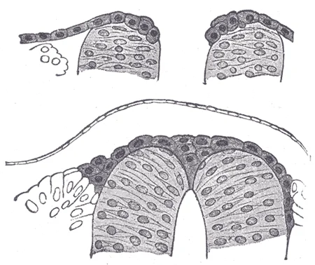
Figure 19Gray644
Long description
Two stages in the development of the neural crest in the human embryo. (Lenhossèk.)

Figure 19: Two stages in the development of the neural crest in the human embryo (Lenhossèk.).
The molecular regulation of nervous system development involves complex signaling pathways that guide the formation and organization of neural tissue. During embryonic development, the neural tube forms from the ectoderm through a process called neurulation, which is regulated by precise molecular signals. The cerebral cortex develops from the dorsal telencephalon and is characterized by its distinctive folded pattern of gyri and sulci, which are determined by genetic programs that control cortical expansion and folding.
Key signaling molecules such as sonic hedgehog (Shh), bone morphogenic proteins (BMPs), and fibroblast growth factors (FGFs) play crucial roles in patterning the neural tube. Shh, secreted by the notochord, ventralizes the neural tube, promoting the development of motor neurons and ventral interneurons. BMPs, expressed in the non-neural ectoderm, dorsalize the neural tube, leading to the formation of sensory neurons and roof plate structures.
The basal nuclei, located deep within the cerebrum, are responsible for comparing cortical processing with the general state of activity in the nervous system to influence movement initiation. These nuclei receive inputs from various cortical areas and modulate motor output through complex neural circuits. The nuclei of the basal forebrain serve as the primary location for acetylcholine production, which modulates overall cortical activity and attention to sensory stimuli. [1]
DefinitionNeurulation
The process by which the neural plate folds to form the neural tube, the precursor to the central nervous system.
The development of specific brain regions is also regulated by molecular signals. The hippocampus and amygdala, medial-lobe structures, develop in response to specific signaling gradients and are involved in long-term memory formation and emotional responses. The parietal lobe, responsible for somatosensation processing, develops from the dorsal telencephalon under the influence of specific morphogens.
This barrier is established during development through molecular interactions between endothelial cells and astrocytes, ensuring proper neural function by regulating the passage of substances.
The nervous system originates from the ectoderm, specifically the neural plate, which folds to form the neural tube. This tube undergoes significant regionalization to produce the central nervous system. The primary vesicles formed are the prosencephalon (forebrain), mesencephalon (midbrain), and rhombencephalon (hindbrain). These further subdivide into secondary vesicles. The telencephalon develops into the cerebrum, which includes the cerebral cortex and subcortical nuclei. The diencephalon forms the thalamus and hypothalamus. The mesencephalon remains undivided and becomes the midbrain, situated between the thalamus and pons. The rhombencephalon divides into the metencephalon (pons and cerebellum) and myelencephalon (medulla oblongata). [1]
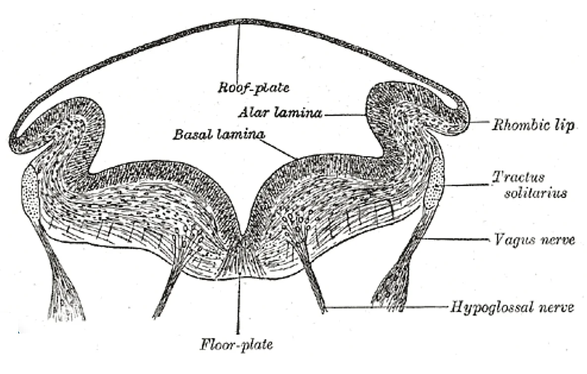
Figure 20Gray648
Long description
Transverse section of medulla oblongata of human embryo. (After His.)

Figure 20: Transverse section of medulla oblongata of human embryo (After His.).
Figure 21Gray647
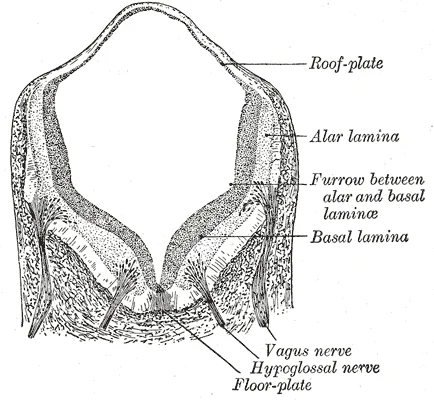
Long description
Transverse section of medulla oblongata of human embryo. X 32. (Kollmann.)

Figure 21: Transverse section of medulla oblongata of human embryo (x 32).
The telencephalon is the largest anterior structure, giving rise to the cerebral hemispheres. Posterior to this is the diencephalon, leading into the midbrain. Posterior to the midbrain is the metencephalon, which forms the pons and cerebellum. The most posterior structure is the myelencephalon, forming the medulla. The spinal cord extends inferiorly from the medulla. Each structure is clearly demarcated, allowing identification of the major adult regions derived from the embryonic vesicles. [2]
Using these landmarks, the cortex can be separated into four major regions, or lobes. [1]
The midbrain is separated into the tectum and tegmentum, from the Latin words for roof and floor, respectively. The cerebral aqueduct passes through the center of the midbrain, such that these regions are the roof and floor of that canal. The brain stem connects the cerebrum and cerebellum to the spinal cord. It consists of the midbrain, pons, and medulla. The cerebellum is located posterior to the brain stem and is involved in coordination and balance. [1]
| Embryonic Structure | Adult Structure |
|---|---|
| Telencephalon | Cerebrum (Cerebral Cortex, Subcortical Nuclei) |
| Diencephalon | Thalamus, Hypothalamus |
| Mesencephalon | Midbrain |
| Metencephalon | Pons, Cerebellum |
| Myelencephalon | Medulla Oblongata |
The development of the nervous system is closely linked to the formation of other embryonic structures. The neural tube also gives rise to the spinal cord. The meninges, including the subarachnoid space filled with circulating CSF, develop from surrounding mesenchyme. This CSF provides a liquid cushion to the brain and spinal cord. The integrity of the blood-brain barrier and the vascular supply is crucial for normal development. A lack of oxygen to the CNS can be devastating, and the cardiovascular system has specific regulatory reflexes to ensure that the blood supply is not interrupted. There are multiple routes for blood to get into the CNS, with specializations to protect that blood supply and to maximize the ability of the brain to get an uninterrupted perfusion. [3]
By the eighth week, all major brain structures are in place. The external genitalia are apparent, but at this point, male and female embryos are indistinguishable. Bone begins to replace cartilage in the embryonic skeleton through the process of ossification. By the end of the embryonic period, the embryo is approximately 3 cm (1.2 in) from crown to rump and weighs approximately 8 g (0.25 oz). [2]
Congenital anomalies of the nervous system arise from disruptions in the complex embryological processes that establish the central and peripheral nervous systems. Understanding these defects requires tracing the lineage from the germ layers through to the mature functional structures.
The neural tube forms from the ectoderm, which gives rise to cell lineages that differentiate to become the central and peripheral nervous systems[1]. Failure of the neural tube to close completely results in major structural anomalies. [2]
In the case of anencephaly, the cranial vault is absent, exposing the rudimentary brain tissue at the top. In spina bifida, the vertebral arches fail to fuse posteriorly, leaving the spinal cord and meninges exposed or protruding. The labels identify the cranium, the defect in the vertebral arches, and the spinal cord.
DefinitionNeural Tube Defect
A congenital anomaly resulting from the failure of the neural tube to close during early embryonic development, leading to exposure of neural tissue.
The ectoderm is responsible for the formation of the nervous system[1]. When this process is disrupted, the resulting anomalies affect higher neurological functions. The cerebrum, comprising the outer gray matter and deep nuclei, is particularly vulnerable. The basal nuclei, responsible for cognitive processing and planning movements, may be affected. The limbic cortex, involved in emotion and memory, may also be compromised.
| Anomaly | Embryological Basis | Key Feature |
|---|---|---|
| Anencephaly | Failure of cranial neural tube closure | Absence of major portions of the brain and skull |
| Spina Bifida | Failure of caudal neural tube closure | Defect in vertebral arches, possible cord exposure |
| Meningocele | Protrusion of meninges through defect | Sac filled with CSF, no neural tissue |
| Myelomeningocele | Protrusion of meninges and cord | Neural tissue in sac, severe neurological deficit |
While not a primary nervous system anomaly, the implantation site is critical for overall embryonic health. Most embryos implant within the body of the uterus[1]. However, in one to two percent of cases, the embryo implants either outside the uterus or in a region that creates complications[1]. [2]
NoteImplantation Context
Understanding normal implantation helps contextualize the high failure rate and potential complications that can affect subsequent development.
A significant percentage (50–75 percent) of blastocysts fail to implant[1]. This high rate of implantation failure is one reason why pregnancy typically requires several ovulation cycles to achieve[1]. Successful implantation is a prerequisite for the development of the nervous system, which begins with the differentiation of the ectoderm[1]. [2]
The choroid plexuses are found in all four ventricles. Observed in dissection, they appear as soft, fuzzy structures that may still be pink, depending on how well the circulatory system is cleared in preparation of the tissue. [3]
DefinitionChoroid Plexus
Specialized structures located in the ventricles responsible for producing cerebrospinal fluid from blood components.
Disruptions in CSF production or flow can lead to hydrocephalus, a condition where excess fluid accumulates in the brain. This can exert pressure on the developing cerebral cortex and subcortical nuclei. The subcortical nuclei, responsible for augmenting cortical functions, receive input from cortical areas and compare it with the general state of the individual.
| Structure | Location | Function |
|---|---|---|
| Choroid Plexus | All four ventricles | Produces CSF from blood components |
| Lateral Ventricles | Within cerebral hemispheres | Initial CSF production site |
| Third Ventricle | Between thalami | CSF passage and production |
| Fourth Ventricle | Between brainstem and cerebellum | CSF passage and production |
The complexity of the cerebrum is different across vertebrate species. In mammals, the cerebrum comprises the outer gray matter that is the cortex and several deep nuclei that belong to three important functional groups. The basal nuclei are responsible for cognitive processing, the most important function being that associated with planning movements. The limbic cortex is the region of the cerebral cortex that is part of the limbic system, a collection of structures involved in emotion, memory, and behavior. [1]
Exam trapFunction vs. Structure
Do not confuse the anatomical location of a structure with its functional role. For example, while the temporal lobe is associated with primary auditory sensation, it is also part of the limbic system and involved in memory.
Different regions of the cerebral cortex can be associated with particular functions, a concept known as localization of function. In the early 1900s, a German neuroscientist named Korbinian Brodmann performed an extensive study of the microscopic anatomy—the cytoarchitecture—of the cerebral cortex and divided the cortex into 52 separate regions on the basis of the histology of the cortex. His work resulted in a system of classification known as Brodmann’s areas, which is still used today to describe the anatomical distinctions within the cortex. The results from Brodmann’s work on the anatomy align very well with the functional differences within the cortex. [1]
The temporal lobe is associated with primary auditory sensation, known as Brodmann’s areas 41 and 42 in the superior temporal lobe. Because regions of the temporal lobe are part of the limbic system, memory is an important function associated with that lobe. Even memories of movement are really the memory of sensory feedback from those movements, such as stretching muscles or the movement of the skin around a joint. Structures in the temporal lobe are responsible for establishing long-term memory, but the ultimate location of those memories is usually in the region in which the sensory perception was processed. [1]
Nuclei beneath the cerebral cortex, known as the subcortical nuclei, are responsible for augmenting cortical functions. The output influences the activity of part of the thalamus that can then increase or decrease cortical activity that often results in changes to motor commands. The limbic system includes deep cerebral nuclei that are responsible for emotion and memory. [1]
Maternal and fetal blood does not commingle because blood cells cannot move across the placenta[1]. Further, it ensures the fetal red blood cells do not enter the pregnant person's circulation and trigger antibody development (if they carry “non-self” antigens)—at least until the final stages of pregnancy or birth[1]. This is the reason that, even in the absence of preventive treatment, an Rh− person doesn’t develop antibodies that could cause hemolytic disease in their first Rh+ fetus[1]. [2]
The nervous system originates from the ectoderm, a layer established during the critical window of gastrulation. This process transforms the two-layered embryonic disc into a three-layered structure, a transition marked by cells shifting from totipotency to multipotency. Understanding this early lineage is essential for interpreting congenital anomalies, as errors in gastrulation can lead to severe structural defects of the central and peripheral nervous systems. [2]
Gastrulation begins in the third week of development, a period of profound morphological change. The embryo assumes the shape of an oval-shaped disc, upon which a distinct indentation forms along the dorsal surface of the epiblast. This indentation is known as the primitive streak, and it serves as the primary site for cell migration. These chemical signals direct cells to multiply and migrate in a coordinated manner. Cells migrate toward and through the primitive streak, subsequently moving laterally to establish two new layers of cells. The first layer formed is the endoderm, which appears as a sheet of cells that displaces the hypoblast and lies adjacent to the yolk sac. The second layer fills in as the middle layer, or mesoderm. Crucially, the cells of the epiblast that remain, having not migrated through the primitive streak, become the ectoderm. It is from this ectodermal layer that the neural plate, and subsequently the entire nervous system, derives. [2]
DefinitionGastrulation
The process by which the two-layered embryonic disc becomes a three-layered disc, establishing the germ layers including the ectoderm, which gives rise to the nervous system.
Defects in the formation of the primitive streak or the subsequent migration of cells can result in significant congenital malformations. For instance, failure of the neural tube to close properly, a process dependent on the integrity of the underlying mesoderm and the overlying ectoderm, leads to neural tube defects such as spina bifida or anencephaly. These conditions highlight the importance of the early embryonic events described. Disruption of this signalling can impair the formation of the notochord and somites, which are critical for inducing neural tube formation.
Clinical pearlKey Concept
The ectoderm, formed by epiblast cells that do not migrate through the primitive streak, is the precursor to the entire nervous system.
Prenatal diagnosis often relies on detecting markers associated with these early developmental errors. Elevated maternal serum alpha-fetoprotein (MSAFP) levels may indicate an open neural tube defect, reflecting a breach in the protective barriers formed during later stages of neural development. While the initial gastrulation events are microscopic, their consequences are macroscopic and clinically significant. The orderly progression from the oval-shaped disc to the three-layered structure is fundamental to normal neurodevelopment. Any deviation in the timing or direction of cell migration through the primitive streak can have lasting effects on the structural integrity of the brain and spinal cord.
| Germ Layer | Primary Derivatives | Relevance to Nervous System |
|---|---|---|
| Ectoderm | Epidermis, Nervous System | Direct precursor to neural plate and tube |
| Mesoderm | Muscle, Bone, Connective Tissue | Provides structural support and induction signals |
| Endoderm | Gut Lining, Respiratory Epithelium | Indirect role via surrounding structures |
- The cerebral cortex surface area is approximately one square meter when laid flat, which is essential for accommodating extensive folding within the limited cranial space[1]. This folding allows for increased surface area without requiring a proportionally larger skull, demonstrating the brain's remarkable adaptation to anatomical constraints.
This structure is crucial for integrating functions between the left and right sides of the brain, such as language processing in the dominant hemisphere and spatial awareness in the non-dominant hemisphere.
These pathways work in opposition to regulate movement initiation and suppression, with the direct pathway facilitating desired movements and the indirect pathway suppressing unwanted movements.
This dual pathway system allows for precise regulation of movement, with dysfunction in either pathway leading to movement disorders such as Parkinson's disease (indirect pathway overactivity) or Huntington's disease (direct pathway impairment).
- The diencephalon consists mainly of the thalamus and hypothalamus, with additional structures like the epithalamus (containing the pineal gland) and subthalamus (including the subthalamic nucleus)[1]. The thalamus serves as the major relay station for sensory information (except olfaction) to the cortex, while the hypothalamus regulates autonomic functions, endocrine control, and homeostasis.
This terminology is especially important in neuroanatomy when describing the location of sensory (dorsal root) and motor (ventral root) components of spinal nerves.
-
The circle of Willis is formed by the internal carotid arteries and branches of the basilar artery, maintaining brain perfusion even if one part is narrowed or blocked through collateral circulation. This arterial anastomosis provides critical backup circulation in cases of occlusion, though complete occlusion of multiple vessels can still lead to ischemic damage. [3]
-
The four brain ventricles develop from the neural tube's central canal: two lateral ventricles connected to the third ventricle via interventricular foramina, then to the fourth ventricle via the cerebral aqueduct. This ventricular system contains cerebrospinal fluid (CSF) that cushions the brain and provides buoyancy, with CSF production occurring primarily in the choroid plexuses of the ventricles. [3]
-
The telencephalon grows into a C-shape within the skull, forming frontal, parietal, occipital, and temporal regions as it expands but is limited by the frontal bone. This C-shaped formation explains the anatomical relationships between different brain regions and their corresponding ventricles, which follow the same curved trajectory. [3]
-
Common trap: Confusing the roles of the direct and indirect pathways in basal nuclei function, as both pathways ultimately converge on the GPi/SNr but have opposite effects on thalamic output [P15-P16]. Remember that the direct pathway facilitates movement by disinhibiting the thalamus, while the indirect pathway suppresses movement by reinforcing thalamic inhibition.
Students frequently confuse the anatomical boundaries of the cerebral lobes, particularly regarding the parietal and occipital regions. A common error is assuming a visible external border exists between these two lobes. In reality, the parieto-occipital sulcus is the obvious landmark on the medial surface that separates them. However, on the lateral surface, there is no obvious anatomical border. This lack of a clear external division is consistent with the fact that the functions of these regions being interrelated. [1]
Another frequent mistake involves the lateralisation of language centres. Students often assume Broca’s area is bilateral or located in the right hemisphere. It is crucial to remember that Broca’s area is responsible for the production of language and controlling movements for speech. In the vast majority of people, it is located only on the left side. Damage to this specific left-sided region results in expressive aphasia, whereas right-sided lesions do not typically cause this specific deficit. [1]
Confusion also arises regarding the role of the thalamus in sensory pathways. A major pitfall is believing that all sensory information passes through the thalamus. The thalamus is a collection of nuclei that relay information between the cerebral cortex and the periphery, spinal cord, or brain stem. It is a requisite synapse in any sensory pathway, except for olfaction. The sense of smell is the sole exception; all other sensory modalities, including vision, hearing, and somatosensation, must synapse in the thalamus before cortical processing. [1]
Students often misinterpret the function of the basal nuclei. They may view them merely as motor initiators. However, the basal nuclei, also referred to as the basal ganglia, play a critical inhibitory role. For example, while a student is sitting in a classroom listening to a lecture, the basal nuclei will keep the urge to jump up and scream from actually happening. They regulate unwanted movements rather than just initiating desired ones. Note that the term ganglia is typically used for peripheral structures, making basal nuclei the preferred term for these central structures to avoid confusion.
The distinction between the superior and inferior colliculi is another area of error. The tectum is composed of four bumps known as the colliculi, which means “little hill” in Latin. The inferior colliculus is the inferior pair and is part of the auditory brain stem pathway. Neurons of the inferior colliculus project to the thalamus, which then sends auditory information to the cerebrum for the conscious perception of sound. Conversely, the superior colliculus combines sensory information about visual space, auditory space, and somatosensory space. Its activity is related to orienting the eyes to a sound or touch stimulus. [1]
Finally, students often misunderstand the purpose of the lumbar puncture and the anatomy of the meninges. The pia mater extends into every convolution of the CNS, lining the inside of the sulci in the cerebral and cerebellar cortices. At the end of the spinal cord, a thin filament extends from the inferior end of CNS at the upper lumbar region of the vertebral column to the sacral end of the vertebral column. Because the spinal cord does not extend through the lower lumbar region of the vertebral column, a needle can be inserted through the dura and arachnoid layers to withdraw CSF. This procedure is called a lumbar puncture and avoids the risk of damaging the central tissue of the spinal cord. Inserting a needle too high risks damaging the cord itself. [3]
Exam trapThalamus Exception
The thalamus is a requisite synapse in any sensory pathway, except for olfaction. Do not assume smell passes through the thalamus.
DefinitionBasal Nuclei
The basal nuclei (or basal ganglia) regulate movement by inhibiting unwanted actions, such as keeping the urge to jump up from happening.
This knowledge is applied at the bedside to localise neurological deficits. If a patient presents with expressive aphasia, the clinician suspects damage to Broca’s area, which is responsible for the production of language. Given that this area is located only on the left side in the vast majority of people, the lesion is likely in the left frontal lobe. Similarly, if a patient has difficulty coordinating eye movements towards a stimulus, the superior colliculus may be involved, as its activity is related to orienting the eyes to a sound or touch stimulus.
In the operating theatre, understanding the meninges is critical for procedures like lumbar puncture. The dura mater is a thick fibrous layer and a strong protective sheath over the entire brain and spinal cord. The arachnoid mater is a membrane of thin fibrous tissue that forms a loose sac around the CNS. Knowing that the spinal cord ends in the upper lumbar region allows surgeons to safely access the subarachnoid space below this level. This avoids the risk of damaging the central tissue of the spinal cord. [3]
On imaging, radiologists use the parieto-occipital sulcus to identify lobar boundaries. From the medial surface, an obvious landmark separating the parietal and occipital lobes is called the parieto-occipital sulcus. This landmark helps in localising tumours or strokes. For instance, a lesion anterior to the central sulcus is in the frontal lobe, which is primarily associated with motor functions. The precentral gyrus is the primary motor cortex. Cells from this region are the upper motor neurons that instruct cells in the spinal cord to move skeletal muscles. [1]
In examinations, students must distinguish between the functions of the brain stem components. The brain stem is composed of the midbrain, pons, and medulla. It controls the head and neck region of the body through the cranial nerves. There are control centers in the brain stem that regulate the cardiovascular and respiratory systems. The pons is the main connection between the cerebellum and the brain stem. The word pons comes from the Latin word for bridge. It is visible on the anterior surface of the brain stem as the thick bundle of white matter attached to the cerebellum. [1]
The cerebellum’s role in motor correction is also tested. The cerebellum is connected to the brain stem, primarily at the pons, where it receives a copy of the descending input from the cerebrum to the spinal cord. It can compare this with sensory feedback input through the medulla and send output through the midbrain that can correct motor commands for coordination. This loop ensures smooth, coordinated movement. [1]
The blood supply to the brain is another practical area. The arterial blood to the brain comes from the internal carotid and vertebral arteries, which both contribute to the unique circle of Willis that provides constant perfusion of the brain even if one of the blood vessels is blocked or narrowed. This anatomical arrangement is crucial for understanding stroke mechanisms and collateral circulation. [3]
Sensory Pathway Logic
- Start: Sensory SignalIdentify the modality
- Question: Is it Olfaction?
Yes
- Bypass ThalamusDirect to cortex
No
- Synapse in ThalamusRelay to cortex
Clinical pearlPia Mater
The pia mater extends into every convolution of the CNS, lining the inside of the sulci in the cerebral and cerebellar cortices.
The parieto-occipital sulcus separates the parietal and occipital lobes on the medial surface. Broca’s area is responsible for language production and is located on the left side in most people. The thalamus relays all sensory information except for olfaction. The basal nuclei inhibit unwanted movements, such as the urge to scream in a classroom. The inferior colliculus is part of the auditory pathway, while the superior colliculus coordinates eye orientation. The pons acts as a bridge between the cerebellum and brain stem. The brain stem controls cranial nerves, cardiovascular, and respiratory functions. The cerebellum corrects motor commands by comparing descending input with sensory feedback.
Answer each one in your head first, then open the answer.
What landmark separates the parietal and occipital lobes on the medial surface?
Show answerHide answer
The parieto-occipital sulcusWhich sensory modality does NOT pass through the thalamus?
Show answerHide answer
Olfaction (smell)Where is Broca’s area typically located?
Show answerHide answer
Only on the left sideWhat is the function of the superior colliculus?
Show answerHide answer
Orienting the eyes to a sound or touch stimulusWhy is a lumbar puncture performed below the upper lumbar region?
Show answerHide answer
To avoid the risk of damaging the central tissue of the spinal cord
- Betts JG, Young KA, Wise JA, et al. The Central Nervous System. OpenStax, Anatomy and Physiology 2e (CC BY-NC-SA 4.0). Accessed .
- Betts JG, Young KA, Wise JA, et al. Embryonic Development. OpenStax, Anatomy and Physiology 2e (CC BY-NC-SA 4.0). Accessed .
- Betts JG, Young KA, Wise JA, et al. Circulation and the Central Nervous System. OpenStax, Anatomy and Physiology 2e (CC BY-NC-SA 4.0). Accessed .
Figure credits
- Figure 1: Henry Vandyke Carter, via Wikimedia Commons, Public domain, Public domain.
- Figure 2: Henry Vandyke Carter, via Wikimedia Commons, Public domain, Public domain.
- Figure 3: Henry Vandyke Carter, via Wikimedia Commons, Public domain, Public domain.
- Figure 4: Henry Vandyke Carter, via Wikimedia Commons, Public domain, Public domain.
- Figure 5: Henry Vandyke Carter, via Wikimedia Commons, Public domain, Public domain.
- Figure 6: Henry Vandyke Carter, via Wikimedia Commons, Public domain, Public domain.
- Figure 7: Henry Vandyke Carter, via Wikimedia Commons, Public domain, Public domain.
- Figure 8: Henry Vandyke Carter, via Wikimedia Commons, Public domain, Public domain.
- Figure 9: Henry Vandyke Carter, via Wikimedia Commons, Public domain, Public domain.
- Figure 10: Henry Vandyke Carter, via Wikimedia Commons, Public domain, Public domain.
- Figure 11: Henry Vandyke Carter, via Wikimedia Commons, Public domain, Public domain.
- Figure 12: Henry Vandyke Carter, via Wikimedia Commons, Public domain, Public domain.
- Figure 13: Henry Vandyke Carter, via Wikimedia Commons, Public domain, Public domain.
- Figure 14: Henry Vandyke Carter, via Wikimedia Commons, Public domain, Public domain.
- Figure 15: Henry Vandyke Carter, via Wikimedia Commons, Public domain, Public domain.
- Figure 16: Henry Vandyke Carter, via Wikimedia Commons, Public domain, Public domain.
- Figure 17: Henry Vandyke Carter, via Wikimedia Commons, Public domain, Public domain.
- Figure 18: Henry Vandyke Carter, via Wikimedia Commons, Public domain, Public domain.
- Figure 19: Henry Vandyke Carter, via Wikimedia Commons, Public domain, Public domain.
- Figure 20: Henry Vandyke Carter, via Wikimedia Commons, Public domain, Public domain.
- Figure 21: Henry Vandyke Carter, via Wikimedia Commons, Public domain, Public domain.
Educational content for medical students and doctors. It is not a substitute for clinical judgement or local guidelines.