Head and neck development
Embryological development of the head, neck, and genitalia from pharyngeal apparatuses.
The head and neck develop from a series of paired structures known as the pharyngeal apparatus. This apparatus consists of pharyngeal arches, pouches, clefts, and membranes. The pharyngeal arches are mesodermal and neural crest-derived structures that form the skeletal and muscular framework. The first pharyngeal arch gives rise to muscles of mastication and the mandible. The second pharyngeal arch forms the muscles of facial expression and the stapes. The third pharyngeal arch contributes to the stylopharyngeus muscle. The fourth and sixth pharyngeal arches form the laryngeal cartilages and intrinsic laryngeal muscles. [1] [2][3]
Pharyngeal pouches are endodermal outpocketings. The first pharyngeal pouch develops into the middle ear cavity and auditory tube. The second pharyngeal pouch contributes to the palatine tonsils. The third pharyngeal pouch gives rise to the inferior parathyroid glands and the thymus. The fourth pharyngeal pouch forms the superior parathyroid glands and the ultimobranchial body. Ectopic tissue can occur if these structures fail to migrate correctly. For example, ectopic parathyroid tissue or thymus may be found along the path of descent.
Pharyngeal clefts are ectodermal grooves. The first pharyngeal cleft forms the external auditory meatus. The other clefts normally disappear, but remnants can persist. A pharyngeal cleft cyst may form if the second cleft fails to obliterate. These cysts are typically located in the anterior triangle of the neck.
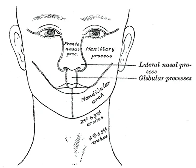
Syndromes arise from defects in arch development. Treacher Collins syndrome involves abnormal development of the first and second arches, leading to mandibular hypoplasia and ear anomalies. Pierre Robin sequence is characterized by micrognathia, glossoptosis, and cleft palate. Orofacial clefts, including cleft lip and cleft palate, result from failure of fusion of the facial processes.
Genital development involves the paramesonephric and mesonephric ducts. In females, the paramesonephric ducts develop into the fallopian tubes, uterus, and upper vagina. The mesonephric ducts regress. In males, the mesonephric ducts persist to form the epididymis and vas deferens, while the paramesonephric ducts regress.
DefinitionPharyngeal Apparatus
A collection of arches, pouches, clefts, and membranes that form the head and neck structures.
| Arch | Muscles | Nerve |
|---|---|---|
| 1st | Muscles of mastication | Trigeminal (V) |
| 2nd | Muscles of facial expression | Facial (VII) |
| 3rd | Stylopharyngeus | Glossopharyngeal (IX) |
| 4th/6th | Laryngeal muscles | Vagus (X) |
Exam trapEctopic Tissue
Ectopic parathyroid and thymus tissue can occur due to abnormal migration of the third and fourth pouch derivatives.
Head and neck development occurs through a series of precisely orchestrated embryonic stages. The process begins with the formation of pharyngeal arches, which are paired structures that develop during the fourth week of gestation. These arches, numbered 1 to 6, give rise to most of the bones, muscles, and connective tissues of the face and neck. Each arch contains a cartilaginous core, an artery, and a cranial nerve that provides innervation. The pharyngeal pouches are endodermal outpouchings that lie between the arches, while the pharyngeal clefts are ectodermal grooves on the external surface. These structures interact to form various components of the head and neck. [1]
Figure 1Gray51
Long description
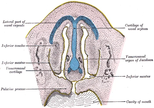
FIG. 51– Frontal section of nasal cavities of a human embryo 28 mm. long. (Kollmann.)

Figure 1: FIG.
Figure 2Gray50
Long description
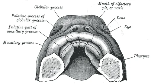
FIG. 50– The roof of the mouth of a human embryo, aged about two and a half months, showing the mode of formation of the palate. (His.)

Figure 2: FIG.
Figure 3Gray45
Long description
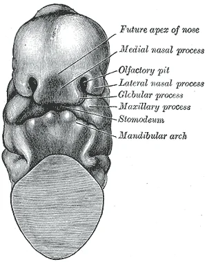
FIG. 45– Head end of human embryo of about thirty to thirty-one days. (From model by Peters.)

Figure 3: FIG.
Figure 4Gray44
Long description
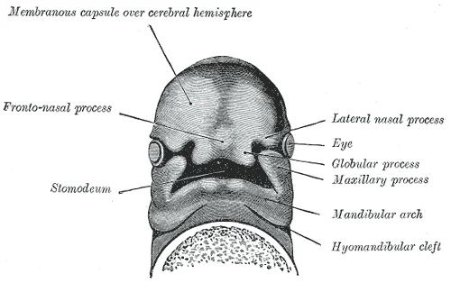
FIG. 44– Under surface of the head of a human embryo about twenty-nine days old. (After His.)

Figure 4: FIG.
Figure 5Gray43
Long description
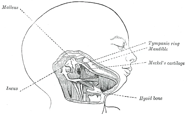
FIG. 43– Head and neck of a human embryo eighteen weeks old, with Meckel’s cartilage and hyoid bar exposed. (After Kölliker.)

Figure 5: FIG.
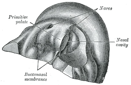
The face develops from the frontonasal prominence and the first pharyngeal arches. By the fifth to eighth weeks, the nasal placodes appear, which will form the nose and palate. The palate begins as the primary palate (formed from the frontonasal prominence) and then develops the secondary palate from the maxillary processes. The secondary palate fuses in the midline during the seventh week, forming the hard and soft palate. The tongue develops from several sources: the body of the tongue arises from the pharyngeal arches 3 and 4, while the anterior two-thirds comes from the first pharyngeal arch. The thyroid gland starts as an outpouching from the floor of the pharynx (the foramen cecum) and descends into its final position in the neck. [2]
Figure 6Gray49
Long description
FIG. 49– Primitive palate of a human embryo of thirty-seven to thirty-eight days. (From model by Peters.) On the left side the lateral wall of the nasal cavity has been removed.

Figure 6: FIG.
Figure 7Gray48
Long description
FIG. 48– Diagram showing the regions of the adult face and neck related to the fronto-nasal process and the branchial arches.

Figure 7: FIG.
The eyes develop from the optic vesicles that project from the diencephalon. These vesicles invaginate to form the optic cup, which will become the retina. The external ear develops from auricular hillocks that appear on the first and second pharyngeal arches. Teeth begin to develop around the sixth week, with the formation of tooth buds from the oral ectoderm and neural crest cells. The first set of teeth (deciduous) begins to mineralize around the 16th week of gestation.
Head and neck development is regulated by complex molecular signalling pathways that coordinate cell proliferation, differentiation, and morphogenesis. These signalling pathways involve various growth factors, cytokines, and transcription factors that interact to shape the developing head and neck structures.
The epidermal growth factor receptor (EGFR) pathway plays a crucial role in head and neck development. EGFR signalling regulates cell proliferation and differentiation in various tissues, including those of the head and neck. Another important pathway involves signal transducer and activator of transcription 3 (STAT3), which is critically altered in head and neck squamous cell carcinoma (HNSCC). STAT3 signalling influences cell survival, proliferation, and immune responses in developing tissues. [2]
Vascular endothelial growth factor receptor (VEGFR) signalling is also significant in head and neck development. VEGF promotes angiogenesis, which is essential for supplying nutrients and oxygen to developing tissues. Hypoxia-inducible factor-1α (HIF-1α) stabilization under hypoxic conditions allows transcription of genes involved in angiogenesis. [2]
Chemokines and their receptors are involved in regulating cell trafficking and organization in developing tissues. These molecules help coordinate the movement of cells to various tissue compartments during development. The interactions between chemokine receptors and their respective ligands are crucial for proper head and neck morphogenesis. [2]
Matrix metalloproteinases (MMPs) such as MMP-9 and MMP-13 are regulated by molecular signalling pathways including CXCR4. These enzymes play important roles in tissue remodeling during development. The ERK signaling pathway is associated with several mechanisms of tumor cell motility mediated by CXCR4, including regulation of the transcriptional levels of MMP-9 and MMP-13. [2]
The molecular signals originate from epithelial and mesenchymal cells and coordinate the complex processes of morphogenesis in the developing head and neck region. [2]
Each arch is associated with a specific cranial nerve, an arterial component, and cartilaginous and muscular derivatives. The clefts, which are ectodermal grooves between the arches, normally obliterate to form the neck, except for the first cleft which forms the external auditory meatus.
The pharyngeal arches are a series of six paired structures that develop in the early embryo. Each arch consists of a core of mesenchyme derived from neural crest cells and mesoderm, covered by ectoderm externally and endoderm internally. The arches are numbered I to VI, although the fifth arch is rudimentary and transient.
| Arch | Nerve | Artery | Cartilage Derivative | Muscle Derivative |
|---|---|---|---|---|
| I | Trigeminal (V) | Maxillary artery | Meckel's cartilage | Muscles of mastication, mylohyoid, anterior belly of digastric |
| II | Facial (VII) | Stapedial artery | Reichert's cartilage | Muscles of facial expression, stapedius, stylohyoid, posterior belly of digastric |
| III | Glossopharyngeal (IX) | Common carotid artery | Greater horn of hyoid | Stylopharyngeus |
| IV | Vagus (X) | Aortic arch (left), Subclavian (right) | Thyroid cartilage | Pharyngeal constrictors, cricothyroid |
| VI | Vagus (X) | Pulmonary arteries | Cricoid, arytenoid, corniculate cartilages | Intrinsic muscles of larynx |
The first arch gives rise to the muscles of mastication, which are innervated by the mandibular division of the trigeminal nerve. The second arch forms the muscles of facial expression, innervated by the facial nerve. The third arch contributes to the stylopharyngeus muscle, supplied by the glossopharyngeal nerve. The fourth and sixth arches contribute to the laryngeal muscles, innervated by the vagus nerve.
The pharyngeal clefts are ectodermal grooves on the external surface of the embryo. The first cleft persists to form the external auditory meatus. The second, third, and fourth clefts normally obliterate, forming the cervical sinus. If they persist, they can lead to branchial cysts or fistulas.
The pharyngeal pouches are endodermal outpocketings into the surrounding mesenchyme. The first pouch forms the middle ear cavity and auditory tube. The second pouch contributes to the palatine tonsil. The third pouch gives rise to the inferior parathyroid glands and the thymus. The fourth pouch forms the superior parathyroid glands and the ultimobranchial body, which contributes to the parafollicular cells of the thyroid gland.
Figure 81117 Muscles of the Neck and Back
Long description
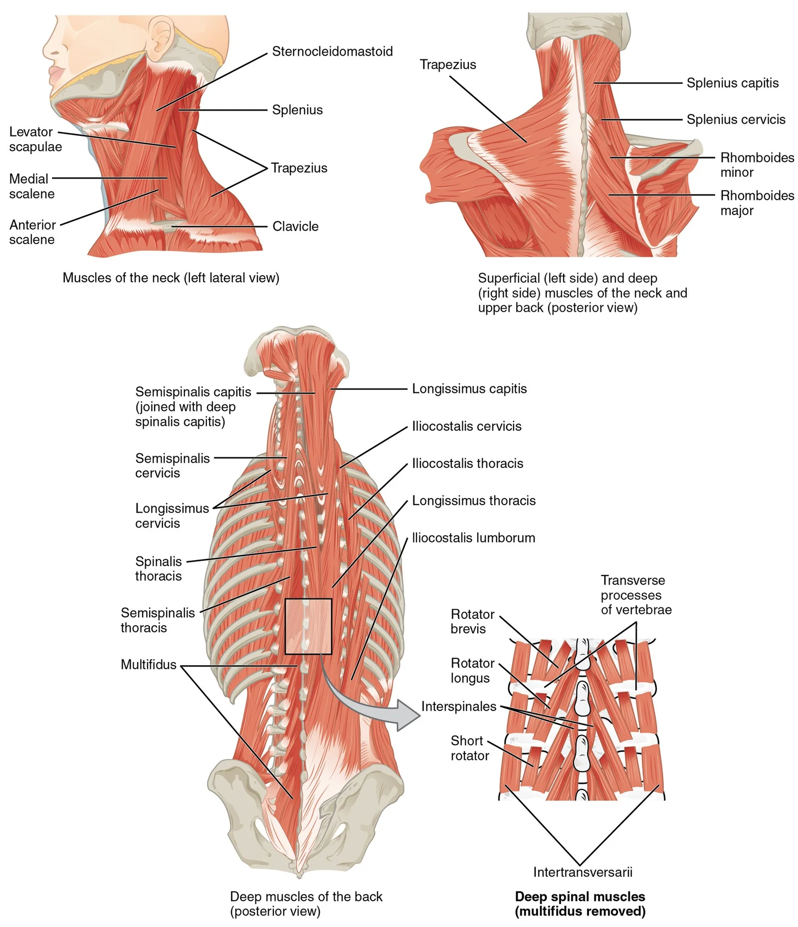
The large, complex muscles of the neck and back move the head, shoulders, and vertebral column.

Figure 8: The large, complex muscles of the neck and back move the head, shoulders, and vertebral column.
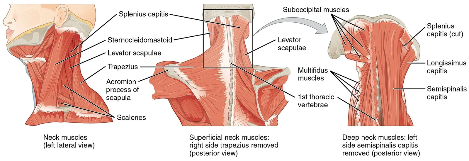
Figure 91111 Posterior and Side Views of the Neck
Long description
The superficial and deep muscles of the neck are responsible for moving the head, cervical vertebrae, and scapulas.

Figure 9: The superficial and deep muscles of the neck are responsible for moving the head, cervical vertebrae, and scapulas.
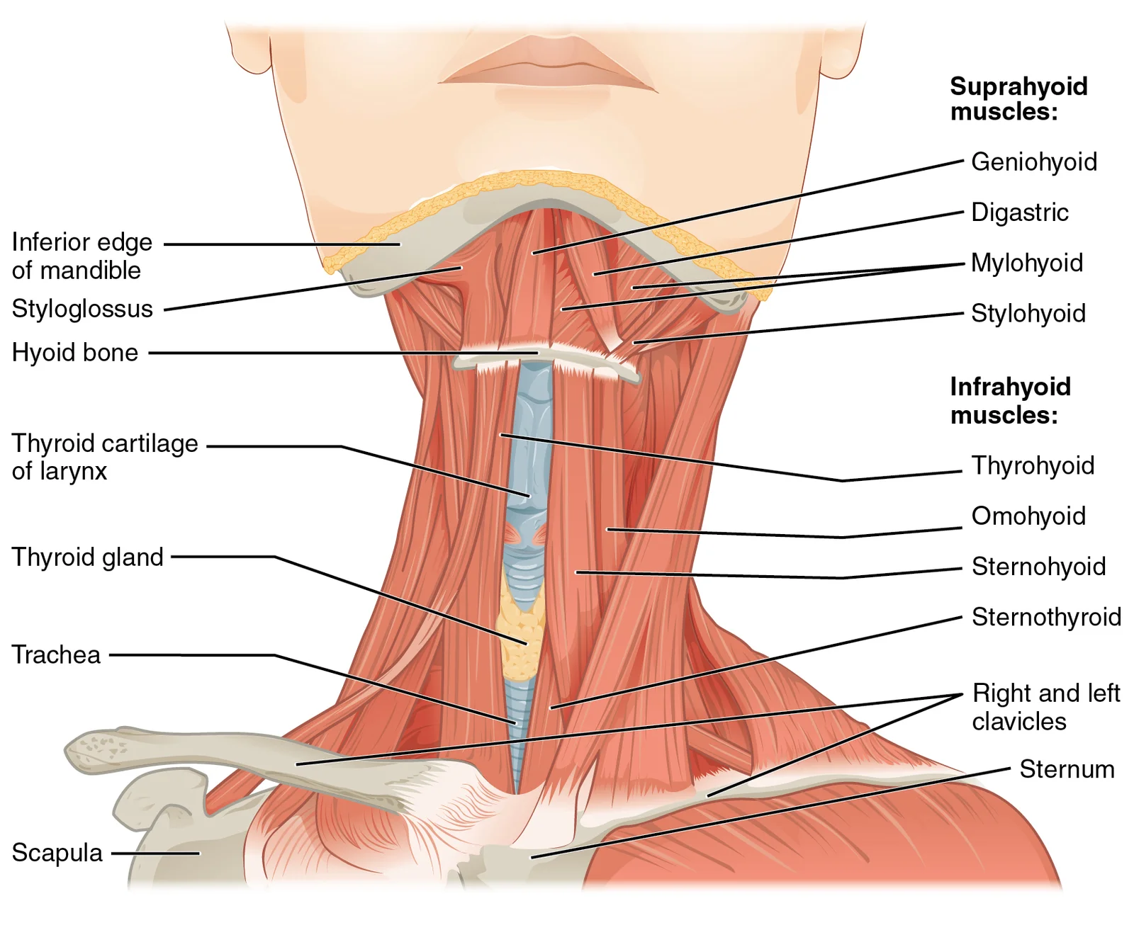
Figure 101110 Muscle of the Anterior Neck
Long description
The anterior muscles of the neck facilitate swallowing and speech. The suprahyoid muscles originate from above the hyoid bone in the chin region. The infrahyoid muscles originate below the hyoid bone in the lower neck.

Figure 10: The anterior muscles of the neck facilitate swallowing and speech.
The muscles of facial expression are unique because their origins are on the surface of the skull, and their insertions are into the skin[1]. This arrangement allows them to move the skin rather than bones, creating facial expressions. The buccinator muscle is a large portion of the face, compressing the cheek to allow whistling, blowing, and sucking, and assisting in chewing[1]. The corrugator supercilii is a small muscle that acts as the prime mover of the eyebrows, allowing for frowning[1].
Chewing, or mastication, requires muscles that can exert significant pressure[1]. The masseter is the main muscle for chewing, elevating the mandible to close the mouth. It is assisted by the temporalis muscle, which retracts the mandible[1]. These muscles are derived from the first pharyngeal arch and are innervated by the trigeminal nerve.
The suprahyoid muscles, including the digastric, stylohyoid, mylohyoid, and geniohyoid, raise the hyoid bone, floor of the mouth, and larynx during swallowing[1]. The digastric has anterior and posterior bellies; the anterior belly depresses the mandible, while the posterior belly elevates the hyoid[1]. The infrahyoid muscles, such as the omohyoid, sternohyoid, thyrohyoid, and sternothyroid, generally depress the hyoid bone and control larynx position[1]. The thyrohyoid elevates the thyroid cartilage, while the sternothyroid depresses it[1].
Congenital anomalies of the head and neck arise from disruptions in the complex embryological processes involving the pharyngeal arches, pouches, and clefts. Understanding these defects requires a clear grasp of normal development, particularly the derivatives of the endodermal pharyngeal pouches. The image displays a lateral view of the developing neck, where the first four pharyngeal pouches are clearly labelled as outpocketings of the foregut endoderm. These pouches lie medial to the corresponding pharyngeal arches and are separated externally by the pharyngeal clefts. The first pouch is situated superiorly, adjacent to the first arch, and is shown expanding towards the first cleft. The second pouch lies inferior to the first, nestled between the second and third arches. The third pouch is located below the second, and the fourth pouch is the most inferior of the labelled group. This anatomical arrangement is critical for identifying the origin of various adult structures.
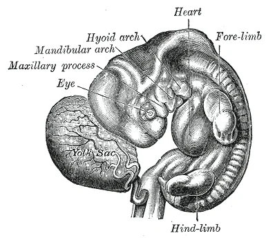
Figure 11Gray52
Long description
FIG. 52– Human embryo from thirty-one to thirty-four days. (His.)

Figure 11: FIG.
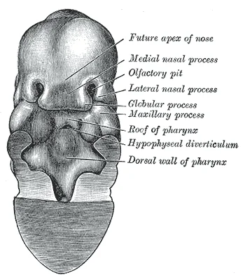
Figure 12Gray46
Long description
FIG. 46– Same embryo as shown in Fig. 45 , with front wall of pharynx removed.

Figure 12: FIG.
The first pharyngeal pouch develops into the tubotympanic recess, which gives rise to the auditory (Eustachian) tube and the middle ear cavity. Anomalies here can lead to congenital deafness or chronic otitis media. The first pharyngeal cleft, in contrast, normally degenerates, but if it persists, it can form a branchial cleft cyst or sinus. The second pharyngeal pouch contributes to the palatine tonsils. Hypoplasia or aplasia of the tonsillar tissue can result in immune deficiencies, although this is less commonly a primary congenital anomaly presentation. The second cleft also normally disappears; persistence leads to branchial cleft cysts, typically located along the anterior border of the sternocleidomastoid muscle.
The third pharyngeal pouch is crucial for immune system development. It differentiates into the inferior parathyroid glands and the thymus. The thymus migrates caudally from the third pouch, while the inferior parathyroids follow a similar but distinct path. Anomalies in this region, such as DiGeorge syndrome, involve the failure of the third and fourth pouches to develop properly, leading to thymic aplasia and hypocalcaemia due to parathyroid defects. The fourth pharyngeal pouch gives rise to the superior parathyroid glands and the ultimobranchial body, which contributes C-cells to the thyroid gland. Defects in fourth pouch development can cause hyperparathyroidism or thyroid dysgenesis.
These embryological relationships explain why congenital anomalies often present as cysts, sinuses, or fistulae along the neck. Branchial cleft cysts, for example, are remnants of the second cleft that failed to obliterate. They present as painless masses in the lateral neck. Understanding the precise origin of each pouch allows clinicians to predict associated anomalies and plan surgical correction.
The development of the head and neck involves the intricate formation of the vertebral column musculature, which is essential for posture and movement. These muscles are complex, overlapping structures that can be divided into five distinct groups. Understanding this anatomy is critical for interpreting congenital anomalies and their clinical presentation.
The splenius group comprises the splenius capitis and the splenius cervicis, which are primarily involved in extending and rotating the head and neck. The erector spinae is a large muscle mass divided into three subgroups: the iliocostalis, longissimus, and spinalis. The iliocostalis group includes the iliocostalis cervicis, iliocostalis thoracis, and iliocostalis lumborum. The longissimus group consists of the longissimus capitis, longissimus cervicis, and longissimus thoracis. The spinalis group includes the spinalis capitis, spinalis cervicis, and spinalis thoracis. [1]
| Group | Components |
|---|---|
| Splenius | Splenius capitis, splenius cervicis |
| Iliocostalis | Iliocostalis cervicis, thoracis, lumborum |
| Longissimus | Longissimus capitis, cervicis, thoracis |
| Spinalis | Spinalis capitis, cervicis, thoracis |
| Transversospinales | Semispinalis capitis/cervicis/thoracis, multifidus, rotatores |
| Segmental | Interspinales, intertransversarii |
| Scalenes | Anterior, middle, posterior scalene |
The transversospinales group includes the semispinalis capitis, semispinalis cervicis, semispinalis thoracis, multifidus, and rotatores. These muscles play a significant role in stabilizing the vertebral column. The segmental muscles include the interspinales and intertransversarii, which provide fine motor control between adjacent vertebrae. Finally, the scalenes include the anterior scalene, middle scalene, and posterior scalene, which are crucial for respiration and neck movement. [1]
DefinitionAnatomical Significance
The muscles of the back and neck that move the vertebral column are complex, overlapping, and can be divided into five groups. [1]
Congenital anomalies in these muscle groups can lead to significant functional deficits. For instance, abnormalities in the scalene muscles may affect respiratory mechanics, while defects in the erector spinae can compromise spinal stability. Prenatal diagnosis often relies on ultrasound imaging to detect structural abnormalities in the developing vertebral column and associated musculature.
It is important to distinguish these developmental structures from pathological conditions such as head and neck squamous cell carcinoma (HNSCC). While HNSCC involves deregulated expression of EGFR and lymphangiogenesis, these are acquired malignancies rather than developmental defects. Similarly, the role of microbiota in cancer progression, such as the association between oral bacteria and colorectal cancer, is unrelated to embryological development.
NoteDistinction from Pathology
Expression of EGFR can be deregulated in many cancers, including HNSCC. [2]
The focus of prenatal diagnosis in head and neck development is on structural integrity and proper formation of the musculoskeletal system. Early detection of anomalies allows for timely intervention and management. The complexity of the muscle groups, including the splenius, erector spinae, and scalenes, underscores the importance of precise embryological development. Any disruption in this process can have lasting clinical consequences.
Exam trapCommon Misconception
Do not confuse congenital muscle anomalies with acquired conditions like HNSCC, which involve factors such as EGFR over-expression and lymphatic infiltration.
In summary, the head and neck musculature is derived from specific embryological precursors that form the splenius, erector spinae, and scalene groups. Understanding this anatomy is fundamental for medical students, as it provides the basis for diagnosing and managing congenital disorders. The clear distinction between developmental anatomy and pathological processes ensures accurate clinical reasoning and patient care.
Clinical pearlp53 mutations
p53 gene mutations occur in over 50% of HNSCC malignancies, leading to loss of growth control and impaired stress response. This tumor suppressor inactivation prevents cells from responding to DNA damage and stress signals. [2]
Exam trapHPV and p53
HPV16 E6 oncoprotein inactivates p53 via ubiquitination, which is a separate mechanism from genetic mutations. Students often confuse viral inactivation with genetic mutations. [2]
Clinical pearlType I collagen effects
Type I collagen stimulates cytokine expression (IL-1α, IL-1β, IL-6, TNF-α, TGF-β) and increases MMP-2 activity in metastatic HNSCC cells more than in primary cells. This differential response contributes to metastatic potential. [2]
Exam trapMAPK phosphorylation
Type I collagen induces phosphorylation of ERK and p38 (not total protein levels) in HNSCC cells, activating MMP production. The phosphorylation status, not protein quantity, is key.
Clinical pearlCytokine crosstalk
Tumor cells release cytokines that induce ECM remodeling, while fibroblasts respond to these signals and can also induce tumor growth. This bidirectional communication creates a pro-tumor microenvironment.
Clinical pearlMMP-TIMP balance
Under normal conditions, the balance between MMPs and TIMPs maintains ECM organization; MMPs digest various substrates including growth factors and cytokines. This balance is disrupted in cancer progression.
Clinical pearlCAFs and MMPs
Cancer-associated fibroblasts require direct cell contact with HNSCC cells to activate MMP-2 and MMP-9 secretion in both cell types. Conditioned media alone is insufficient for full activation. [2]
Exam trapIntegrin signaling
Type I collagen activates MMP-2 and MMP-9 through α2β1 integrin, triggering tyrosine kinases, PKC, and MAPK pathways. This integrin-ECM interaction is crucial for invasion. [2]
Clinical pearlMicrobiota diversity
Gastric cancer patients show significantly increased overall microbial diversity in saliva and plaque. A screening system based on 11 different genera demonstrated high sensitivity (97%). [3]
Clinical pearlMicrobiota screening
An oral and fecal microbiota test can discriminate healthy controls from CRC patients with 76% sensitivity and 94% specificity. This shows diagnostic potential for microbiota analysis. [3]
Clinical pearlPeriodontal pathogens
Higher levels of T. denticola and P. intermedia are significantly associated with increased CRC risk. Additionally, 11 common and 16 rare taxa showed associations with cancer risk, particularly among African-American and female populations. [3]
A common pitfall is confusing the embryological origins of head and neck structures. The mandibular and hyoid arches grow rapidly, telescoping the posterior arches and forming the bilateral cervical sinus, which is bounded anteriorly by the hyoid arch and posteriorly by the thoracic wall before obliteration via wall fusion [2]. The first pharyngeal cleft gives rise to the concha and external acoustic meatus, while surrounding mesenchymal swellings on the mandibular and hyoid arches form the auricle [2]. The first pharyngeal pouch elongates dorsally to form the auditory tube and tympanic cavity, and the pharyngeal membrane between the first and second arches, invaded by mesoderm, becomes the tympanic membrane [2]. In mammals, pharyngeal membranes do not perforate. The mandibular arch, positioned between the first cleft and stomodeum, produces the lower lip, mandible, muscles of mastication, and anterior tongue [2]. Its cartilaginous core (Meckel’s cartilage) yields the malleus dorsally and contributes to the mandibular body containing the incisors ventrally [2]. The third arch cartilage forms the greater cornu of the hyoid, while fused ventral ends of the second and third arches create the hyoid body and posterior tongue [2]. The fourth and sixth arch cartilages form the thyroid, cricoid, arytenoid, and tracheal cartilages [2].
Another pitfall is misinterpreting the role of the extracellular matrix (ECM). While the ECM provides a scaffold for tissue organisation, the passages highlight its role in tumour invasion rather than its developmental function. Students should not assume that the mechanisms of proteolytic degradation described for cancer cells apply to normal morphogenesis without specific embryological evidence, which is absent here. The passages state that invasion involves changes in tumour cell adhesion and proteolytic degradation of the ECM. This is a pathological process, not a developmental one.
Furthermore, students often overlook the distinction between primary tumour sites and metastatic sites. The passages indicate that the distinct pattern of chemokine receptor expression by cancer cells determines the site of metastatic spread. This is a feature of disease progression, not embryological patterning. Similarly, the up-regulation of CXCR4 and CCR7 induces migration and invasion in cancer cells. This mechanism is exploited by tumour cells, not used by developing tissues to form struct
The role of inflammation in cancer is also frequently misunderstood. The presence of leukocytes in tumours was initially interpreted as an aborted attempt by the immune system to reject the tumour. However, this is a pathological response, not a developmental signal. The hypothesis that cancer originates at sites of chronic inflammation is a theory of carcinogenesis, not a principle of embryology. Students must not confuse the stromal reactions promoting cancer progression, such as angiogenesis, with the angiogenic processes that occur during normal organogenesis. [2]
Finally, the concept of dysbiosis in the oral cavity is often mistakenly applied to normal development. Dysbiosis is characterised by the loss of beneficial microbes, expansion of pathogenic microbes, and general loss of microbial diversity. This ecological imbalance is linked to cancer development. It is not a feature of healthy oral development. The use of saliva to detect dysbiotic trends is a diagnostic tool for pathology, not a developmental marker. Students must strictly separate the embryological facts of head and neck formation from the pathological mechanisms of HNSCC described in the passages. [3]
Knowledge of the molecular interactions in HNSCC is critical at the bedside for understanding disease progression and prognosis. The correlation between COX-2 overexpression and the number of tumour-infiltrating Foxp3+ regulatory T cells suggests a mechanism for immune evasion. Clinicians use this information to interpret immunohistochemical findings and potentially guide immunotherapy strategies. The up-regulation of CXCR4 expression in HNSCC cells is associated with increased migration and invasion. This knowledge helps in assessing the risk of metastasis, particularly to lymph nodes, as the crosstalk between CXCL12 and its receptor is involved in lymph node metastasis. [2]
In the operating theatre, understanding the stromal-tumour interactions is vital for surgical planning. The interaction between HNSCC cells and periodontal ligament fibroblasts leads to the up-regulation of MMPs, including MMP-2, MMP-1, and MMP-13. This suggests that the local microenvironment actively contributes to tumour invasion. Surgeons must be aware that tumour cells may exploit these pathways to invade surrounding tissues, necessitating wider margins or more extensive resections. The activation of MMP-2 requires MT1-MMP or αv integrins, which are up-regulated in both tumour cells and fibroblasts. This crosstalk highlights the importance of considering the stromal component in surgical outcomes.
On imaging, the understanding of chemokine-mediated metastasis aids in the interpretation of spread patterns. The distinct pattern of chemokine receptor expression determines the site of metastatic spread. Radiologists can use this knowledge to anticipate potential sites of metastasis, such as lymph nodes, based on the expression profile of the primary tumour. The role of hypoxia in enhancing CXCR4 expression via HIF-1α may also correlate with imaging features of hypoxic tumours, which are often associated with poorer prognosis. [2]
In examinations, students are tested on their ability to distinguish between normal developmental processes and pathological mechanisms. Questions may ask about the role of specific molecules in HNSCC progression. For example, the role of PGE2 in increasing the production of angiogenic factors, promoting invasion and metastasis, is a key fact. Similarly, the fact that CXCL5 stimulates cell proliferation and invasion in metastatic HNSCC cells is a specific detail that may be tested. Students must be able to recall that pro-MMP-9 is produced by HNSCC cells, not by PDL fibroblasts, and is activated on the surface of tumour cells. This level of detail is essential for demonstrating a thorough understanding of the molecular pathology of HNSCC.
HNSCC Metastasis Mechanisms
- Primary HNSCC CellExpresses CXCR4 and COX-2
- Question: Microenvironment Interaction?
Yes
- Stromal CrosstalkPDL fibroblasts up-regulate MMP-2
- Chemokine SignalingCXCL12 binds CXCR4
- Question: Result?
Invasion
- EMT and Migration
Metastasis
- Lymph Node Spread
No
- Local Growth Only
- Invasion is a three-step process involving changes in adhesion, proteolytic degradation of the ECM, and migration. [2]
- COX-2 overexpression is positively correlated with tumour-infiltrating Foxp3+ regulatory T cells.
- CXCR4 up-regulation enhances migration and invasion in HNSCC cells. [2]
- The CXCL12-CXCR4 interaction induces epithelial-mesenchymal transition (EMT). [2]
- Pro-MMP-2 is secreted by PDL fibroblasts, while pro-MMP-9 is produced by HNSCC cells. [2]
- Dysbiosis is characterised by loss of beneficial microbes and expansion of pathogenic microbes.
- Saliva allows early detection of overall dysbiotic trends in the oral cavity. [3]
- Angiogenesis is a stromal reaction promoting cancer progression. [2]
Answer each one in your head first, then open the answer.
What is the three-step process of invasion?
Show answerHide answer
Changes in tumour cell adhesion, proteolytic degradation of the ECM, and migration of tumour cells in a proteolytically modified ECMHow does COX-2 relate to regulatory T cells?
Show answerHide answer
COX-2 overexpression is positively correlated with the number of tumour-infiltrating Foxp3+ regulatory T cellsWhich chemokine receptor up-regulation enhances migration?
Show answerHide answer
CXCR4 expression up-regulation enhances migration and invasionWho produces pro-MMP-9?
Show answerHide answer
Pro-MMP-9 is produced by HNSCC cells, not by PDL fibroblastsWhat characterises dysbiosis?
Show answerHide answer
Loss of beneficial microbes, expansion of pathogenic microbes, and general loss of microbial diversity
- Betts JG, Young KA, Wise JA, et al. Axial Muscles of the Head, Neck, and Back. OpenStax, Anatomy and Physiology 2e (CC BY-NC-SA 4.0). Accessed .
- Koontongkaew S. The tumor microenvironment contribution to development, growth, invasion and metastasis of head and neck squamous cell carcinomas. Journal of Cancer 2013;4(1):66-83. doi:10.7150/jca.5112. Accessed .
- Mascitti M, Togni L, Troiano G, et al. Beyond Head and Neck Cancer: The Relationship Between Oral Microbiota and Tumour Development in Distant Organs. Frontiers in cellular and infection microbiology 2019;9:232. doi:10.3389/fcimb.2019.00232. Accessed .
Figure credits
- Figure 1: Henry Vandyke Carter, via Wikimedia Commons, Public domain, Public domain.
- Figure 2: Henry Vandyke Carter, via Wikimedia Commons, Public domain, Public domain.
- Figure 3: Henry Vandyke Carter, via Wikimedia Commons, Public domain, Public domain.
- Figure 4: Henry Vandyke Carter, via Wikimedia Commons, Public domain, Public domain.
- Figure 5: Henry Vandyke Carter, via Wikimedia Commons, Public domain, Public domain.
- Figure 6: Henry Vandyke Carter, via Wikimedia Commons, Public domain, Public domain.
- Figure 7: Henry Vandyke Carter, via Wikimedia Commons, Public domain, Public domain.
- Figure 8: OpenStax, via Wikimedia Commons, CC BY 4.0, CC BY 4.0. resized.
- Figure 9: OpenStax, via Wikimedia Commons, CC BY 4.0, CC BY 4.0. resized.
- Figure 10: OpenStax, via Wikimedia Commons, CC BY 4.0, CC BY 4.0. resized.
- Figure 11: Henry Vandyke Carter, via Wikimedia Commons, Public domain, Public domain.
- Figure 12: Henry Vandyke Carter, via Wikimedia Commons, Public domain, Public domain.
Educational content for medical students and doctors. It is not a substitute for clinical judgement or local guidelines.