Gametogenesis
Gametogenesis is the process of forming haploid sperm and eggs from diploid germ cells via mitosis, meiosis, and differentiation.
Gametogenesis is the specialised production of haploid gametes from diploid germ cells. It ensures genetic diversity and maintains chromosome number across generations. The process involves mitosis, meiosis, and maturation.
Male fertility relies on the production by the testis of large numbers of normal spermatozoa. This process is known as spermatogenesis, which can be divided in three major steps. The first step is mitosis with the multiplication of the spermatogonia. The second step is meiosis to reduce the number of chromosomes from diploid to haploid cells. The cells are then called primary spermatocytes that then divide to form secondary spermatocytes. These will divide and undergo meiosis to form the round spermatids. The third step is spermiogenesis, which refers to the successful maturation of round spermatids into spermatozoa. All of these steps are central in the spermatogenic process. Any defect during the spermatogenesis can result in the reduction or absence of sperm production. [2]
Figure 1Gray5
Long description
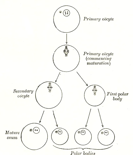
Diagram showing the reduction in number of the chromosomes in the process of maturation of the ovum.

Figure 1: Diagram showing the reduction in number of the chromosomes in the process of maturation of the ovum.
Figure 2Gray4
Long description
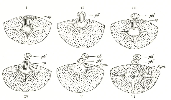
Formation of polar bodies in Asterias glacialis. (Slightly modified from Hertwig.) In I the polar spindle ( sp ) has advanced to the surface of the egg. In II a small elevation ( pb 1 ) is formed which receives half of the spindle. In III the elevation is constricted off, forming the first polar bod

Figure 2: Formation of polar bodies in Asterias glacialis.
While spermatogenesis is continuous, oogenesis is cyclic and arrested. The only CTA proteins functionally established in gametogenesis are SCP-1, involved in chromosome pairing during meiosis, OY-TES-1, SPO11, and BORIS. [1]
The ovarian cycle regulates oocyte maturation. Fertilisation triggers the zona reaction to prevent polyspermy. Cleavage follows, forming a morula and then a blastocyst. The first week proceeds day by day from zygote to implantation. Abnormal fertilisation and IVF in outline are clinical considerations.
DefinitionSpermiogenesis
The successful maturation of round spermatids into spermatozoa.
| Stage | Description |
|---|---|
| Mitosis | Multiplication of spermatogonia. |
| Meiosis I | Primary spermatocytes to secondary spermatocytes. |
| Meiosis II | Secondary spermatocytes to round spermatids. |
| Spermiogenesis | Maturation into spermatozoa. |
Epigenetic mechanisms regulate gene expression during gametogenesis. DNA methylation, histone modifications, ncRNA, and chromatin structure are the epigenetic mechanisms required to regulate the gene expression. Therefore, no development or biological process can be understood without the integration of epigenetics and genetics. This is the case for normal developmental processes like spermatogenesis and oogenesis. [2]
NoteEpigenetics
Environmental factors such as nutrition, toxicants, or stress to impact developmental processes like gametogenesis involves alterations in the epigenetics. [2]
Since the gametogenesis process produces the gametes, sperm and eggs, alterations in gametogenesis impact the future generations physiology, phenotype, and gametes. This non-genetic form of inheritance termed epigenetic transgenerational inheritance requires manipulation during gametogenesis to obtain epigenetically modified sperm and eggs. [2]
Gametogenesis Pathway
- Diploid Germ CellSpermatogonia or Oogonia
- Question: Mitosis?
Yes
- Multiply Germ Cells
No
- Enter Meiosis
- MeiosisReduce to haploid
- MaturationSpermiogenesis or Ootid
Gametogenesis is the biological process by which diploid precursor cells undergo meiosis to form haploid gametes. In males, this process is called spermatogenesis, occurring in the seminiferous tubules of the testes. In females, it is called oogenesis, taking place within the ovaries.
Figure 3Gray7
Long description
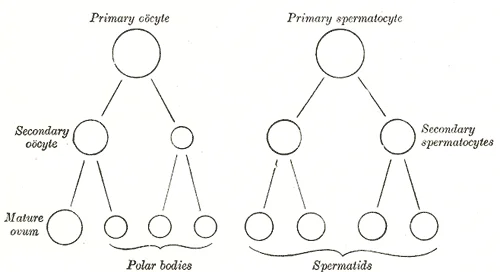
Scheme showing analogies in the process of maturation of the ovum and the development of the spermatids (young spermatozoa).

Figure 3: Scheme showing analogies in the process of maturation of the ovum and the development of the spermatids (young spermatozoa).
Spermatogenesis begins with spermatogonia, which are mitotically active cells. As development progresses, PLU-1 expression reduces during early prophase I stages (leptotene, zygotene) but reappears at pachytene, remaining detectable in diplotene cells. This protein is diffusely located over the nucleus and may play a role in regulating meiotic transcription during specific meiotic stages. [1]
Figure 4Gray6
Long description
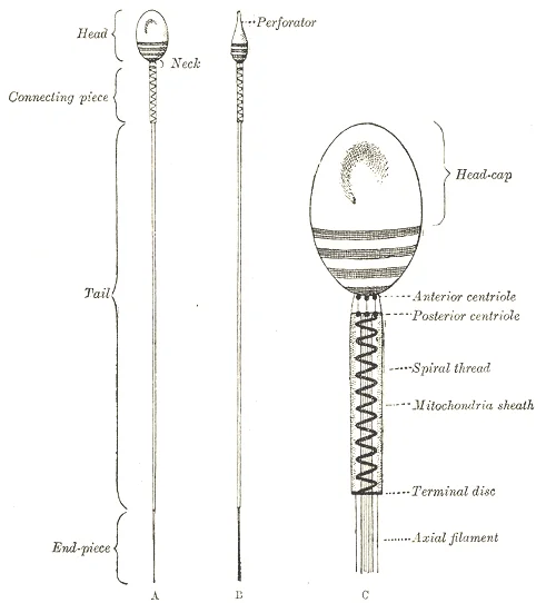
Human spermatozoön. Diagrammatic. A. Surface view. B. Profile view. In C the head, neck, and connecting piece are more highly magnified.

Figure 4: Human spermatozoön (diagrammatic).
Androgen plays an essential role in the development and maintenance of spermatogenesis. This loss can be reversed by testosterone treatment. [2]
During spermatogenesis, a cascade of epigenetic changes occurs, with the most dramatic changes happening during early developmental stages. Environmentally induced changes in histone retention also occur during different stages of spermatogenic development. [2]
Oogenesis begins with primordial follicles in the ovaries. When a primordial follicle begins developing, the surrounding flattened pre-granulosa cells become cuboidal and start proliferating, with surrounding stromal cells destined to become theca cells. The follicle is then termed a primary follicle.
Subsequent development into secondary and pre-antral follicles involves continued proliferation of granulosa cells to form multiple layers and the initial development of a theca cell layer. Follicles with multiple layers of granulosa cells gain sensitivity to follicle stimulating hormone (FSH) and are regulated to grow in cyclic waves coordinated with the estrous cycle.
As granulosa and theca cells proliferate, a fluid-filled space or antrum forms, dividing granulosa cells into cumulus granulosa surrounding the oocyte and mural granulosa around the follicle's periphery. Extensive cell-cell communication and growth factor signaling occur between the oocyte and somatic cells at all stages of oogenesis. Most developing follicles undergo atresia and regress rather than reaching ovulation.
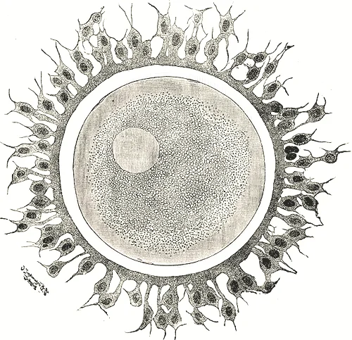
Figure 5Gray3
Long description
Human ovum examined fresh in the liquor folliculi. (Waldeyer.) The zona pellucida is seen as a thick clear girdle surrounded by the cells of the corona radiata. The egg itself shows a central granular deutoplasmic area and a peripheral clear layer, and encloses the germinal vesicle, in which is seen

Figure 5: Human ovum examined fresh in the liquor folliculi.
Epigenetic regulation is crucial in oogenesis. Primordial follicle assembly, transition to primary follicle, and subsequent oogenesis stages are regulated by small non-coding RNA expression. DNA methylation occurs at differentially imprinted sites between male and female gametes, allowing mono-allelic expression of imprinted genes in offspring. In oocytes of antral stage follicles and later, H3K4Me3 histone methylation increases, which is important for normal oocyte function and establishing DNA methylation patterns in mature oocytes. [2]
Gametogenesis is regulated by a complex network of molecular signals and epigenetic modifications. Cancer-testis antigens (CTAs) are a group of genes that play diverse roles during spermatogenesis, with different CTAs expressed at various stages of this process[1]. These antigens are involved in multiple functions, including regulation of mitotic cycling in spermatogonia, association with the meiotic cycle in spermatocytes, and acrosome maturation in sperm[1]. The expression
Notably, PRDM14 targets are not conserved between mouse and human, highlighting evolutionary divergence in the molecular network for primordial germ cell specification. [2]
Epigenetic reprogramming is essential for gametogenesis. Primordial germ cells (PGCs) undergo extensive epigenetic reprogramming, including a genome-wide loss of approximately 90% of 5-methylcytosine (5mC). This process requires a set of germline reprogramming responsive (GRR) genes, which have unique promoter sequence characteristics with high levels of both 5mC and 5-hydroxymethylcytosine (5hmC). The loss of DNA methylation combined with polycomb repressive complex 1 (PRC1) repression is uniqu [2]
Epigenetics refers to ‘the molecular factors and processes around the DNA that regulate genome activity independent of DNA sequence, and that are mitotically stable’. These epigenetic mechanisms are required for stem cells to develop into differentiated cell types and are integral to normal biology. Non-coding RNA-directed DNA methylation and DNA methylation-directed histone retention may play a role in the environmentally induced epigenetic tra [2]
Gametogenesis is the process by which germ cells develop into mature gametes. It begins in early embryogenesis with the formation and migration of primordial germ cells (PGCs) into the gonadal ridge. These signals induce the expression of PRDM1 (BLIMP1), PRDM14, and AP2γ, which form a transcriptional network that establishes germ cell fate. [2]
In the developing gonad, PGCs undergo epigenetic reprogramming. This reorganization is critical for resetting the epigenetic landscape before meiosis begins. [2]
DefinitionPrimordial Germ Cells
Primordial germ cells are the precursors of sperm and oocytes, specified early in embryogenesis and migrating to the gonadal ridges to undergo gametogenesis.
Surrounding these are somatic support cells. The labels indicate the direction of migration and the subsequent differentiation into spermatogonia or oogonia.
| Embryonic Structure | Adult Structure |
|---|---|
| Primordial Germ Cells (PGCs) | Spermatogonia / Oogonia |
| Spermatogonia | Spermatozoa (in males) |
| Oogonia | Oocytes (in females) |
| Somatic Support Cells | Sertoli Cells / Granulosa Cells |
In males, spermatogenesis continues post-natally. The chromatin in developing sperm undergoes significant reorganisation. Histones are replaced by protamines, allowing the DNA to be packaged into a highly compact structure. This process involves hyperacetylation of histones, followed by eviction and replacement with transition proteins, and finally protamines. The resulting toroid structure of protamines packages DNA tenfold more compactly than somatic heterochromatin. [2]
Estrogens also play a role in testicular function. In neonates, estrogen-dependent ERα signaling is required for normal adult spermatogenesis. In adult males, estrogens can stimulate spermatogenesis by increasing FSH release, whereas exogenous estrogens may inhibit it by suppressing LH and testosterone. [2]
NoteClinical Note
Disruption in the transcriptional network of PRDM1, PRDM14, and AP2γ leads to defects in PGC specification, highlighting their dominant role in germ cell fate determination.
The process is tightly regulated by various signalling pathways. For instance, Wnt signalling is involved in cell fate determination and patterning in early embryos. The suppression of Wnt signals by proteins like SGY-1 helps control transcription activation of genes such as c-myc and cyclin D1.
Gametogenesis Overview
- SpecificationBMP/WNT signals induce PRDM1, PRDM14, AP2γ in epiblast
- Question: Migration?
Yes
- PGCs migrate to gonadal ridge
No
- Defect in specification
Gametogenesis is the process by which germ cells develop into mature spermatozoa or oocytes. Errors in this intricate sequence can lead to congenital anomalies of the reproductive system. These anomalies often stem from disruptions in the early specification of primordial germ cells (PGCs) or failures in the subsequent meiotic and post-meiotic maturation phases. Understanding the embryological basis requires tracing the journey from the genital ridge to the fully formed gamete.
The central axis represents the timeline from PGC migration to spermiation.
The embryological basis of many congenital anomalies lies in the failure of germ cells to establish genomic stability. In prospermatogonia, prepachytene piRNAs are necessary for silencing mobile elements through guiding the de novo DNA methylations of transposable elements in order to guarantee genome stability[1]. If this silencing fails, transposable elements may activate, causing DNA damage that can lead to apoptosis or, if repaired incorrectly, to chromosomal abnormalities. This mechanism is crucial for preventing the transmission of genetic errors to the next generation. [2]
Meiosis represents another critical window for congenital anomalies. During meiosis of the gametogenesis process, germ cells stop their progress at the prophase stage to allow parental genomes to exchange genetic information through meiosis recombination[1]. At this stage, chromosomes pair in a homologous manner, and large pieces of the chromosomes can be exchanged through crossover events. These crossover events are necessary to maintain euploidy in gametes. An absence of crossover events has been linked to infertility and aneuploidy in the offspring. When crossover events fail to occur or are misplaced, the resulting gametes may possess an incorrect number of chromosomes (aneuploidy), leading to conditions such as Klinefelter syndrome or Turner syndrome in the offspring. [2]
DNA methylation patterns also play a vital role in the epigenetic programming of gametes. Similar observations were made with ancestral vinclozolin exposure . This shows that transgenerational changes in DNA methylation arise throughout gametogenesis[1]. Disruptions in these methylation windows can lead to imprinting disorders, which are a significant category of congenital anomalies affecting growth and development. [2]
DefinitionMeiotic Crossover
A physical exchange of genetic material between homologous chromosomes during prophase I of meiosis, essential for proper chromosome segregation.
Exam trapAneuploidy Risk
Failure of crossover events does not just cause infertility; it directly increases the risk of aneuploidy in offspring, such as Down syndrome, due to nondisjunction.
| Stage | Key Epigenetic Event | Consequence of Failure |
|---|---|---|
| Prospermatogonia | Silencing of mobile elements via DNA methylation | Genomic instability, transposon activation |
| Pachytene Spermatocyte | Homologous recombination and crossover | Aneuploidy, infertility |
| Round Spermatids | Histone-to-protamine transition | Abnormal sperm morphology |
The expression pattern of CTAs during spermatogenesis is of special interest. Functional analysis of these genes during gametogenesis might well give some clues about their possible role in tumours. Their expression is restricted exclusively to spermatogenic germ cells with other tubular cells (e.g. Leydig and Sertoli) being negative. This fits well with the findings of Yuasa et al. who demonstrated that CTAs have much higher expression frequencies in the germ cell cancers (seminomas) than non-seminomas. While primarily discussed in the context of cancer, the aberrant re-expression of these genes can be seen as a deviation from the normal, tightly regulated gametogenic program, highlighting the importance of precise gene silencing during development. [1]
Gametogenesis represents the fundamental biological process by which diploid germ cells undergo meiosis to produce haploid gametes, a mechanism critical for sexual reproduction and genetic diversity. In clinical embryology, understanding the timeline and cellular differentiation of these processes is essential for diagnosing congenital anomalies and infertility. The journey begins with the migration of primordial germ cells to the developing gonads. In the mammalian testis, this migration occurs during fetal development. Once the primordial germ cells arrive, they associate with mesenchymal cells. These mesenchymal cells subsequently differentiate into Sertoli cells, which provide structural and nutritional support. At this stage, the sex cords are formed. The primordial germ cells then differentiate into prospermatogonia. These cells remain centrally positioned within the cords. They are surrounded by immature Sertoli cells during this initial phase. [2]
DefinitionProspermatogonia
The precursor cells to spermatogonia, found centrally in the sex cords during fetal development.
Following a period of proliferation, the prospermatogonia undergo a critical migration. In humans, however, this migration is prenatal. The cells move to the basement membrane of the sex cords. Here, they divide to form type A spermatogonia. This distinction in timing between species is vital for interpreting experimental data and understanding human developmental pathology. The type A spermatogonia serve as the stem cell population for spermatogenesis. Depending on the species, morphological di [2]
In contrast, the rodent testis exhibits a different classification. Multiple type A spermatogonia, intermediate forms, and type B spermatogonia are reported. Their appearance in the testis is temporally controlled. This temporal control ensures the continuous production of sperm throughout adult life. Disruptions in this timeline can lead to infertility or germ cell tumours. For instance, failure of prospermatogonia to migrate or differentiate properly can result in cryptorchidism or impaired spermatogenesis. [2]
Prenatal diagnosis of disorders affecting gametogenesis often relies on ultrasound and genetic screening. While direct visualization of germ cell differentiation is not possible prenatally, markers such as testicular volume and hormonal profiles can provide indirect evidence. This approach aims to provide a more "complete" immune response. However, vaccines designed to prime the immune system against tumours expressing various CTA have so far shown only partial clinical success. This highlights the complexity of immune interactions in germ cell pathology. [1]
Exam trapImmune Response
Vaccines targeting CTA-expressing tumours have shown only partial clinical success, not complete immunity.
Understanding the precise origin, course, and termination of germ cell migration is crucial. The origin is the yolk sac endoderm. The course involves migration through the hindgut to the genital ridges. The termination is the basement membrane of the sex cords. Any deviation in this pathway can have significant clinical consequences. Therefore, medical students must master the detailed timeline of spermatogonial development to accurately diagnose and manage related conditions.
Gametogenesis involves the formation of haploid gametes from diploid germ cells, a process strictly regulated by epigenetic mechanisms. A critical phase occurs during primordial germ cell (PGC) migration. One central epigenetic change in pre-migratory and early migratory mouse PGCs is the loss of H3K9me2. This event is followed by an accumulation of H3K27me3 signal. [2]
DefinitionEpigenetic Reprogramming
The global erasure of DNA methylation and histone modifications in primordial germ cells to reset developmental potential.
Environmental factors can induce sperm DNA methylation changes.
Exam trapHuman vs Murine Timing
Human PGCs lose H3K27me3 after week 9 of gestation, whereas murine models show dynamic changes after entry into the genital ridge.
These proteins may function as transcriptional repressors. This translocation results in the fusion of the synovial sarcoma translocation gene (SYT) on chromosome 18 to one of the SSX genes on chromosome X. The encoded hybrid proteins are probably responsible for transforming activity. [1]
| Gene | Chromosome | Associated Translocation | Function |
|---|---|---|---|
| SSX1 | X | t(X;18) | Transcriptional repressor |
| SSX2 | X | t(X;18) | Transcriptional repressor |
| SSX4 | X | t(X;18) | Transcriptional repressor |
NoteNY-ESO-1 Expression
NY-ESO-1 expression ceases in well-progressed tumours, making it a marker for early testicular tumorigenesis progression.
From these, 19 families are testis-restricted, and 11 show additional expression in one or two somatic tissues. Nine are expressed in 3–6 tissue types besides testis, and 5 are ubiquitously somatically expressed. [1]
Clinical pearlCTA Expression Patterns
19 of 44 CTA families are testis-restricted; others show lower expression in pancreas or ubiquitous somatic tissues.
Such upregulated expression has been reported for several MAGE members – LAGE-1, SSX-2, CAGE, NY-ESO-1. For MAGE-A1, demethylation is necessary and sufficient for gene expression, suggesting demethylation is the primary mechanism of transcription control. [1]
Definition5-aza-2-deoxycytidine (5DC)
A demethylating agent that entraps DNA methyltransferases, leading to loss of DNA methylation and CTA upregulation.
It removes imprinting from genes during the last mitotic division of spermatocytes. Several lines of evidence indicate that expression of some CTAs is dependent not only on demethylation, but on other transcriptional mechanisms. [1]
Exam trapBoris Function
Boris regulates methylation during spermatogenesis by removing imprinting during the last mitotic division of spermatocytes.
The program sounds more like an ontogenetic (life-cycle) one. [1]
NoteOntogenetic Theory
Cancer theories including gametic, parthenogenetic, and trophoblastic models are viewed as embryonal or ontogenetic theories.
DefinitionPloidy Cycles
Cyclic increase and reduction of chromosome number involving homologous pairing and segregation, omitting one DNA replication round.
This event is followed by an accumulation of H3K27me3 signal.
Clinical pearlH3K27me3 Dynamics
Human PGCs lose H3K27me3 after week 9 of gestation, mirroring porcine PGC patterns.
Exam trapEnvironmental Inducers
BPA, phthalates, vinclozolin, DDT, glyphosate, and atrazine all induce sperm DNA methylation changes.
NoteTransgenerational Epimutations
Epimutations occur in F3 generation rat sperm after ancestral toxicant exposure, raising questions about timing of origin.
Following a period of proliferation, the prospermatogonia undergo a critical migration. In humans, however, this migration is prenatal. The cells move to the basement membrane of the sex cords. Here, they divide to form type A spermatogonia. This distinction in timing between species is vital for interpreting experimental data and understanding human developmental pathology. The type A spermatogonia serve as the stem cell population for spermatogenesis. Depending on the species, morphological di
Another significant pitfall involves the molecular classification of specific CTA proteins. Students often misattribute enzymatic functions without considering structural homology. For instance, TSP50 is protease-like, CT17 is phospholipase-like, and CT15 is metalloproteinase-like. Specifically, the CT15 gene encodes ADAM domain 2, a member of the ADAM protein family. These proteins are membrane-anchored and structurally related to snake venom disintegrins, implicating them in cell-cell and cell [1]
Confusion also arises regarding the epigenetic regulation of germ cells. A common mistake is believing that DNA demethylation is complete and uniform across all loci during primordial germ cell (PGC) migration. However, despite this near-complete erasure, some genome loci remain methylated and are referred to as ‘escapees’. Additionally, studies indicate that a loss of TET1 and TET2 in germ cells does not necessarily impact infertility, and few loci show altered epigenetic states. [2]
Students may also incorrectly assume that hypomethylation in PGCs leads to genomic instability. Usually, loss of DNA methylation in somatic cells induces ectopic retrotransposon expression, anarchic proliferation, and apoptosis. However, PGCs develop normally despite this hypomethylated state. The hypothesis is that chromatin reorganization enhances genome stability, ensures proper chromosome alignment and segregation during mitosis, and maintains global transcriptional quiescence during this developmental period. This process is conserved across species, with human PGCs following similar demethylation patterns to rodents. [2]
Finally, there is confusion regarding the role of BORIS and epigenetic inheritance. The chromosome region 20q13.2 containing the BORIS gene is commonly amplified or exhibits moderate gains in many human cancers, strengthening the idea that this region contains a major oncogene. Theoretically, aberrant expression of amplified BORIS may demethylate CTA genes on the X-chromosome, activating a large body of CTA genes in malignancies. However, this does not explain the activation of other gametogenesis-related CTA genes from other chromosomes, suggesting amplification may not be random. Furthermore, epigenetic transgenerational inheritance is defined as germline-mediated inheritance of epigenetic information between generations without continued direct environmental influences. Changes in the F1 generation due to direct exposure in the F0 parent are not transgenerational inheritance; true transgenerational inheritance requires changes to persist in the unexposed F2 or F3 generations. [1] [2]
Exam trapEpigenetic Erasure
DNA demethylation in PGCs is near-complete but not total; 'escapee' loci remain methylated, and loss of TET enzymes does not always cause infertility.
Knowledge of gametogenesis and associated molecular markers is utilised at the bedside, in the operating theatre, on imaging, and in examinations through the identification of specific protein domains and epigenetic markers. In clinical diagnostics, understanding the structural homology of CTAs aids in identifying potential therapeutic targets. For example, recognizing that CT15 is a subunit of fertilin helps explain its role in sperm-egg interactions and its potential involvement in tumour evasion. This structural knowledge is critical when interpreting molecular profiles of tumours that aberrantly express these genes.
In the operating theatre and during surgical pathology, the recognition of amplified genomic regions is vital. The amplification of the 20q13.2 region containing the BORIS gene is a significant finding in many human cancers. Surgeons and pathologists must be aware that this amplification may indicate the presence of a major oncogene, potentially influencing prognosis and treatment decisions.
On imaging and in laboratory investigations, the limited information on normal epigenetic changes during oogenesis is a practical constraint. Evaluating developing oocytes in ovaries is difficult due to the small number of oocytes available for isolation compared to male germ cells. This limitation affects the ability to establish comprehensive epigenomic baselines for female germ cells, impacting the interpretation of epigenetic disorders related to oogenesis. [2]
In examinations, students are tested on the precise definitions and mechanisms of epigenetic inheritance. It is essential to distinguish between direct multigenerational exposure and true epigenetic transgenerational inheritance. If a male or non-pregnant female is exposed to an environmental factor, epimutations arise in the F0 generation and their germ cells. The F1 generation is directly exposed, so changes seen in F1 are not transgenerational inheritance. True transgenerational inheritance occurs if changes are passed to the unexposed F2 generation. Similarly, if a pregnant female is exposed, the F1 embryo and its germ cells (forming F2) are directly exposed; changes must be seen in the F3 generation or later to qualify as transgenerational inheritance. [2]
DefinitionTransgenerational Inheritance
Germline-mediated inheritance of epigenetic information between generations without continued direct environmental influence, requiring effects to appear in unexposed F2 or F3 generations.
Epigenetic Inheritance Pathways
- Environmental ExposureToxicant or factor
- Question: Parent Generation (F0)
Male or Non-Pregnant Female
- Epimutations in F0 Germ CellsDirect exposure
- Question: Next Generation (F1)
Directly Exposed
- Not TransgenerationalMultigenerational exposure only
Unexposed F2
- True Transgenerational InheritanceGermline-mediated inheritance
Pregnant Female
- F1 Embryo Directly ExposedIncluding F1 Germ Cells
- Question: Next Generation (F2)
Directly Exposed
- Not TransgenerationalMultigenerational exposure only
Unexposed F3
- True Transgenerational InheritanceGermline-mediated inheritance
CTAs are restricted to male germ cells and malignancies, but not all have confirmed antigenic properties. CT15 is an ADAM domain protein, a subunit of fertilin, involved in sperm-egg interactions and tumour metastasis. TET1 and TET2 drive DNA demethylation in PGCs between E10.5 and E12.5, but 'escapee' loci remain methylated. Loss of TET1/2 does not necessarily cause infertility, and PGCs maintain stability despite hypomethylation,. BORIS gene amplification at 20q13.2 is common in cancers and may activate CTA genes. True epigenetic transgenerational inheritance requires phenotypic changes in unexposed F2 or F3 generations. Oogenesis epigenetics is less understood due to the difficulty of isolating small numbers of oocytes. CTA gene functions are often inferred from homology, such as TSP50 being protease-like.
Answer each one in your head first, then open the answer.
Which protein family does CT15 belong to and what is its role in fertilization?
Show answerHide answer
CT15 belongs to the ADAM protein family and is a subunit of fertilin, playing an important role in sperm-egg interactions.What is the definition of epigenetic transgenerational inheritance?
Show answerHide answer
It is germline-mediated inheritance of epigenetic information between generations in the absence of continued direct environmental influences.Why is the term 'Cancer/Testis Associated' preferred over the initial definition for CTAs?
Show answerHide answer
Because many members still need to be proven as possessing antigenic properties in cancer patients.What happens to DNA methylation in PGCs between E10.5 and E12.5?
Show answerHide answer
Further demethylation erasure takes place, with DNA methylation at its lowest level due to TET1 and TET2 activity.When is epigenetic transgenerational inheritance observed if a pregnant female is exposed?
Show answerHide answer
When epigenetic and phenotypic changes are seen in the F3 generation or later.
- Kalejs M, Erenpreisa J. Cancer/testis antigens and gametogenesis: a review and 'brain-storming' session. Cancer cell international 2005;5(1):4. doi:10.1186/1475-2867-5-4. Accessed .
- Ben Maamar M, Nilsson EE, Skinner MK. Epigenetic transgenerational inheritance, gametogenesis and germline development†. Biology of reproduction 2021;105(3):570-592. doi:10.1093/biolre/ioab085. Accessed .
Figure credits
- Figure 1: Henry Vandyke Carter, via Wikimedia Commons, Public domain, Public domain.
- Figure 2: Henry Vandyke Carter, via Wikimedia Commons, Public domain, Public domain.
- Figure 3: Henry Vandyke Carter, via Wikimedia Commons, Public domain, Public domain.
- Figure 4: Henry Vandyke Carter, via Wikimedia Commons, Public domain, Public domain.
- Figure 5: Henry Vandyke Carter, via Wikimedia Commons, Public domain, Public domain.
Educational content for medical students and doctors. It is not a substitute for clinical judgement or local guidelines.