Fertilisation
Fertilisation is the fusion of sperm and egg to form a zygote.
Fertilisation typically occurs in the uterine tubes, the most common site for sperm to encounter the oocyte[1]. The journey from the vagina to the uterine tubes takes approximately 30 minutes to 2 hours, facilitated by uterine contractions[1]. Sperm can survive in the uterine tubes for 3-5 days, allowing fertilisation to occur if intercourse takes place a few days before ovulation[1].
During their journey, sperm undergo capacitation, a process that improves their motility and depletes cholesterol molecules from the sperm head membrane, thinning it to facilitate enzyme release[1]. Capacitation is essential for sperm to have the "capacity" to fertilise an oocyte; sperm that reach the oocyte before capacitation is complete cannot penetrate its outer layers[1].
When the oocyte encounters capacitated sperm, chemical attractants from the corona radiata cells guide the sperm toward it[1]. To fertilise the oocyte, sperm must penetrate two protective layers.
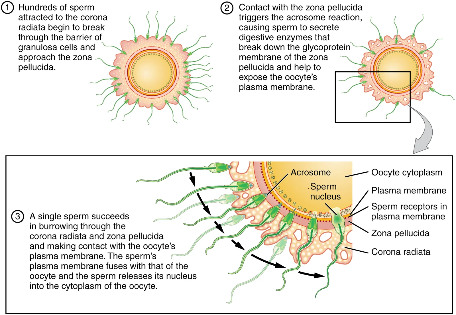
Upon contact with the zona pellucida, sperm bind to receptors and initiate the acrosomal reaction, releasing enzymes that clear a path through the zona pellucida[1]. A single sperm then makes contact with sperm-binding receptors on the oocyte's plasma membrane and fuses with it, entering the oocyte interior[1].
Figure 12901 Sperm Fertilization
Long description
Before fertilization, hundreds of capacitated sperm must break through the surrounding corona radiata and zona pellucida so that one can contact and fuse with the oocyte plasma membrane.

Figure 1: Before fertilization, hundreds of capacitated sperm must break through the surrounding corona radiata and zona pellucida so that one can contact and fuse with the oocyte plasma membrane.
When the first sperm fuses with the oocyte, two mechanisms prevent polyspermy. The fast block involves an immediate change in sodium ion permeability, depolarising the membrane and preventing additional sperm fusion[1]. This is followed by the slow block (cortical reaction), where cortical granules release zonal inhibiting proteins that destroy sperm receptors and prevent further binding[1]. Fertilisation is complete when the haploid nuclei of the sperm and oocyte unify, form
Fertilisation is the process that concludes with the formation of a single-celled diploid zygote. This zygote contains the complete set of genetic instructions required for development into an adult. [1]
The pre-embryonic stage encompasses the first 2 weeks of prenatal development. This period is defined by rapid cell division, migration, and differentiation. [2]
During this stage, the zygote develops into a blastocyst. At the end of the first week, the blastocyst adheres to and embeds itself within the uterine lining using trophoblast cells, a process known as implantation. Implantation typically occurs on the posterior wall or in the fundus of the uterus. This event signals the conclusion of the pre-embryonic stage. [2]
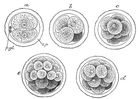
Figure 2Gray9
Long description
First stages of segmentation of a mammalian ovum. Semidiagrammatic. (From a drawing by Allen Thomson.) z.p. Zona striata. p.gl. Polar bodies. a. Two-cell stage. b. Four-cell stage. c. Eight-cell stage. d, e. Morula stage.

Figure 2: First stages of segmentation of a mammalian ovum (semidiagrammatic).
NoteImplantation Failure
Between 50% and 75% of blastocysts fail to implant and are shed during menses. [2]
The zygote is the single-cell starting point. As it divides, it forms a blastocyst, which consists of an inner cell mass and an outer layer of trophoblast cells. The trophoblast cells are the outermost layer that directly contacts the uterine wall to facilitate embedding.
The embryonic stage occurs from week 3 to week 8. By the end of this period, all organ systems have formed in a rudimentary, though often nonfunctional or semi-functional, state. [2]
Key developmental processes during this stage include:
- Gastrulation: The differentiation of embryonic cells into an embryonic disc consisting of three primary germ layers: the endoderm, mesoderm, and ectoderm. [2]
- Neurulation: The initiation of central nervous system development.
- Organogenesis: The establishment of the basic structural plan for all organ systems. [2]
- Embryonic Folding: The process by which the fetus begins to take its physical shape.
The chorion is the outermost membrane, surrounding all others. [2]
The fetal stage begins at the ninth week of gestation and continues until birth. [2]
Implantation usually occurs in the uterine body, but in 1% to 2% of cases, it occurs in abnormal locations. An ectopic pregnancy occurs when implantation is outside the uterus. If implantation occurs in the inferior portion of the uterus, the placenta may cover the cervical opening, resulting in placenta previa.
Exam trapFolate Deficiency
Maternal folate deficiency in the first weeks of pregnancy can lead to neural tube defects, such as spina bifida or anencephaly.
Fertilisation is a tightly regulated sequence of molecular events that ensures a single sperm fuses with a single oocyte. The process is divided into several stages, each controlled by specific signalling molecules and receptors.
Sperm capacitation occurs in the female reproductive tract and is a prerequisite for fertilisation. It involves the removal of cholesterol from the sperm plasma membrane, which increases membrane fluidity and exposes receptors. This process is mediated by bicarbonate ions, calcium, and albumin in the oviductal fluid. Capacitation also leads to the phosphorylation of tyrosine residues on sperm proteins, which is essential for the acrosome reaction.
The acrosome reaction is triggered when the capacitated sperm binds to the zona pellucida. The zona pellucida is composed of four glycoproteins: ZP1, ZP2, ZP3, and ZP4. This binding causes an influx of calcium ions into the sperm head, leading to the fusion of the acrosomal membrane with the overlying plasma membrane. The acrosome then releases its stored digestive enzymes. [1]
Sperm–oocyte membrane fusion occurs after the sperm has traversed the zona pellucida. This binding is species-specific and triggers the fusion of the two membranes. The sperm nucleus and its centriole then enter the oocyte cytoplasm. [1]
Cortical reaction and the block to polyspermy are critical for preventing multiple sperm from fertilising the oocyte. Upon sperm entry, intracellular calcium oscillations are triggered in the oocyte. These oscillations cause cortical granules to migrate to the oocyte periphery and fuse with the plasma membrane, releasing their contents into the perivitelline space. The e [1]
Oocyte activation is the final molecular event. The calcium oscillations also trigger the resumption of meiosis, the completion of the second meiotic division, and the formation of the female pronucleus. The sperm nucleus decondenses to form the male pronucleus. Both pronuclei replicate their DNA and migrate towards each other, and their nuclear envelopes break down, allowing the maternal and paternal chromosomes to align on the first mitotic spindle. This completes fertilisation and initiates the first cleavage division.
The process of fertilisation initiates a sequence of rapid cellular divisions and structural reorganisations that transform a single-celled zygote into a complex embryo. The initial products of these divisions are blastomeres. By approximately 3 days after fertilisation, these cells form a compacted 16-cell mass known as the morula. This structure subsequently develops into a blastocyst, which consists of a fluid-filled cavity called the blastocoel, an outer layer of trophoblasts, and an inner cell mass. [1]
This potency is transient, lasting only a few days before cells are committed to specific lineages. The inner cell mass is fated to become the embryo itself. [2]
Supporting the embryo are four essential embryonic membranes:
- The amnion: A sac that sits atop the flat embryo and eventually envelops it during embryonic folding. * The yolk sac: Formed by the hypoblast extending into the blastocyst cavity. It provides primitive blood circulation during the second and third weeks and later serves as the source of germ cells and blood cells. * The chorion: Formed by the syncytiotrophoblast, cytotrophoblast, and extraembryonic mesoderm. It envelops the conceptus and develops chorionic villi. [2]
The fetal portion of the placenta is derived from these chorionic villi, which burrow into the maternal decidua basalis. Placentation is completed by weeks 14–16. [2]
The blastocoel is the central fluid-filled space that separates the inner cell mass from the trophoblast. The trophoblast is the outer ring of cells that will eventually contribute to the chorion and placenta.
| Embryonic Structure | Adult Structure |
|---|---|
| Inner cell mass | Embryo |
| Allantois | Part of the urinary bladder [2] |
| Yolk sac stalks and allantois stalks | Umbilical cord [2] |
| Chorionic villi | Fetal portion of the placenta |
| Decidua basalis | Maternal portion of the placenta |
| Primitive gut (endoderm-lined tube) | Gastrointestinal system [2] |
Congenital anomalies result from disruptions in the normal process of fertilisation and early embryonic development. These abnormalities can affect any structure or system in the developing embryo and may be caused by genetic factors, environmental influences, or a combination of both.
DefinitionCongenital Anomaly
A structural or functional abnormality present at birth that develops prenatally during the embryonic or fetal period.
One common category of congenital anomalies is related to errors in fertilisation itself. Polyspermy, the fertilisation of an oocyte by more than one sperm, typically results in non-viable embryos due to abnormal chromosome numbers. Another anomaly is the formation of a complete hydatidiform mole, where an empty egg is fertilised by one or two sperm without maternal chromosomes, leading to abnormal placental development without a viable fetus. [1]
Chromosomal abnormalities represent a significant group of congenital anomalies. Down syndrome (trisomy 21) occurs in approximately 1 in 700 live births and is characterised by intellectual disability, characteristic facial features, and congenital heart defects in about 50% of cases. Turner syndrome (45,X) affects and is associated with short stature, ovarian dysgenesis, and cardiovascular abnormalities. [1]
Neural tube defects (NTDs) are among the most common serious congenital anomalies, affecting about 1-2 per 1,000 pregnancies in the UK. These defects result from failure of the neural tube to close properly between days 21 and 28 of gestation. [1]
| Anomaly | Embryological Basis | Prevalence |
|---|---|---|
| Down syndrome (trisomy 21) | Nondisjunction of chromosome 21 | 1 in 700 live births |
| Turner syndrome (45,X) | Monosomy X | 1 in 2,500 female births [1] |
| Neural tube defects | Failure of neural tube closure (days 21-28) | 1-2 per 1,000 pregnancies |
| Complete hydatidiform mole | Fertilisation of empty egg by sperm without maternal chromosomes | Variable |
The grape-like vesicles represent abnormal chorionic villi without fetal tissue. This condition results from fertilisation of an empty egg by one or two sperm without maternal chromosomes, leading to abnormal placental development without a viable fetus. The uterine wall appears distended by the molar tissue, which can cause complications such as persistent trophoblastic disease if not properly managed.
Fertilisation normally occurs in the ampulla of the uterine tube within 24 hours of ovulation. When this timing or site is disturbed, clinical problems arise. The most important correlate is ectopic pregnancy, where the embryo implants outside the uterine cavity, most often in the ampullary region of the tube. Risk factors include previous tubal surgery, pelvic inflammatory disease, and assisted reproduction. A ruptured tubal ectopic is a surgical emergency causing haemoperitoneum and shock; diagnosis is suggested by a positive pregnancy test with pelvic pain and is confirmed by transvaginal ultrasound showing no intrauterine sac with an adnexal mass. [2]
Another correlate is multiple pregnancy, which occurs when more than one oocyte is fertilised (dizygotic, from multiple ovulations) or when one zygote splits (monozygotic, producing identical twins). The clinical importance lies in preterm birth, pre-eclampsia, and discordant growth; monozygotic twins may also suffer twin-to-twin transfusion syndrome if they share a placenta.
Prenatal diagnosis begins with screening for aneuploidy, not with fertilisation itself. However, the conceptus’s chromosomal status is determined at fertilisation: non-disjunction in meiosis I or II of the oocyte or sperm produces trisomies. The most common live-born trisomy is Down syndrome (trisomy 21), whose risk rises with maternal age: at age 20 the risk is about 1 in 1500, at age 35 about 1 in 350, and at age 40 about 1 in 100. Screening in the first trimester combines maternal serum pregnancy-associated plasma protein-A (PAPP-A) and free beta-human chorionic gonadotrophin (β-hCG) with fetal nuchal translucency on ultrasound; this detects about 85–90% of trisomy 21 cases at a 5% false-positive rate. Definitive diagnosis requires chorionic villus sampling (CVS) at 11–13 weeks or amniocentesis after 15 weeks; both carry a procedure-related miscarriage risk of about 0.5–1%.
Preimplantation genetic testing (PGT) is a prenatal diagnostic tool applied before implantation. PGT-A (aneuploidy) screens for chromosome copy number; PGT-M (monogenic) detects specific inherited mutations. The main limitation is mosaicism, where not all cells share the same karyotype, so a biopsied cell may not represent the whole embryo. In low-resource settings, prenatal diagnosis relies on ultrasound alone, which cannot detect chromosomal abnormalities but can identify structural defects such as anencephaly or spina bifida from the second trimester.
Special populations deserve attention. In older mothers, the higher aneuploidy risk justifies offering invasive testing directly. In pregnancies from donor oocytes, the risk of aneuploidy follows the donor’s age, not the recipient’s. In multiple pregnancies, nuchal translucency and serum markers are less accurate, and invasive testing carries higher risks, so counselling must be individualised. In women with recurrent miscarriage, parental karyotyping may reveal a balanced translocation that predisposes to unbalanced offspring; prenatal diagnosis or PGT-SR (structural rearrangement) is then offered.
-
The oocyte is surrounded by two layers: the outer corona radiata (follicular granulosa cells) and the inner zona pellucida, a thick transparent glycoprotein membrane. The corona radiata consists of actual cells that were released with the oocyte from the ovarian follicle, while the zona pellucida is an acellular glycoprotein matrix that plays a crucial role in species-specific sperm binding. [1]
-
At fertilisation, the secondary oocyte is still arrested in metaphase of meiosis II; only sperm entry triggers completion of meiosis and ejection of the second polar body. This represents a unique cellular pause point in female gametogenesis, where the oocyte remains in suspended animation until activated by the sperm's entry. [1]
-
After sperm entry, the oocyte becomes an ovum; the male and female haploid nuclei are called pronuclei, which decondense, replicate DNA, migrate together, and intermingle their genetic material. This process transforms the oocyte from a arrested gamete into a developing zygote with restored diploid chromosome number. [1]
-
Dizygotic (fraternal) twins arise from two eggs fertilised by two sperm and occur in approximately 1 percent of ovulation cycles. These twins develop from two separate zygotes and are no more genetically similar than siblings born at different times, sharing on average 50% of their genetic material. [1]
-
A common trap: dizygotic twins are no more genetically similar than siblings born at different times—they are not identical. Unlike monozygotic twins who originate from a single fertilised egg, dizygotic twins result from two separate fertilisation events. [1]
-
Another trap: the zona pellucida is not a cellular layer; it is an acellular glycoprotein membrane, whereas the corona radiata is cellular. This distinction is important when understanding the mechanisms of sperm penetration and polyspermy prevention. [1]
-
Do not confuse the pronuclei with the polar bodies: polar bodies are discarded haploid remnants, not participating in the zygote genome. The first polar body may divide to form a second polar body, but neither contributes genetic material to the embryo. [1]
-
The sperm contributes the centriole and activates the oocyte; the oocyte contributes the mitochondria—mitochondrial DNA is maternally inherited. This explains why mitochondrial diseases are passed from mother to child exclusively. [1]
-
Fertilisation restores the diploid chromosome number and determines the chromosomal sex of the embryo. The presence of the Y chromosome from the sperm determines male development, while its absence results in female development. [1]
-
A trap: fertilisation does not occur in the uterus or ovary; it must occur in the distal uterine tube before the oocyte degenerates. Implantation occurs later in the uterus, but fertilisation must precede it within the correct timeframe and location. [1]
-
The second polar body is ejected only after fertilisation, so its presence confirms that sperm entry has occurred. This cellular event is a reliable marker that fertilisation has successfully taken place and meiosis has been completed. [1]
Exam trapConfusing fertilization with implantation
Fertilization is the fusion of sperm and oocyte nuclei to form a zygote, which occurs in the ampulla of the Fallopian tube, not in the uterus. Implantation happens later when the blastocyst attaches to the endometrium. [1]
Exam trapMisunderstanding the conceptus
The term conceptus refers to the zygote and its associated membranes as they travel to the uterus, not just the embryo after implantation.
Exam trapIncorrectly describing the fertilization membrane
The fertilization membrane consists of mucopolysaccharides coating the zygote combined with the hardened zona pellucida, not just the zona pellucida alone.
Exam trapConfusing trophoblast functions
The trophoblast cells develop into the chorionic sac and fetal portion of the placenta, not the maternal portion. They also secrete hCG to maintain the corpus luteum. [2]
Exam trapMisrepresenting hCG production
Human chorionic gonadotropin (hCG) is secreted by the trophoblast after implantation, not by the corpus luteum or endometrium. [2]
Exam trapOverlooking the timeline of development
The zygote undergoes five or six rapid mitotic cell divisions during its journey to the uterus, and organogenesis occurs within the first 8 weeks of gestation, not immediately after fertilization. [2]
Exam trapConfusing germ layer derivatives
The ectoderm forms the nervous system and epidermis, mesoderm forms muscles and connective tissue, and endoderm forms the gastrointestinal lining and organs like the liver and pancreas, not interchangeable structures.
NoteClinical assessment of pregnancy
The detection of hCG in maternal blood or urine is the basis for pregnancy tests. This hormone is produced by the trophoblast after implantation, typically occurring 6-12 days after fertilization.
Pregnancy Testing Timeline
- FertilizationSperm and oocyte nuclei fuse in Fallopian tube
- Cleavage divisionsZygote undergoes 5-6 mitotic divisions
- Blastocyst formationCells differentiate into trophoblast and inner cell mass
- Question: Implantation?
Success
- hCG productionTrophoblast secretes hCG
- Pregnancy testDetectable in blood/urine ~10-14 days after fertilization
Failure
- MenstruationLoss of conceptus without pregnancy establishment
DefinitionAmniotic fluid composition
Early in development, amniotic fluid consists almost entirely of a filtrate of maternal plasma, but by approximately the eighth week, fetal kidneys begin to add urine to the volume. [2]
NoteSubstance teratogenicity
The placenta is permeable to lipid-soluble fetotoxic substances including alcohol, nicotine, barbiturates, antibiotics, and certain pathogens. This knowledge guides counseling of pregnant patients about avoiding harmful exposures. [2]
Clinical pearlUnderstanding FASD
Alcohol consumption during pregnancy can result in fetal alcohol spectrum disorders (FASD), including organ and facial malformations, as well as cognitive and behavioral disorders.
| Germ Layer | Derived Structures |
|---|---|
| Ectoderm | Central and peripheral nervous systems, sensory organs, epidermis, hair, nails |
| Mesoderm | Skeleton, muscles, connective tissue, heart, blood vessels, kidneys |
| Endoderm | Epithelial lining of gastrointestinal tract, liver, pancreas, lungs |
NoteUmbilical cord development
Fetal mesenchymal cells derived from mesoderm differentiate into blood vessels, including the three umbilical blood vessels that connect the embryo to the developing placenta. This knowledge is essential for understanding umbilical cord anatomy and function. [2]
Fertilization is the fusion of haploid sperm and oocyte to form a diploid zygote The conceptus undergoes 5-6 mitotic divisions during transport to the uterus Trophoblast cells develop into the chorionic sac and fetal portion of the placenta hCG secreted by trophoblast maintains the corpus luteum and is detectable in pregnancy tests The amnion forms by the end of the second week and contains protective amniotic fluid The three germ layers (ectoderm, mesoderm, endoderm) form all embryonic structures The placenta allows exchange of substances but is permeable to harmful substances Organogenesis establishes all rudimentary organ structures within the first 8 weeks
Answer each one in your head first, then open the answer.
Where does fertilization typically occur?
Show answerHide answer
In the ampulla of the Fallopian tubeWhat hormone is secreted by the trophoblast after implantation?
Show answerHide answer
Human chorionic gonadotropin (hCG)How many mitotic divisions does the zygote undergo during its journey to the uterus?
Show answerHide answer
Five or sixWhich germ layer gives rise to the nervous system?
Show answerHide answer
EctodermWhen does organogenesis occur in embryonic development?
Show answerHide answer
Within the first 8 weeks of gestation
- Betts JG, Young KA, Wise JA, et al. Fertilization. OpenStax, Anatomy and Physiology 2e (CC BY-NC-SA 4.0). Accessed .
- Betts JG, Young KA, Wise JA, et al. Embryonic Development. OpenStax, Anatomy and Physiology 2e (CC BY-NC-SA 4.0). Accessed .
Figure credits
Educational content for medical students and doctors. It is not a substitute for clinical judgement or local guidelines.