Cleavage and blastocyst formation
Cleavage and blastocyst formation in early embryonic development.
The pre-embryonic stage covers the first 2 weeks of prenatal development. Cleavage begins immediately after fertilization, with the zygote undergoing rapid mitotic divisions. By day 2-3, the zygote becomes a 4-8 cell stage called a morula. The morula continues to divide and differentiates into an outer layer of cells (trophoblast) and an inner cell mass by day 4-5. [1]
By day 5, the blastocyst forms, consisting of the trophoblast, inner cell mass, and a fluid-filled blastocoel cavity. During this stage, the trophoblast excretes enzymes that degrade the zona pellucida in a process called "hatching," allowing the blastocyst to break free. This hatching process is crucial for the next stage of development as it enables the blastocyst to make contact with the uterine lining. [1]
At the end of the first week (around day 6-7), the blastocyst comes in contact with the uterine wall and adheres to it via trophoblast cells, beginning implantation. The blastocyst typically implants in the fundus of the uterus or on the posterior wall. However, 50-75 percent of blastocysts fail to implant and are shed with the endometrium during menses. This high failure rate explains why pregnancy typically requires several ovulation cycles to achieve, as not every fertilized egg successfully implants. [1]
| Implantation anomalies | Abnormal implantation of the blastocyst | [1] |
Figure 12908 Germ Layers-02
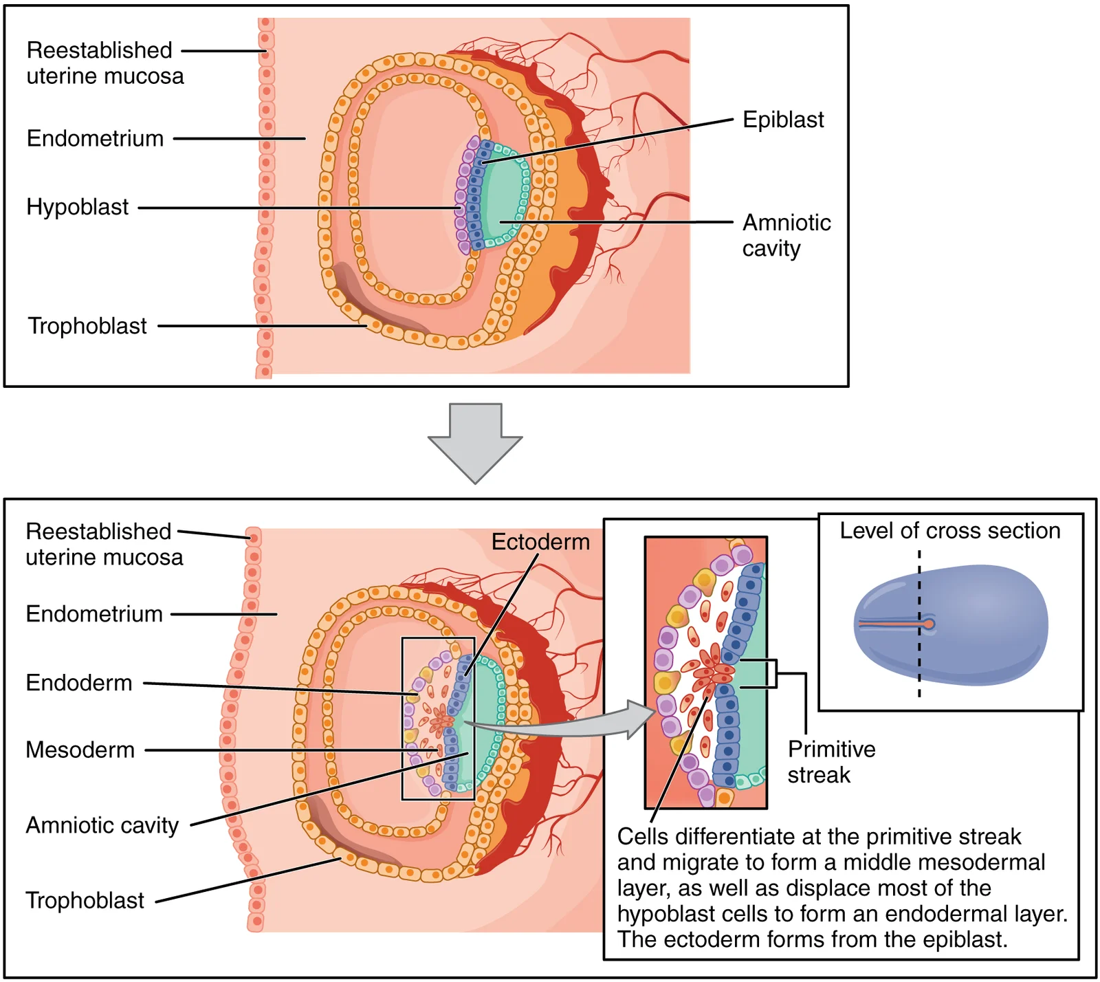
Long description
Formation of the three primary germ layers occurs during the first 2 weeks of development. The embryo at this stage is only a few millimeters in length.

Figure 1: Formation of the three primary germ layers occurs during the first 2 weeks of development.
Following fertilization, the single-celled diploid zygote carries all the genetic instructions needed to develop into an adult[1]. It begins its journey toward the uterus, propelled by peristalsis and the beating cilia of the epithelial cells lining the Fallopian tube[1]. During this transit, the zygote undergoes five or six rapid mitotic cell divisions[1]. Each division produces smaller daughter cells called blastomeres, and although the number of cells increases, the total volume of the conceptus does not change[1].
Approximately 3 days after fertilization, the conceptus, now composed of 16 cells, reaches the uterus[1]. These cells, previously loosely grouped, become compacted into a solid mass resembling a mulberry; this structure is named the morula[1]. Once inside the uterine cavity, the morula floats freely for several days, continuing to divide until it forms a ball of roughly 100 cells[1]. During this time, it consumes nutritive endometrial secretions known as uterine milk, while the uterine lining thickens in preparation for implantation[1].
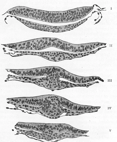
Figure 2Gray15
Long description
Series of transverse sections through the embryonic disk of Tarsius. (After Hubrecht.) Section I passes through the disk, in front of Hensen’s knot and shows only the ectoderm and entoderm. Sections II, III, and IV pass through Hensen’s knot, which is seen in V tapering away into the primitive strea

Figure 2: Series of transverse sections through the embryonic disk of Tarsius.
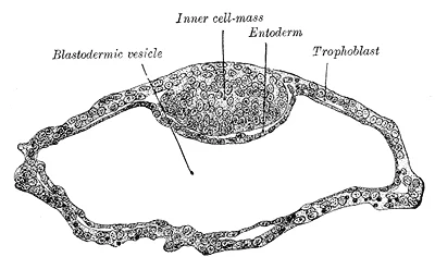
The tightly bound ball of cells now begins to secrete fluid and organizes itself around a central, fluid-filled cavity called the blastocoel[1]. At this stage, the conceptus is termed a blastocyst[1]. Within the blastocyst, a distinct group of cells forms the inner cell mass, which is fated to become the embryo, while the outer layer of cells, the trophoblast, will contribute to the placenta[1].
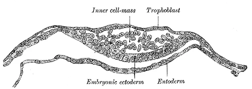
Figure 3Gray11
Long description
Section through embryonic disk of Vespertilio murinus. (After van Beneden.)

Figure 3: Section through embryonic disk of Vespertilio murinus (After van Beneden.).
Figure 4Gray10
Long description
Blastodermic vesicle of Vespertilio murinus. (After van Beneden.)

Figure 4: Blastodermic vesicle of Vespertilio murinus (After van Beneden.).
The blastocyst then implants into the uterine wall, where the trophoblasts fuse to form a syncytiotrophoblast, and the conceptus becomes enveloped by the endometrium[1]. In most cases, implantation occurs within the body of the uterus in a location that can support growth; however, in one to two percent of cases, the embryo implants outside the uterus (ectopic pregnancy) or in a region that creates complications, such as placenta previa when the placenta grows over the cervical opening[1].
At the beginning of the second week, the inner cell mass forms a two-layered disc of embryonic cells, and the amniotic cavity opens up between it and the trophoblast[1]. Cells from the upper layer, the epiblast, extend around this cavity, creating a membranous sac that becomes the amnion by the end of the second week[1]. The amnion fills with amniotic fluid, initially almost entirely a filtrate of maternal plasma, but from approximately the eighth week fetal urine adds to its volume[1]. Floating in this fluid, the embryo is protected from trauma and rapid temperature changes[1]. The chorion is the outermost membrane, surrounding all the others, and its villi extend into the endometrium to form the fetal portion of the placenta[1]. The placenta supplies oxygen and nutrients and removes carbon dioxide and other wastes, while maternal and fetal blood never commingle, preventing the mother’s cytotoxic T cells from destroying the fetus and avoiding antibody formation against fetal red cells[1].
Figure 5Gray14
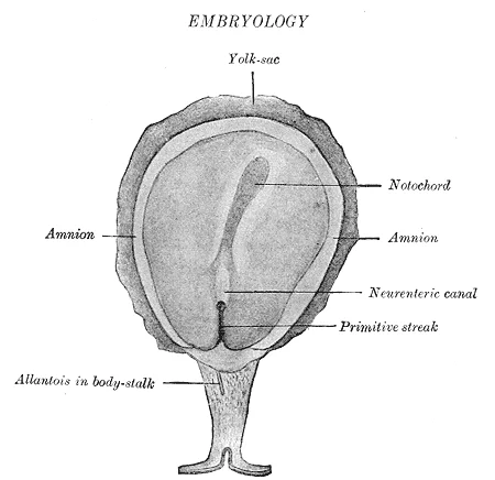
Long description
Surface view of embryo of Hylobates concolor. (After Selenka.) The amnion has been opened to expose the embryonic disk.

Figure 5: Surface view of embryo of Hylobates concolor.
Figure 6Gray12
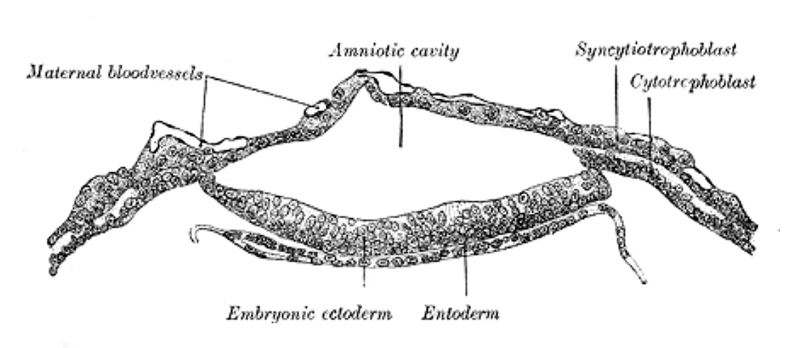
Long description
Section through embryonic area of Vespertilio murinus to show the formation of the amniotic cavity. (After van Beneden.)

Figure 6: Section through embryonic area of Vespertilio murinus to show the formation of the amniotic cavity (After van Beneden.).
The process of cleavage and blastocyst formation is tightly regulated by a complex network of molecular signals and gene expression patterns. These molecular mechanisms ensure proper cell division, differentiation, and formation of the blastocyst structure with its distinct cell lineages.
The initial cleavage divisions are driven by maternally derived mRNAs and proteins stored in the oocyte. As development progresses, zygotic genome activation occurs, marking the transition from maternal to embryonic control of development. This typically occurs at the 4-8 cell stage in humans. [1]
Several key signaling pathways are involved in regulating cleavage and blastocyst formation. The Wnt signaling pathway plays a crucial role in establishing cell polarity and regulating asymmetric cell divisions during early cleavage stages. Similarly, the BMP (Bone Morphogenetic Protein) signaling pathway is essential for proper differentiation of the trophectoderm and inner cell mass. [1]
The Hippo signaling pathway regulates cell proliferation and apoptosis during blastocyst formation. When active, it promotes apoptosis and restricts cell proliferation, while its inactivation allows for cell proliferation and blastocyst expansion. [1]
Transcription factors such as Oct4, Nanog, and Sox2 are critical for maintaining pluripotency in the inner cell mass. These factors form a regulatory network that ensures the self-renewal of embryonic stem cells. [1]
Cell adhesion molecules, particularly E-cadherin, play a vital role in maintaining cell-cell contacts during cleavage divisions. [1]
The derivatives of cleavage and blastocyst formation are fundamental to understanding early embryonic development. Totipotency lasts for only a few days before the cells' fates are set as being the precursors to a specific lineage of cells. [1]
The placenta develops from both maternal and embryonic tissues. The maternal portion of the placenta develops from the deepest layer of the endometrium, the decidua basalis. The embryonic portion forms when the syncytiotrophoblast and cytotrophoblast cells proliferate along with extraembryonic mesoderm cells to form the chorionic membrane. This membrane envelops the entire conceptus as the chorion and develops finger-like structures called chorionic villi that burrow into the endometrium. These villi, along with the cytotrophoblast cells that perforate them and remodel maternal blood vessels, constitute the fetal portion of the placenta. [1]
The embryo undergoes significant transformation during development. Beginning as a flat sheet of cells, it acquires a cylindrical shape through embryonic folding. The embryo folds laterally and at both ends, forming a C-shape with distinct head and tail ends. During this process, the embryo envelops a portion of the yolk sac, which protrudes with the umbilical cord from what will become the abdomen. This folding creates a tube called the primitive gut, lined by endoderm. The amniotic sac, which initially sits atop the flat embryo, envelops the embryo as it folds. [1]
By the end of the embryonic period, the embryo is approximately 3 cm (1.2 in) from crown to rump and weighs approximately 8 g (0.25 oz). During the sixth week, uncontrolled fetal limb movements begin, and the intestines temporarily loop into the umbilical cord due to rapid gastrointestinal development. Paddle-shaped hands and feet develop fingers and toes through apoptosis, which disintegrates the tissues between the digits. By week 7, the facial structure becomes more complex with nostrils, outer ears, and lenses. By the eighth week, the head is nearly as large as the rest of the embryo's body, all major brain structures are in place, and the external genitalia are apparent (though still indistinguishable between sexes). Bone begins to replace cartilage in the embryonic skeleton through ossification. [1]
| Embryonic Structure | Adult Derivative |
|---|---|
| Inner cell mass | All tissues of the body |
| Trophoblast cells | Placental fetal portion |
| Extraembryonic mesoderm | Chorionic membrane |
| Chorionic villi | Fetal part of placenta |
| Decidua basalis | Maternal part of placenta |
| Yolk sac | Part of umbilical cord |
| Primitive gut | Gastrointestinal tract |
| Amniotic sac | Amnion |
The yolk sac protrudes through the umbilical cord, and the amniotic sac envelops the folded embryo.
Congenital anomalies related to cleavage and blastocyst formation represent critical disruptions in the earliest stages of human development, occurring during the first week after fertilization. These anomalies, though relatively rare, have profound implications for embryonic viability and pregnancy outcomes. The precise timing and mechanisms of these disruptions determine their clinical manifestations and severity. [1]
Exam trapComplete hydatidiform mole
A condition where the blastocyst develops abnormally due to the absence of maternal chromosomes, resulting in abnormal trophoblastic proliferation and no embryonic tissue. This anomaly occurs in approximately 1 in 1,500 pregnancies and is characterized by diffuse vesicular changes in the placenta. [1]
DefinitionPartial hydatidiform mole
An anomaly where the blastocyst has both maternal and paternal chromosomes but with an abnormal number, typically 69 chromosomes instead of the normal 46, leading to abnormal development. This condition affects about 1 in 1,000 pregnancies and may contain some fetal tissues with triploid karyotype.
| Anomaly | Embryological Basis | Clinical Features |
|---|---|---|
| Complete hydatidiform mole | Absence of maternal chromosomes; abnormal trophoblastic proliferation | Uterine enlargement (greater than dates for gestational age), vaginal bleeding (in 50% of cases), elevated hCG levels (often >100,000 mIU/mL) [1] |
| Partial hydatidiform mole | Abnormal chromosome number (69 chromosomes instead of 46) | Uterine size appropriate for dates, vaginal bleeding, preeclampsia in 20% of cases, persistent trophoblastic disease in 5% [1] |
| Blastocyst arrest | Failure of the blastocyst to develop further after formation | [1] |
| Implantation anomalies | Abnormal implantation of the blastocyst | [1] |
The partial hydatidiform mole demonstrates some disorganized embryonic tissue alongside abnormal trophoblastic changes, with focal vesicular patterns.
NoteClinical significance
These anomalies highlight the critical importance of normal cleavage and blastocyst formation for subsequent embryonic development and pregnancy success. Disruptions during this early period can lead to complete pregnancy failure or the development of gestational trophoblastic disease, which requires specialized management.
Cleavage and blastocyst formation represent critical early developmental stages that form the foundation for prenatal diagnostic approaches and understanding early pregnancy complications. The blastocyst, typically formed by day 5 after fertilization, consists of an inner cell mass that will develop into the embryo and a trophoblast that contributes to the placenta. This developmental timeline is crucial for interpreting various prenatal tests and understanding when certain abnormalities might first become detectable. [1]
NoteImplantation timing
Implantation of the blastocyst normally occurs between days 6 and 10 after fertilization. [1]
Prenatal diagnosis often relies on assessing embryonic development at specific time points corresponding to these early stages. Ultrasound technology has advanced to visualize early pregnancy structures with remarkable precision. Transvaginal ultrasound can identify the gestational sac as early as 4.5-5 weeks menstrual age, which corresponds to the late blastocyst or early embryonic stage. The absence of a gestational sac at this time, when the beta-human chorionic gonadotropin (β-hCG) level is [1]
The presence of a yolk sac, visible by transvaginal ultrasound at approximately 5 weeks menstrual age, confirms an intrauterine pregnancy and indicates successful implantation. The yolk sac serves as an important landmark, as its absence when a gestational sac is larger than 8mm is associated with pregnancy failure. By 5-6 weeks, the embryonic pole should be visible, and its presence confirms a viable pregnancy. The detection of fetal heart motion, typically visible by 6 weeks menstrual age, is [1]
| Time (menstrual age) | Developmental feature | Significance | Normal parameters |
|---|---|---|---|
| 4.5-5 weeks | Gestational sac | Earliest sign of pregnancy | Mean sac diameter should match gestational age (MSD in mm = weeks + 3) |
| 5 weeks | Yolk sac | Confirms intrauterine pregnancy | Should be visible when sac is >8mm |
| 5-6 weeks | Embryonic pole | Presence confirms viable pregnancy | CRL should increase by 1mm/day |
| 6 weeks | Fetal heart motion | Indicator of embryonic viability | FHR should be 110-160 bpm |
Chromosomal abnormalities can be detected through procedures like chorionic villus sampling (CVS), which samples trophoblast cells at 10-13 weeks gestation. This analysis is particularly relevant as the trophoblast is the first tissue to differentiate and carries the same genetic makeup as the embryo. CVS can detect chromosomal abnormalities such as trisomy 21 (Down syndrome), trisomy 18, and trisomy 13 with high accuracy. This technique allows for the selection of euploid embryos, reducing the [1]
Prenatal diagnostic timeline
- 4.5-5 weeksGestational sac visible on ultrasound
- 5 weeksYolk sac appears
- Question: Concern about genetic abnormalities?
Yes
- 10-13 weeksChorionic villus sampling (CVS)
IVF treatment
- Blastocyst stagePreimplantation genetic testing
- Cleavage is the process of rapid mitotic cell divisions following fertilization, producing smaller cells called blastomeres. [1]
- The zygote undergoes cleavage as it travels down the uterine tube, forming a morula by approximately 3 days. [1]
- The morula develops into a blastocyst, consisting of an inner cell mass and a trophoblast. [1]
- The blastocyst implants into the uterine endometrium, with the trophoblast differentiating into cytotrophoblast and syncytiotrophoblast. [1]
- Gastrulation occurs during the third week, transforming the bilaminar disc into a trilaminar disc with ectoderm, mesoderm, and endoderm. [1]
- The primitive streak forms along the dorsal surface of the epiblast. [1]
- Cells migrate through the primitive streak to form the mesoderm and displace the hypoblast to form the endoderm. [1]
- Neurulation begins during the fourth week, with the neural plate folding to form the neural tube, which will develop into the central nervous system. [1]
- The notochord, derived from mesoderm, induces neural tube formation and later becomes the nucleus pulposus of intervertebral discs. [1]
- Somites form alongside the neural tube and differentiate into the axial skeleton, skeletal muscle, and dermis. [1]
- Fertilization typically occurs in the distal uterine tube within 24 hours after ovulation. [2]
- The secondary oocyte is surrounded by the corona radiata and zona pellucida, which sperm must penetrate to achieve fertilization. [2]
- The acrosomal reaction is triggered when sperm bind to receptors in the zona pellucida, releasing enzymes to digest this barrier. [2]
- Hundreds of sperm undergo the acrosomal reaction to create a path for one sperm to fertilize the oocyte. [2]
- Polyspermy is prevented by two mechanisms after the first sperm fuses with the oocyte, as triploidy is incompatible with life. [2]
- The oocyte completes meiosis II upon fertilization, ejecting a second polar body and becoming a haploid ovum. [2]
- The male and female pronuclei fuse to form the diploid zygote, initiating embryonic development. [2]
- Dizygotic twins occur when two eggs are released and fertilized in the same cycle, happening in approximately 1 percent of ovulation cycles. [2]
Exam trapConfusing fertilization with cleavage
Fertilization is the fusion of sperm and oocyte nuclei to form a zygote, while cleavage is the series of mitotic divisions that follow, producing smaller cells called blastomeres.
Exam trapMisunderstanding the zona pellucida's role
The zona pellucida is the glycoprotein layer surrounding the oocyte that sperm must penetrate. After fertilization, cortical granules release enzymes that harden this layer, forming the fertilization membrane that prevents polyspermy. [2]
Exam trapAssuming all sperm can fertilize immediately
Sperm must undergo capacitation in the female reproductive tract before gaining the ability to fertilize an oocyte. This process takes several hours and involves membrane changes necessary for enzyme release.
Exam trapOverestimating sperm survival time
While sperm can survive in the uterine tubes for 3-5 days, only a few thousand out of hundreds of millions released during ejaculation typically reach the site of fertilization. [2]
Exam trapConfusing trophoblast with embryonic tissue
The trophoblast forms the outer shell of the blastocyst and develops into the chorionic sac and fetal portion of the placenta, not the embryo proper. [1]
Exam trapMisrepresenting the three germ layers
The three germ layers (ectoderm, mesoderm, endoderm) each give rise to specific structures: ectoderm forms nervous system and epidermis; mesoderm forms skeleton and muscles; endoderm forms gut lining and organs.
NoteClinical assessment of pregnancy
Understanding blastocyst formation is crucial when assessing early pregnancy viability. The blastocyst implants approximately 6-7 days after fertilization, and failure of proper implantation can lead to early pregnancy loss.
NoteTeratogen counseling
Knowledge of embryonic development informs counseling about teratogen exposure. The period of organogenesis (weeks 3-8) is particularly sensitive, as substances like alcohol can cause fetal alcohol spectrum disorders.
NotePlacental function assessment
Understanding the development of the chorionic villi and their role in placental function is essential when evaluating placental insufficiency or abnormal placentation.
Early Embryonic Development
- FertilizationSperm penetrates oocyte, forming zygote
- CleavageRapid mitotic divisions producing smaller blastomeres
- Question: Blastocyst formation?
Yes
- Blastocyst cavity formsFluid-filled cavity with inner cell mass and trophoblast
- ImplantationBlastocyst attaches to uterine lining ~6-7 days post-fertilization
No
- Development arrestMay lead to failed pregnancy
| Event | Time Post-Fertilization | Significance |
|---|---|---|
| Fertilization | Day 0 | Formation of zygote with full genetic complement |
| Cleavage begins | Day 1-2 | Rapid mitotic divisions producing smaller cells |
| Morula formation | Day 3-4 | Solid ball of 16-32 cells |
| Blastocyst formation | Day 5-6 | Formation of blastocyst cavity with inner cell mass and trophoblast |
| Implantation | Day 6-7 | Attachment to endometrium |
| Placental development | Week 2-3 | Trophoblast differentiates into chorionic villi |
- Fertilization occurs when sperm and oocyte nuclei fuse, forming a diploid zygote
- Cleavage involves rapid mitotic divisions producing smaller cells called blastomeres
- The blastocyst consists of an inner cell mass (embryo proper) and trophoblast (future placenta)
- Capacitation prepares sperm for fertilization by improving motility and thinning the membrane
- Polyspermy is prevented by fast block (membrane depolarization) and slow block (cortical reaction) [2]
- The three germ layers (ectoderm, mesoderm, endoderm) each give rise to specific structures
- Trophoblast cells develop into the chorionic sac and fetal portion of the placenta [1]
Answer each one in your head first, then open the answer.
What is the primary function of the trophoblast?
Show answerHide answer
To form the chorionic sac and fetal portion of the placentaHow long can sperm survive in the uterine tubes?
Show answerHide answer
3-5 daysWhat prevents polyspermy after fertilization?
Show answerHide answer
Fast block (membrane depolarization) and slow block (cortical reaction)Which germ layer gives rise to the nervous system?
Show answerHide answer
EctodermWhat is the significance of the zona pellucida hardening after fertilization?
Show answerHide answer
It forms the fertilization membrane that prevents additional sperm from penetrating
- Betts JG, Young KA, Wise JA, et al. Embryonic Development. OpenStax, Anatomy and Physiology 2e (CC BY-NC-SA 4.0). Accessed .
- Betts JG, Young KA, Wise JA, et al. Fertilization. OpenStax, Anatomy and Physiology 2e (CC BY-NC-SA 4.0). Accessed .
Figure credits
- Figure 1: OpenStax College, via Wikimedia Commons, CC BY 3.0, CC BY 3.0. resized.
- Figure 2: Ambrosius Hubrecht, via Wikimedia Commons, Public domain, Public domain.
- Figure 3: Henry Vandyke Carter, via Wikimedia Commons, Public domain, Public domain.
- Figure 4: Henry Vandyke Carter, via Wikimedia Commons, Public domain, Public domain.
- Figure 5: Henry Vandyke Carter, via Wikimedia Commons, Public domain, Public domain.
- Figure 6: Henry Vandyke Carter, via Wikimedia Commons, Public domain, Public domain.
Educational content for medical students and doctors. It is not a substitute for clinical judgement or local guidelines.