Implantation
Embryonic implantation involves trophoblast invasion and hCG signalling during the third week of gestation.
Week 1
The process begins with the formation of the embryonic disc. On the ventral side of the embryonic disc, opposite the amnion, cells in the lower layer of the embryonic disk (the hypoblast) extend into the blastocyst cavity and form a yolk sac. [1]
DefinitionTrophoblast Layers
The trophoblast differentiates into the inner cytotrophoblast and the outer syncytiotrophoblast, which are critical for implantation and hormone production.
Trophoblast Cytology and Syncytiotrophoblast
Implantation is driven by specific hormonal signals produced by the trophoblast. It has long been speculated that hCG may have a role in implantation of pregnancy [-]. However, hCG is an endocrine. Hyperglycosylated hCG is an autocrine or paracrine and has been shown to directly promote implantation of pregnancy. This is seeming what was considered the hCG implantation function. [2] [3]
Clinical pearlhCG Variants
Hyperglycosylated hCG is the principal variant of hCG produced in early pregnancy. Hyperglycosylated hCG levels then dwindles to <1% of total hCG during the 2nd and 3rd trimesters of pregnancy. This is consistent with hyperglycosylated hCG having a function in promoting implantation in early pregnancy. [2]
hCG and Ectopic Implantation
The proportions of hyperglycosylated hCG molecules rapidly decline in the weeks following implantation, averaging 68% and 49% in urine and serum samples at the time of missing menses, 50% and 36% at 5 weeks of gestation, 25% and 21% at 6 weeks of gestation, and < 1.5% in the second and third trimesters of pregnancy (Table ). [3]
Exam trapEctopic Risk
Ectopic implantation can disrupt normal hCG dynamics. Total hCG concentration rises from pregnancy implantation (3rd week of gestation) to a peak at 10 weeks of gestation. hCG levels rise exponentially during the first 7 weeks of pregnancy, increasing approximately 12-fold every week or 1.52-fold every day or 2.3-fold every two days. [4] [3]
Hydatidiform Mole
Abnormal trophoblast proliferation leads to hydatidiform mole. Patients with a hydatidiform mole commonly present with unusually high serum hCG results (Table ). Regular hCG is the predominant measurable hCG that is secreted by the villous syncytiotrophoblast tissue. Hyperglycosylated hCG accounts for 4.9 ± 2.1% of the measured hCG. It is assumed that this is coming from the extravillous invasive cytotrophoblast cells on anchoring villi. Only 3.6 ± 1.7% of the total hCG of partial moles were hyperglycosylated hCG. [3]
NoteMole Complications
The biggest danger with hydatidiform mole is persistence of disease or invasion by extravillous invasive cytotrophoblast cells which can invade the uterus and other organs. [3]
The blastocyst consists of an inner cell mass and an outer layer called the trophoblast. The trophoblast differentiates into two layers: the cytotrophoblast and the syncytiotrophoblast. The syncytiotrophoblast is responsible for invading the uterine lining. This invasion is critical for establishing blood flow to the developing embryo.
Implantation begins with the apposition and attachment of the blastocyst to the endometrium. The endometrium undergoes decidualisation, a process where stromal cells enlarge and accumulate glycogen. This prepares the uterus to support the embryo. The syncytiotrophoblast erodes maternal capillaries, creating lacunae that fill with maternal blood. This establishes the hemochorial placenta.
Human chorionic gonadotropin (hCG) is produced by the trophoblast. In the first week of pregnancy, hyperglycosylated hCG is the principal or sole form of hCG produced in the first week of pregnancy, following implantation of the fetus. This variant drives extravillous invasion. [3]
| Variant | Timing | Significance |
|---|---|---|
| Hyperglycosylated hCG | First week | Principal form; drives invasion |
| Regular hCG | Later pregnancy | Dominant form; supports corpus luteum |
| β-core fragment | 7 weeks to term | Predominates in urine |
The hCG doubling test has been widely used as an indication of pregnancy failure, miscarriage or ectopic pregnancy between 4 and 7 weeks of gestation. The test involves measuring serum hCG once and them measuring hCG 48 hours later and determining if serum concentration has doubled during this 48 hour period. This is used in individuals with worry of miscarriage due to older age for pregnancy (> 40), previous history of miscarriage or infertility problems. [3]
A hydatidiform mole is a gestational trophoblastic disease. While minimally present in hydatidiform mole cases, rising proportions of hyperglycosylated hCG mark invasive hydatidiform mole (invasive mole) and development and advancement of choriocarcinoma. The remaining one third of failures are due to hydatidiform mole or genetic abnormalities. [2] [3]
In ectopic pregnancy, the blastocyst implants outside the uterus. After the hCG doubling test confirmation methotrexate is used to destroy and abort the ectopic pregnancy or a salpingectomy is performed. [3]
Management of Suspected Ectopic Pregnancy
- Suspect EctopicPatient with pain/bleeding, 4-7 weeks gestation
- Question: Ultrasound Result?
Visible Ectopic
- Confirm Diagnosis
Not Visible
- Perform hCG Doubling TestMeasure serum hCG at 0 and 48 hours
- Question: hCG Doubles?
Yes
- Likely Viable IntrauterineContinue monitoring
No/Slow Rise
- Confirm EctopicUse ultrasound and clinical context
- TreatmentMethotrexate or Salpingectomy
NoteIVF Context
Hyperglycosylated hCG is the principal form of hCG produced in the first weeks of pregnancy. If hyperglycosylated hCG is used as a pregnancy test it will only detect endogenous hyperglycosylated hCG independent of administered regular hCG. [3]
Unlike standard hCG, hyperglycosylated hCG acts as a potent promoter of cytotrophoblast cell invasion. This process mirrors the invasive behaviour seen in choriocarcinoma, where hyperglycosylated hCG specifically promotes the invasion in implantation of pregnancy, and the deep implantation of the villous placental structures that is driven by extravillous cytotrophoblast cells. This deep invasion is executed by extravillous cytotrophoblast cells. Conversely, the presence of hyperglyc [2][4]
The myometrium is the muscle layer surrounding the uterine endometrium. This deep penetration is facilitated by hyperglycosylated hCG-induced cytotrophoblastic metalloproteinases and collagenase activities. These enzymes degrade the extracellular matrix, allowing the trophoblast to invade the uterine wall. [4]
While pregnancy is traditionally dated from the last menstrual period, this introduces significant variability. For instance, in a standard 28-day cycle, implantation can occur from 12 days prior to a missed period to 2 days after. If pregnancies are dated to the day of implantation rather than the last menstrual period, the variation in hormone levels decreases significantly. Urine hCG levels at the fourth week since the last menstrual period can range from less than 0.1 ng/ml to 213 ng/ml, a variation of over 2130-fold.
Exam trapDating Variability
Dating pregnancy to the last menstrual period causes high variability in hCG interpretation; dating to implantation reduces this variation significantly.
Failure of implantation is a major cause of early pregnancy loss. Pituitary hCG may also be detected alongside LH in the menstrual cycle, but it is the placental hyperglycosylated hCG that driv [3][4]
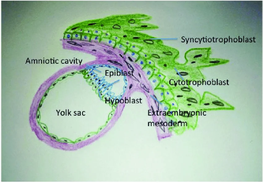
Figure 1Bilaminar-embryonic-disc-at-14-days-in-the-implantation-site-in-endometrium
Long description
Newly formed embryonic disc at site of implantation in the endometrium

Figure 1: Newly formed embryonic disc at site of implantation in the endometrium.
| Embryonic Structure | Adult Derivative |
|---|---|
| Ectoderm | Epidermis, nervous system, hair, nails, lens of eye, enamel of teeth |
| Mesoderm | Muscles, bones, connective tissue, cardiovascular system, urinary system, reproductive system |
| Endoderm | Lining of digestive tract, respiratory tract, liver, pancreas, bladder |
This disc consists of two layers: the epiblast (superficial layer) and the hypoblast (deep layer). The epiblast gives rise to all three germ layers (ectoderm, mesoderm, and endoderm) through the process of gastrulation, while the hypoblast contributes to extraembryonic structures like the yolk sac.
The disc is positioned between the amniotic cavity superiorly and the primary yolk sac inferiorly. The epiblast forms the upper layer of the disc, composed of columnar cells, while the hypoblast forms the lower layer, made of cuboidal cells. Between these two layers is the basement membrane, known as the membrane of Rauber. The disc is surrounded by the cytotrophoblast and syncytiotrophoblast, which will develop into the placenta. The bilaminar disc represents the beginning of embryonic proper, with all future body structures developing from these two layers through the processes of folding and organogenesis.
The process of embryonic folding transforms the flat bilaminar disc into a cylindrical embryo, establishing the basic body plan. This folding occurs in both the horizontal (transverse) and vertical (sagittal) planes, bringing the embryo away from the yolk sac and creating the primitive gut tube. The folding process is essential for the proper formation of organ systems and marks the transition from the embryonic to the fetal period of development.
Implantation itself is a critical event where the blastocyst adheres to the uterine wall via trophoblast cells, embedding itself in the uterine lining. This process typically occurs in the fundus of the uterus or on the posterior wall. However, if the endometrium is not fully developed, the blastocyst may detach and seek a better site. This high rate of failure explains why pregnancy often requires several ovulation cycles to achieve. [1]
Following implantation, embryonic cells undergo gastrulation, in which they differentiate and separate into an embryonic disc and establish three primary germ layers (the endoderm, mesoderm, and ectoderm). Through the process of embryonic folding, the fetus begins to take shape. Neurulation starts the process of the development of structures of the central nervous system and organogenesis establishes the basic plan for all organ systems. [1]
The heart also begins its development in the embryo as a tube-like structure, connected via capillaries to the chorionic villi. Cells of the primitive tube-shaped heart are capable of electrical conduction and contraction. During weeks 4–5, the eye pits form, limb buds become apparent, and the rudiments of the pulmonary system are formed.
Clinical pearlClinical Correlation
Implantation can be accompanied by minor bleeding, which may be mistaken for menstruation.
NoteDiagnostic Note
Hyperglycosylated hCG free β is produced at the time of implantation in very early pregnancy, and at very low proportions throughout the length of pregnancy. [3]
DefinitionGastrulation
The process by which the single-layered blastocyst is reorganized into a multilayered structure known as the gastrula.
Implantation Outcomes
- Blastocyst contacts uterine wallAdhesion via trophoblast cells
- Question: Is endometrium fully developed?
Yes
- Implantation succeedsEmbryo embeds in lining
No
- Blastocyst detachesFinds better spot or sheds
Congenital anomalies of implantation are rare but profound disruptions in the establishment of the uteroplacental unit. To understand these anomalies, one must first grasp the normal sequence: trophoblast differentiation, invasion, and vascular remodelling. In this sagittal section of the early placenta, the chorionic plate lies at the top, facing the amniotic cavity. Below it, the villous tree extends into the intervillous space. The key labelled structure is the syncytiotrophoblast, which forms the outer multinucleated layer of the floating villi, directly bathing in maternal blood. Beneath this lies the cytotrophoblast shell, which anchors the villi to the decidua. The spiral arteries, originating from the myometrium, are shown penetrating the decidua and opening into the intervillous space. This arrangement ensures that fetal villi are bathed in maternal blood, facilitating efficient gas and nutrient exchange. The orientation of the figure is anterior-posterior, with the maternal side on the right and the fetal side on the left.
Just a few days after implantation, the trophoblast has secreted enough hCG for an at-home urine pregnancy test to give a positive result. However, not all hCG is identical. This molecule is dramatically different from regular hCG and is essential for deep invasion. [1][3]
Anomalies arise when this invasion is either excessive or insufficient. Excessive invasion leads to placenta accreta spectrum disorders, where the trophoblast fails to stop at the decidua basalis. Insufficient invasion results in pre-eclampsia and intrauterine growth restriction, due to shallow implantation and poor spiral artery remodelling. The variation in hCG production is critical here. That is hCG daily increase rate in the first 4 weeks following implantation. hCG daily increase rate was measured in 82 women (all results from women with biochemical or miscarried pregnancies removed) with term outcome pregnancies, from day of implantation for 28 days. The increase rate per day was averaged over the 28 days. The increase rate per day ranged from 1.52-fold per day to 2.92-fold per day among the 82 women. If this is considered over 7 days then it is equivalent to 1.527 and 2.927 or to an increase of 18.7-fold vs. an increase of 1810-fold. Individual hCG daily amplification rate is also a major cause of variation. [2]
The structural basis for this functional difference lies in the glycosylation of the hCG molecule. Variations are found in the N-linked and O-linked oligosaccharide structures on hCG with differences in cellular metabolism and different expression of glycosyltransferase activities (18,19,36). There are 2 principal types of O-linked oligosaccharides (Figure ), trisaccharide and hexasaccharide. While first trimester normal pregnancy urine hCG contained 12.3 to 19% of the hexasaccharide sugar structure (regular hCG, n = 6 individuals, mean = 15.6%), choriocarcinoma urine hCG contains 60 to 100% hexasaccharide structures (hyperglycosylated hCG, n = 6 individuals, mean = 74.2%), 5-fold more than first trimester pregnancy. A smaller difference was observed with the 4 N-linked oligosaccharide structures on pregnancy and choriocarcinoma molecules. While 6 first trimester pregnancy samples contained an average of 10.3% triantennary structures at the four N-linked sites on hCG, 5 choriocarcinoma cases contained 33% of the triantennary structures (3-fold more than first trimester pregnancy). The largest difference between regular hCG and hyperglycosylated hCG is at the O-linked oligosaccharides. [3]
Researchers found a greater fucose content on N-linked oligosaccharides in cancer cases. They demonstrated site-specific difference in O-linked oligosaccharides. They showed the constant presence of predominantly hexasaccharide structures in pregnancy and choriocarcinoma at Serine residue 121 [68–89%], with variable structures on Ser 127, 132 and 138, primarily trisaccharide structures on regular hCG in pregnancy and hexasaccharide structures on hyperglycosylated hCG in choriocarcinoma. [3]
In first trimester pregnancy serum, 9% of hCG dimer molecules are nicked, all free β molecules are also nicked, in the last 2 months of gestation 21% of dimer molecules are nicked and all free β molecules are nicked. In pregnancy urine samples, β-subunit core fragment concentration or the degradation end product concentration is relatively small in the first weeks of gestation, it equals hCG dimer concentration at 6–7 weeks of pregnancy. β-core fragment concentrations then exceeds dimer concentrations in urine thereafter, 7 weeks to term. [3]
The clinical utility of these distinctions is evident in screening. In the late 1990s serum markers were first used for first trimester Down syndrome screening, with chorionic villous sampling used to determine cytogenetics in those indicated at risk. Use of hCG free β-subunit was found preferable to regular hCG. The combination of ultrasound nuchal translucency with laboratory measurement of free β-subunit and pregnancy-associated plasma protein-A (PAPP-A) became a standard combination of tests to assess risk for Down Syndrome fetus. In 2003, hyperglycosylated hCG was used to replace free β-subunit and to improve the sensitivity of the test combination. [3]
The evolutionary origin of this system highlights its importance. I first named it "invasive trophoblast antigen" because we knew at that time that it independently drove implantation of pregnancy and invasion by choriocarcinoma cells. Within 2 years I received an official letter from the World Health Organization instructing me to rename it a molecule containing the name hCG, because it contains hCG amino acid sequence. I renamed it hyperglycosylated hCG based on its structure. Can we prove that hCG and hyperglycosylated hCG drive hemochorial placentation in humans. No, the research would be unethical. The only evidence that hCG related molecules are the driving force of hemochorial placentation is the first appearance of invasive hemochorial placentation in early simian primates. Primates first evolved 80 million years ago, early simian primates evolved just 37 million years. Interestingly, chorionic gonadotropin (CG) and hyperglycosylated CG first evolved in this same species. The logical reason that hemochorial placentation developed is the parallel evolution of its driving signal CG and hyperglycosylated CG. [4]
However, the lack of pure or clinically useful preparations of [3]
Human chorionic gonadotropin (hCG) is secreted by the syncytiotrophoblast, which forms when superficial trophoblast cells fuse after the blastocyst adheres to the endometrium. This hormone directs the corpus luteum to survive and produce progesterone and estrogen, suppressing menses. Serum hCG concentrations rise logarithmically from implantation to a peak at 10 weeks of gestation, then fall to about one fifth of peak levels and remain there to term. Hyperglycosylated hCG is very prominent in the first 2–3 weeks following implantation but becomes a relatively minor component thereafter. [3] [4]
As preeclampsia is the biggest cause of maternal death in pregnancy, new hyperglycosylated hCG prenatal screening tests may identify women at high risk for these conditions in the first and second trimesters. [4]
Problems exist with current standards for hCG measurement. While standards are available for nicked hCG, no standard was made available by the WHO for hyperglycosylated hCG, a key placental molecule, due to the absence of a reliable source. [3]
1. Implantation Site and Complications Most embryos implant within the body of the uterus, a location capable of supporting growth. However, in one to two percent of cases, the embryo implants either outside the uterus (an ectopic pregnancy) or in a region of uterus that can create complications for the pregnancy. [1]
2. hCG Structure and Subunits The hormone hCG comprises an α-subunit and a β-subunit. The β-subunit of hCG, while structurally somewhat similar to the β-subunit of LH, differentiates hCG, hyperglycosylated hCG, and pituitary hCG from other molecules. [2]
Receptor Binding and Half-Life** Both hCG and LH bind and function through a common hCG/LH receptor. [2]
4. Trophoblast Differentiation hCG and hyperglycosylated hCG seemingly act to together to promote the growth and differentiation of trophoblast cells or formation of the placenta villous structures. They seemingly start their action early with blastocyst signaling of the endometrium of forthcoming implantation. Hyperglycosylated hCG then promotes implantation and growth of cytotrophoblast cells. hCG promotes the differentiation of cytotrophoblast cells to syncytiotrophoblast cells and so the villous structures which are mixture of the two cell types are formed. [2] [4]
5. Vascular and Maternal Effects hCG also promotes the uterine vasculature to maximally provide blood to the hemochorial placentation structure. Preeclampsia is one of the most life-endangering complication of pregnancy and occurs uniquely in humans with the fetal demand for deep implantation and the oxygen and nutritional mechanisms needed to support brain development. [2] [3]
6. Hyperglycosylated hCG and Apoptosis This exposes other receptor binding site on hyperglycosylated hCG. Common regions include a transforming growth factor beta (TGFβ)/platelet-derived growth factor (PDGF)/Nerve growth factor (NGF) common cystine-knot related structure. [2]
**7. It is inferred that the high incidence of pregnancy failures, in humans, is a consequence of deficient hyperglycosylated hCG. [3]
8. Diagnostic Testing Principles The principal of the immunometric test is one antibody (called the capture antibody) binding one site on hCG and immobilizing it and a second antibody labeled with enzyme tracer (called a tracer antibody) binding a distant site on hCG labeling the immobilize complex. These mechanisms are the basis of the automated tests used in all commercial laboratories today. [3]
**9. There is also no requirement to detect free β-subunit or its degradation derivatives. [3]
10. Molecular Uniqueness This is a unique biological combination, 3 molecules differing in carbohydrate structure. A common question is why are the 5 independent variants of hCG all called hCG. This is because they all share a common α-subunit and ß-subunit amino acid sequence. [3] [4]
Students frequently conflate the different molecular variants of human chorionic gonadotrophin (hCG), leading to errors in interpreting clinical markers and understanding placental physiology. A primary error is assuming all hCG acts as a systemic hormone. Regular hCG is produced by differentiated syncytiotrophoblast cells and functions as a hormone to promote progesterone production by ovarian corpus luteal cells. In contrast, hyperglycosylated hCG is not a hormone but an autocrine factor acting on cytotrophoblast cells to promote cell growth and invasion during implantation. Confusing these roles leads to incorrect assumptions about how the placenta establishes itself. [2]
Another significant pitfall involves the interpretation of early pregnancy tests. Students often assume a positive test at the time of missed menses confirms a viable intrauterine pregnancy. These failures cause a brief incline of serum and urine hCG concentrations which may cause a pregnancy test to misleadingly be positive at the time of missing menses. This transient hCG can in some cases extend to 4 day after the time of missing menses. Relying on a single early test ignores the possibility of a false positive due to early loss or a false negative due to late implantation. [3]
The utility of the hCG doubling test is another area of frequent misunderstanding. The concept arose when tests detected all variants equally, but modern automated tests poorly detect hyperglycosylated hCG and free β-subunit. Consequently, the hCG doubling test has become less appropriate, yet it remains widely used. The USA hCG Reference Service considers this test a very vague indication (1 in 4 detection of 1 in 5.5 pregnancy detections). Students should not treat rising hCG levels as a definitive marker of viability without considering the limitations of the assay.
Differentiating between trophoblastic diseases is critical for prognosis. Choriocarcinoma is characterised by predominant cytotrophoblast cells producing hyperglycosylated hCG. This variant is a potent marker for these malignancies. Conversely, placental site trophoblastic tumour (PSTT) is a malignancy of intermediate trophoblast cells and does not produce hyperglycosylated hCG. PSTT is very rare, accounting for 1 in approximately 40,000 pregnancies. It carries a poorer prognosis, with a five year survival rate for PSTT is 74% , compared with 91% for choriocarcinoma. Mistaking PSTT for choriocarcinoma leads to incorrect prognostic counselling and management. [3]
Finally, students often overlook the biological waste products of trophoblast activity. Extravillous invasive cytotrophoblast cells make an excess of α-subunit which is secreted as free α-subunit and O-glycosylated free α-subunit. It is noteworthy that free α-subunit has no known biological function, it is a biological waste product. Attributing clinical significance to free α-subunit levels is erroneous. Similarly, hCG free β-subunit has a circulating 1/2-life of just seconds before it is degraded. Its detection in serum is limited to 30% of cases despite production by advanced cancers.
Knowledge of hCG variants is applied at the bedside to interpret early pregnancy outcomes accurately. The clinician should explain that transient hCG can in some cases extend to 4 day after the time of missing menses. This prevents unnecessary anxiety when a subsequent scan shows no gestational sac.
In the context of gynaecological oncology, identifying the specific hCG variant guides diagnosis and monitoring. Hyperglycosylated hCG is a potent marker for choriocarcinoma and testicular germ cell malignancies. In Down syndrome pregnancies, trisomy 21 limits the differentiation of villous cytotrophoblast cells leading to an accumulation of these cells and hyperglycosylated hCG. This is apparently due to a failure of regular hCG-promoted differentiation. It is for this reason that hyperglycosylated hCG is a marker of Down syndrome in the first and second trimester of pregnancy. Serum screening protocols utilise this marker to assess risk. [3]
Imaging and surgical planning benefit from distinguishing between trophoblastic pathologies. PSTT presents as a tumor in the uterus, which is readily removed by hysterectomy. Unlike choriocarcinoma, PSTT does not produce hyperglycosylated hCG. Recognising this absence helps differentiate PSTT from other gestational trophoblastic diseases on biochemical profiling. The poorer prognosis of PSTT, with a five year survival rate for PSTT is 74% , compared with 91% for choriocarcinoma, necessitates careful surgical assessment. [3]
Examinations frequently test the physiological role of hCG variants in implantation. Regular hCG promotes myometrial spiral artery angiogenesis, aiding nutrient supply to the placenta. Hyperglycosylated hCG controls placental invasion and implantation. Super-hCG raised the circulating 1/2-life to an incredible 36 hours, or to a biopotency of 109X over prosimian LH. It is this super hCG and super-hyperglycosylated hCG that drove implantation to as deep as 1/3rd the thickness of the myometrium. This deep implantation drove hemochorial placentation to the extreme of efficiency. [4]
DefinitionHyperglycosylated hCG
A sugar variant of hCG made by root cytotrophoblast cells or extravillous cytotrophoblast cells as pregnancy progresses.
| Variant | Source Cell | Primary Function | Clinical Marker For |
|---|---|---|---|
| Regular hCG | Syncytiotrophoblast | Promotes progesterone production | Normal pregnancy maintenance |
| Hyperglycosylated hCG | Extravillous cytotrophoblast | Autocrine growth and invasion | Choriocarcinoma, Down syndrome |
| Free α-subunit | Extravillous cytotrophoblast | None (biological waste) | Not clinically significant |
| Free β-subunit | Advanced cancers | Invasion promotion | Detected in 30% of malignancies |
Research now shows that there are at least 4 independent variants of hCG, each produced by different cells with separate biological functions. This complexity means that hCG is not a single entity but a group of molecules each with different functions. Regular hCG is a hormone produced by syncytiotrophoblast cells which promotes myometrial spiral artery angiogenesis. Hyperglycosylated hCG is an autocrine factor made by extrauterine invasive cytotrophoblast cells in pregnancy. This form of hCG controls placental invasion and implantation.
The finding that the Siemen's assay is the most appropriate is strictly the result of blind unbiased testing at multiple laboratories. This highlights the importance of assay selection when measuring specific variants. For instance, modern automated hCG test that poorly detect hyperglycosylated hCG and free β-subunit. This limitation affects the reliability of the hCG doubling test.
Clinical pearlPSTT Prognosis
The five year survival rate for PSTT is 74% , compared with 91% for choriocarcinoma.
Regular hCG is produced by differentiated syncytiotrophoblast cells and promotes progesterone production. Hyperglycosylated hCG is an autocrine factor promoting cell growth and invasion. [2]
Transient hCG can in some cases extend to 4 day after the time of missing menses. Hyperglycosylated hCG is a marker of Down syndrome in the first and second trimester of pregnancy. PSTT does not produce hyperglycosylated hCG. The five year survival rate for PSTT is 74% , compared with 91% for choriocarcinoma. Super-hCG drove implantation to as deep as 1/3rd the thickness of the myometrium.
Answer each one in your head first, then open the answer.
Which cell type produces regular hCG that promotes progesterone production?
Show answerHide answer
Differentiated syncytiotrophoblast cellsWhat is the biological function of free α-subunit?
Show answerHide answer
It has no known biological function, it is a biological waste productWhy is hyperglycosylated hCG a marker for Down syndrome?
Show answerHide answer
Trisomy 21 limits the differentiation of villous cytotrophoblast cells leading to an accumulation of these cells and hyperglycosylated hCGWhat is the five year survival rate for PSTT?
Show answerHide answer
74%How deep did super-hCG drive implantation?
Show answerHide answer
1/3rd the thickness of the myometrium
- Betts JG, Young KA, Wise JA, et al. Embryonic Development. OpenStax, Anatomy and Physiology 2e (CC BY-NC-SA 4.0). Accessed .
- Cole LA. Biological functions of hCG and hCG-related molecules. Reproductive biology and endocrinology : RB&E 2010. Accessed .
- Cole LA. New discoveries on the biology and detection of human chorionic gonadotropin. Reproductive biology and endocrinology : RB&E 2009. Accessed .
- Cole LA. hCG, the wonder of today's science. Reproductive biology and endocrinology : RB&E 2012. Accessed .
Figure credits
Educational content for medical students and doctors. It is not a substitute for clinical judgement or local guidelines.