Gastrulation
The process by which the bilaminar disc transforms into a trilaminar embryo with three distinct germ layers.
Gastrulation is the pivotal embryological event that establishes the body plan by converting the bilaminar embryonic disc into a trilaminar structure. This process is initiated by the appearance of the primitive streak on the epiblast surface. The timeline of this critical phase typically spans from the beginning of the third week to the end of the third week of human development. During this period, the notochord forms as a midline structure that serves as the primary axis for subsequent neurulation. The primary outcome is the generation of the three definitive germ layers: the ectoderm, mesoderm, and endoderm. These layers give rise to all tissues and organs in the adult body.
DefinitionGastrulation
The process by which the single-layered blastula is reorganized into a multilayered structure known as the gastrula, establishing the three primary germ layers.
The primitive streak appears as a linear thickening of the epiblast cells. Cells within the epiblast undergo an epithelial-to-mesenchymal transition (EMT) and ingress through the primitive streak. The first cells to ingress displace the underlying hypoblast to form the definitive endoderm. Subsequent ingressing cells fill the space between the epiblast and the newly formed endoderm to create the mesoderm. The epiblast cells that remain behind become the ectoderm. This sequential ingress ensures the proper spatial arrangement of the germ layers.
NoteClinical Correlation
Abnormalities in primitive streak regression can lead to sacrococcygeal teratoma, a tumour arising from pluripotent cells left behind at the caudal end.
The notochord develops from cells that ingress through the primitive node, the thickened anterior end of the primitive streak. These cells migrate cranially to form a solid cord of cells known as the notochordal process. The notochord induces the overlying ectoderm to thicken and form the neural plate, initiating neurulation. It also serves as a mechanical axis for the developing embryo.
Exam trapTeratoma Origin
Sacrococcygeal teratomas arise from remnants of the primitive streak, not from the notochord or neural tube.
While gastrulation establishes the germ layers, other structures have distinct embryological origins. For instance, craniopharyngiomas are tumours derived from remnants of Rathke’s pouch. Rathke’s pouch is an upward invagination of the oral ectoderm (stomodeum) that eventually forms the anterior pituitary gland. This is distinct from the neural ectoderm formed during gastrulation. Understanding these distinct origins is crucial for differential diagnosis.
| Germ Layer | Primary Derivatives |
|---|---|
| Ectoderm | Epidermis, nervous system, lens of eye |
| Mesoderm | Muscle, bone, circulatory system, kidneys |
| Endoderm | Lining of gut, respiratory tract, liver, pancreas |
The formation of the mesoderm is not uniform. Cells ingressing at the anterior end of the streak form the prechordal plate and head mesoderm. Cells ingressing at the middle form the paraxial mesoderm, which gives rise to somites. These somites differentiate into sclerotome (vertebrae), myotome (muscle), and dermatome (dermis). Cells ingressing at the lateral and posterior regions form the lateral plate mesoderm and extraembryonic mesoderm. The extraembryonic mesoderm contributes to the formation of the chorion and the connecting stalk, supporting the embryo’s attachment to the placenta.
Clinical pearlNotochord Function
The notochord induces neural tube formation and later degenerates, with remnants persisting as the nucleus pulposus of intervertebral discs.
The precision of gastrulation is vital. Errors in the timing or location of cell ingression can lead to severe congenital anomalies. The primitive streak must regress completely by the end of the fourth week. Failure of this regression results in caudal dysgenesis or teratomas. The ectoderm, once the other layers have formed, thickens to form the neural plate, marking the transition from gastrulation to neurulation. This seamless transition ensures the continuity of embryonic development.
Figure 1Gastrulation
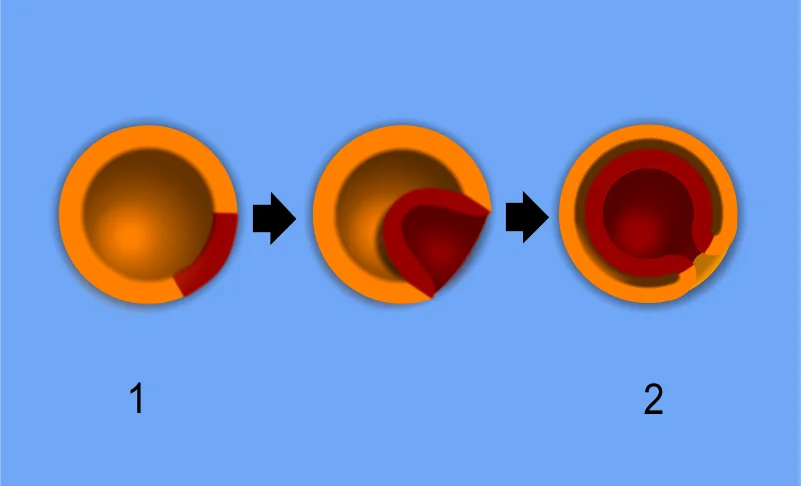
Long description
A scheme of embryo gastrulation. Labelled illustration from Wikimedia Commons, used under its open licence.

Figure 1: A scheme of embryo gastrulation.
The outermost layer is the epiblast, a pluripotent epithelial cell layer. Beneath it lies the yolk mass, which provides mechanical and topological constraints. The central focus is the primitive streak, a linear thickening on the epiblast surface. Cells within the epiblast undergo ingression, moving through the streak to form internal layers. The anterior end of the streak marks the future head region, while the posterior end extends towards the tail.
Gastrulation is a fundamental morphogenetic programme where the embryo changes from a single one-dimensional epithelial cell layer, known as the blastula, into an organised three-dimensional gastrula. This structure consists of the germ layers: ectoderm, mesoderm, and endoderm in triploblastic species. The process arises from coordinating characteristic epithelial cell behaviours, including division, shape change, and cell rearrangement, as well as cell ingression or abandonment of the layer. [2]
Early embryonic development involves the separation of embryonic and extraembryonic tissues and the establishment of anterior-posterior polarity. These initial events occur before the egg is laid and are less accessible to direct experimentation. The determination of avian embryonic territories and their patterning happens before gastrulation begins. Despite progress, mechanistic questions remain regarding the signals controlling differentiation and the molecular mechanisms establishing polarity. [1]
The topology of germ layers is specified and reorganised during this phase. Surprisingly, gastrulation morphology is highly variable across the animal kingdom, indicating it is not rigidly constrained by evolution. Experimental results show that alternative gastrulation modes can be generated in single organisms, such as in early cnidarian, arthropod, and vertebrate embryos. This plasticity suggests gastrulation is a highly evolvable process. [2]
In species with large yolk volumes, mechanical constraints alter the process. The increase in yolk necessitates adaptations, leading to meroblastic cleavages and discoidal embryos that sit on top of the yolk. This large yolk impedes complete internalisation by epiboly, blocking blastopore closure. Increases in yolk size are a significant evolutionary force, constraining the embryo into a two-dimensional discoidal shape with extra-embryonic tissues outside. These constraints disappear after gastrulation, allowing for the conservation of the pharyngula stage among distinct phyla. [2]
Cell dynamics within the primitive streak are critical. From initial stages, individual cell ingressions occur throughout the epiblast, potentially contributing to tissue fluidisation. Later, during extension stages (HH2–3), ingressions become prominent in the streak, creating a tissue sink that pulls more cells. Cell proliferation is fast, with a cell cycle of around 6 hours during early gastrulation. Most of the molecular toolkit for epithelial layers is present in sponges, preceding metazoan evolution. However, true gastrulation with segregated embryonic layers appears in eumetazoans. Genetic evidence indicates that robust phenotypes in gastrulation allow for greater genotypic diversity and evolvability. [1]
Gastrulation is driven by a complex interplay of signalling pathways that establish cell competence and induce differentiation. The formation of the primitive streak is not random but depends on specific molecular cues. The competence of the marginal zone (MZ) to respond to GDF3 is determined by WNT8A expression. Immediately after laying, WNT8A is initially expressed in the area opacum but later is restricted to the posterior marginal zone and the primitive streak as the mesendoderm differentiates. The co-expression of GDF3 and WNT1 can initiate the formation of the primitive streak even far from the marginal zone, suggesting that WNT signalling makes the cells competent for the formation of the primitive streak, while TGFβ signalling works as an inducer. The posterior marginal zone does not contribute to the embryonic tissues, only having a role inducing the mesoderm which arises where a TGFβ and WNT signalling converge. These findings suggest that the posterior marginal zone is analogous to the Nieuwkoop centre of amphibians. [1]
Fibroblast Growth Factor (FGF) signalling plays a prominent role in mesendoderm differentiation. At the early stages of development, FGF4/8 is expressed in the posterior marginal zone and the hypoblast; whereas later, FGF4/8 is produced by cells in the streak. The inhibition of FGF signalling prevents the formation of the mesendoderm and blocks gastrulation movements, while ectopic FGF can induce an extra streak. Furthermore, the ubiquitous addition of FGF results in the differentiation of a ring of mesendoderm precursors all along the marginal zone, presumably where FGF coincides with WNT secreted by the area opacum. [1]
DefinitionPlanar Cell Polarity
The non-canonical WNT-dependent Planar Cell Polarity (PCP) signalling pathway has been implicated into the control of convergent-extension behaviour during gastrulation in amphibians and fish. [1]
PDGF signalling is also required for functional N-cadherin expression, through regulation at the posttranslational level, suggesting that PDGF signalling facilitates the collective movement of mesoderm cells through the control of the expression of N-cadherin on migrating cells. [1]
The area opacum is located anteriorly and expresses WNT8A initially. The posterior marginal zone is located at the posterior end and expresses WNT8A later. The primitive streak forms in the posterior marginal zone. The hypoblast lies beneath the epiblast and expresses FGF4/8 early on. The mesendoderm differentiates from the primitive streak. The labels clearly identify the area opacum, posterior marginal zone, primitive streak, hypoblast, and mesendoderm.
NoteEvolutionary Context
Paradoxically, avian gastrulation presents more similarities with the mammals than with the other sauropsids (reptiles) probably as a result of convergent evolution. Reptiles, instead, internalise the mesendoderm through the blastoporal canal, where the cells roll-in under the epiblast (amphibian-like involution) in the anterior part, and ingress as individual cells in the posterior part, the primitive plate (similar to the primitive streak in birds and mammals). [1]
At the posterior midline, the primitive streak forms as a macroscopic convection-like flow involves up to 50,000 epithelial cells in the epiblast. Mesendoderm precursors are located into the midline of the epiblast to form the primitive streak. These cells acquire a mesenchymal phenotype and ingress into the embryo. [1]
The three germ layers arise from this process. Gastrulation is a morphogenetic programme that needs to accomplish three objectives: the generation of the main body axis, the patterning of the germ layers and the internalisation of the mesodermal and endodermal precursors[1]. In amniotes, mesoderm precursors acquire a mesenchymal phenotype and ingress into the embryo as individual cells[1]. After ingression, cells migrate laterally to form the mesoderm and endoderm. [2]
| Embryonic Structure | Adult Structure / Derivative |
|---|---|
| Ectoderm (Surface) | Epidermis, hair, nails, lens of eye |
| Neuroectoderm | Central nervous system, retina |
| Neural Crest | Peripheral nervous system, melanocytes, adrenal medulla |
| Mesoderm (Axial) | Notochord (disappears, contributes to nucleus pulposus) |
| Mesoderm (Paraxial) | Somites (skeletal muscle, vertebrae, dermis) |
| Mesoderm (Intermediate) | Kidneys, gonads |
| Mesoderm (Lateral Plate) | Somatic splanchnic mesoderm (body wall, limb bones); Splanchnic mesoderm (gut wall, heart) |
| Endoderm | Epithelial lining of gut and respiratory tract |
| Primitive Streak remnants | Sacrococcygeal teratoma |
|
The notochord is derived from the axial mesoderm. It serves as a primary axis for the embryo. The comparison of morphogenetic events and cell fate between embryos of different species suggests that embryonic mesoderm (axial, paraxial, intermediate and lateral plate) is internalised through the blastoporal canal[1]. The extra-embryonic mesoderm is internalised by cell ingression in the blastoporal plate[1]. [2]
Sacrococcygeal teratoma (SCT) arises from remnants of the primitive streak. The sacrococcygeal region also corresponds to the primitive streak and Hensen’s node in early embryonic development. The pathogenesis of SCT should be understood in the broad context of GCT development. All GCTs have been thought to be derived from primitive germ cells (germ cell hypothesis). The hypothesis explains the peculiar anatomical distribution of extragonadal GCTs along the midline of the body. Primordial germ cells (PGCs) migrate from the yolk sac to their final destinations in the gonadal ridges by passing the hindgut where SCT can arise. However, research on embryos and embryonic stem (ES) cells revealed that teratomas could be derived from ES cells, making ES cells candidate cells for developing into SCT. Stem cells residing in extraembryonic tissues can also induce teratoma formation, adding other candidate cells to the possible origins of SCT. [3]
Clinical pearlClinical Pearl
Sacrococcygeal teratomas originate from pluripotent cells remaining at the site of the primitive streak, typically presenting as a mass at the base of the coccyx.
The neural crest cells migrate extensively. They derive from the neuroectoderm at the border of the neural plate. These cells give rise to diverse structures including the peripheral nervous system and melanocytes. The surface ectoderm remains on the exterior, forming the epidermis and its appendages. The endoderm forms the lining of the gastrointestinal and respiratory tracts. The mesoderm subdivides into paraxial, intermediate, and lateral plate regions, each with distinct fates. Paraxial mesoderm forms somites. Intermediate mesoderm forms the urogenital system. Lateral plate mesoderm splits into somatic and splanchnic layers, forming the body wall and gut-associated structures respectively.
The ectoderm is dorsal. The endoderm is ventral. The mesoderm lies between them. The notochord is central. The primitive streak is posterior. Understanding these derivatives is essential for diagnosing congenital anomalies and tumours such as sacrococcygeal teratoma.
Gastrulation establishes the three primary germ layers, and errors in this process can lead to significant congenital anomalies. The most common anomalies involve the urogenital system, such as hydronephrosis, which can be considered as a consequence of compression by the tumor mass. Although the proportion of congenital anomalies is higher than that of the normal population (3–4%), causative associations are rare except for the Currarino triad. [3]
The Currarino triad is a very rare syndrome and consists of sacral bony defects, anorectal anomalies, and a presacral mass. Approximately 40% of the presacral masses in the Currarino triad are benign teratomas. The Currarino triad is an autosomal dominant syndrome linked to mutations in the HLXB9 gene. [3]
Clinical pearlCurrarino Triad
Remember the triad: Sacral defect, Anorectal malformation, and Presacral mass. It is linked to HLXB9 mutations.
Anterior to the sacrum lies the presacral mass, which may appear as a cystic or solid structure.
| Anomaly | Embryological Basis | Key Feature |
|---|---|---|
| Sacrococcygeal Teratoma | Abnormal primitive streak activity | Presacral mass |
| Currarino Triad | HLXB9 mutation | Sacral defect, anorectal anomaly, presacral mass |
| Hydronephrosis | Compression by tumor mass | Urogenital system involvement |
| Syndrome | Inheritance | Genetic Link |
|---|---|---|
| Currarino Triad | Autosomal dominant | HLXB9 gene mutation |
DefinitionSacrococcygeal Teratoma
A tumour arising from remnants of the primitive streak, located at the base of the coccyx.
The embryological basis for these anomalies lies in the regulation of the primitive streak. The primitive streak is the site of cell ingression during gastrulation. Abnormalities in the formation or regression of the primitive streak can lead to the persistence of pluripotent cells, resulting in teratomas. The Currarino triad highlights the importance of genetic regulation in sacral development. The HLXB9 gene plays a crucial role in the development of the sacrum and anorectal region. Mutations in this gene disrupt the normal patterning, leading to the characteristic triad of defects.
Understanding these anomalies requires knowledge of the normal gastrulation process. The primitive streak forms at the posterior end of the embryo. Cells ingress through the streak to form the mesoderm and endoderm. If this process is disrupted, cells may remain in the presacral region, forming a teratoma. The association with urogenital anomalies suggests a broader impact on midline development. Hydronephrosis, for example, may result from mechanical compression by the growing mass, rather than a direct developmental defect of the kidney itself.
Exam trapCommon Misconception
Do not assume all presacral masses are malignant. In the context of Currarino triad, 40% are benign teratomas.
The clinical presentation of these anomalies varies. Patients with SCT may present with a visible mass at the base of the spine. Larger masses can cause urinary or bowel obstruction, leading to hydronephrosis or constipation. The Currarino triad may be diagnosed prenatally via ultrasound or postnatally due to the presence of a sacral defect or anorectal malformation. Genetic counselling is important for families with a history of Currarino triad due to its autosomal dominant inheritance.
Management depends on the type and size of the anomaly. Surgical resection is often required for SCT to prevent complications such as malignancy or obstruction. In Currarino triad, management is multidisciplinary, addressing the sacral defect, anorectal malformation, and presacral mass. Early diagnosis and intervention are crucial for improving outcomes.
The sacral defect is located posteriorly, while the presacral mass lies anterior to it. The anorectal anomaly involves the distal bowel, which is displaced from its normal position. Understanding these relationships is essential for surgical planning and radiological interpretation.
NoteClinical Relevance
Always screen for associated urogenital anomalies in patients with presacral masses.
In summary, congenital anomalies of gastrulation are rare but significant. They arise from errors in the formation and regression of the primitive streak. The Currarino triad is a key example, linking genetic mutations to specific structural defects. Recognition of these anomalies and their associated features is essential for appropriate management and genetic counselling.
Gastrulation represents a critical window in early human development where the pluripotent epiblast is restructured into the three primary germ layers: ectoderm, mesoderm, and endoderm[1]. Disruptions during this phase can lead to severe congenital anomalies, although many are not directly visible on routine prenatal scans unless they manifest as structural defects in the formed organs. However, understanding the cellular mechanics of gastrulation provides context for conditions such as sacrococcygeal teratomas (SCTs), which arise from pluripotent cells that have failed to differentiate or migrate correctly during early embryogenesis.
Sacrococcygeal teratomas are the most common congenital tumours in newborns, originating from remnants of the primitive streak, a structure formed during gastrulation. Approximately 25–50% of SCTs are diagnosed in utero before birth (i.e., true congenital neoplasms) during routine ultrasonographic surveillance[1]. This prenatal diagnosis is crucial because it allows for vigilant fetal monitoring. The presence of an SCT carries higher risks of polyhydroamniosis, fetal hydrops, or placentomegaly, which may lead to premature delivery or fetal demise[1]. [3]
The timing of diagnosis significantly impacts clinical management. Early detection enables obstetricians to plan for potential complications such as fetal dystocia and rupture of tumors during labor. Since SCT is a hypervascular tumor, tumor rupture and massive hemorrhage are possible during labor[1]. Consequently, cesarean section may reduce the risks accompanying labor and delivery[1]. However, babies with a small SCT (<5 cm) can be safely delivered vaginally[1]. [3]
Not all posterior masses are teratomas. SCTs are usually diagnosed immediately after birth by the presence of a protruding mass[1]. Direct inspection and physical palpation can lead to the appropriate diagnosis[1]. It is essential to distinguish SCTs from anomalies of spinal dysraphism, such as cystic limited dorsal myeloschisis, terminal myelocele, and terminal myelocystocele[1]. [3]
Anomalous masses of spinal dysraphism are usually characterized by a higher location at sacrum and coccyx, skin stigmata (hair or hemangioma) over the mass, or accompanying neurological deficits such as lower extremity weakness and bladder dysfunction[1]. Recognizing these distinctions prevents misdiagnosis and ensures appropriate surgical intervention. [3]
The formation of the primitive streak and the subsequent migration of mesoderm and endoderm cells are driven by coordinated cell behaviours including cell division, differentiation, shape changes, and movements[1].
Errors in these precise migratory pathways can result in the retention of pluripotent cells in abnormal locations. These cells may later form teratomas containing tissues from all three germ layers. Understanding the normal biochemical patterning and cell-cell signalling mechanisms that guide gastrulation helps clinicians appreciate the origin of such congenital neoplasms and the importance of early prenatal surveillance[1].
DefinitionSacrococcygeal Teratoma
A germ cell tumor arising from totipotent stem cells concentrated in the Hensen node, commonly found in the sacrococcygeal region.
Long-term survivors face significant morbidity. A questionnaire survey indicated that long-term survivors complain of impaired bowel function (46%), urinary incontinence (31%), and unacceptable scarring (40%). Chronic constipation, fecal incontinence, neurogenic bladder, and vesicoureteral reflux are common sequelae. Extensive pelvic surgery in early life also tends to lead to sexual dysfunction in adulthood. Therefore, long-term surveillance and evaluation of functional status are important for survivor programs. [3]
The development of the embryo proper involves the differentiation of radial embryonic and extraembryonic territories from a stratified epithelium. [1]
Recurrence is driven by incomplete resection and biological aggressiveness. These variations highlight the heterogeneous nature of SCT pathophysiology and the importance of complete en bloc resection of the coccyx to prevent recurrence. [4]
Students frequently confuse the mechanisms of cell movement during gastrulation, particularly regarding the role of the extracellular matrix. A common error is assuming that mesendoderm precursor cells migrate actively over large distances by crawling on the matrix. In reality, antibody labelling shows that the extracellular matrix moves along with the cells, implying that cells get little or no traction from the matrix and do not move actively over large distances. Instead, movement is driven by cell-cell contacts within a cellular scaffold, where individual cells get traction from neighbours, analogous to epithelial intercalation. [1]
Another frequent mistake involves the regulation of cadherins during ingression. Students often believe that the loss of E-cadherin or P-cadherin is the primary trigger for cells to ingress through the primitive streak. However, the onset of ingression is not linked with the loss of E-cadherin or P-cadherin at the protein level. The process likely requires the developmentally controlled expression of N-cadherin in mesendoderm precursor cells rather than the downregulation of other cadherins. [1]
Students also misinterpret the role of SNAI2 in cell ingression. It is a trap to assume that SNAI2 overexpression is sufficient to induce ingression or that its inactivation is strictly necessary. While ectopic expression of SNAI2 induced ingression in some studies, several other studies failed to show ingression by the overexpression of SNAI2. Furthermore, blocking of ingression by the inactivation of SNAI2 has also been reported, suggesting a complex regulatory network rather than a simple on-off switch. [1]
Confusion also arises regarding the directionality of cell movement relative to growth factors. It is incorrect to state definitively that FGF8 acts solely as a chemoattractant or chemorepellent in a simple gradient. Based on the directionality of the cell trajectories, it was proposed that the cells moved by chemo repulsion. However, FGF8 may act as a chemokinetic factor, where higher concentrations result in faster movement, creating a biased random walk away from the streak. [1]
Finally, students may misunderstand the mechanical cause of primitive streak elongation. It is a pitfall to think that lengthening is directly selected for by genetic programming. The buckling at the tip of the extending streak is caused by a failure of the streak epiblast to accommodate all the tissue flowing in from the mesoderm wings at the base of the streak. This suggests that lengthening is a consequence of cell ingression and the mechanical environment, not a directly selected trait. [2]
Exam trapCadherin Switch
Ingression does not require the loss of E/P-cadherin but requires the expression of N-cadherin.
Understanding gastrulation principles is critical for interpreting congenital anomalies and surgical approaches to sacrococcygeal teratomas (SCT), which arise from remnants of the primitive streak. In clinical practice, the anatomical characteristics of SCTs make biopsy difficult.
Recent reports have shown that a small number of SCTs can be safely resected laparoscopy, and some surgeons have also used endoscopy to remove tumors through the sacral approach. Resectability depends on the pathological type and extent of the tumor. [4]
In examinations, students must distinguish between canonical and non-canonical Wnt signalling roles. Except for the ligand, all other components of this signalling pathway are highly conserved in vertebrates. In Xenopus, it has been shown that Wnt5 might be the ligand needed for apical contraction and bottle cell formation. There is some indication that inhibition of noncanonical WNT signalling through WNT5A/B and cWNT11 inhibits ingression of mesendoderm precursor cells in the streak of the chick embryo. [1]
DefinitionSCT Biopsy
Avoid preoperative biopsy if malignancy is suspected due to recurrence risk.
Surgical Approach for SCT
- Diagnose SCTUltrasound/MRI confirmation
- Question: Malignancy suspected?
Yes
- Avoid biopsyRisk of metastasis
No
- Plan ResectionSurgical resection preferred
- Question: Tumour Location?
High lesion
- TransabdominalExposes back of rectum
Low pelvis
- Question: Size ≤10cm & Below S3?
Yes
- TranssacralPreferred for small/low
No
- Transabdominal-SacralComplex resection
Gastrulation involves conserved cell behaviours and signalling pathways across vertebrates. Ingression is driven by N-cadherin expression, not loss of E/P-cadherin. Cell movement is often matrix-independent, relying on cell-cell traction. FGF8 may act as a chemokinetic factor creating a biased random walk. Primitive streak elongation is a mechanical consequence of tissue flow. SCT biopsy is difficult and risky if malignancy is suspected. Ultrasound is the primary safe modality for screening and characterisation. Surgical approach depends on lesion height, size, and relation to sacrum.
Answer each one in your head first, then open the answer.
What is the preferred treatment for sacrococcygeal teratoma?
Show answerHide answer
Surgical resectionWhich cadherin is required for mesendoderm ingression?
Show answerHide answer
N-cadherinWhat is the maximum size for transsacral approach?
Show answerHide answer
10 cmWhich vertebra is the landmark for transsacral approach?
Show answerHide answer
Third sacral vertebraWhat is the risk of preoperative biopsy in suspected malignancy?
Show answerHide answer
Recurrence or metastasis
- Serrano Nájera G, Weijer CJ. Cellular processes driving gastrulation in the avian embryo. Mechanisms of development 2020;163:103624. doi:10.1016/j.mod.2020.103624. Accessed .
- Serrano Nájera G, Weijer CJ. The evolution of gastrulation morphologies. Development (Cambridge, England) 2023;150(7):dev200885. doi:10.1242/dev.200885. Accessed .
- Phi JH. Sacrococcygeal Teratoma : A Tumor at the Center of Embryogenesis. Journal of Korean Neurosurgical Society 2021;64(3):406-413. doi:10.3340/jkns.2021.0015. Accessed .
- Guo JX, Zhao JG, Bao YN. Adult sacrococcygeal teratoma: A review. Medicine 2022;101(52):e32410. doi:10.1097/md.0000000000032410. Accessed .
Figure credits
Educational content for medical students and doctors. It is not a substitute for clinical judgement or local guidelines.