Lower limb: arterial supply
Anatomical and clinical overview of lower limb arterial supply, venous drainage, and associated pathologies.
The assessment of pulses is a direct clinical application of this anatomy.
DefinitionFemoral Triangle
A triangular space in the upper thigh containing the femoral nerve, artery, vein, and deep inguinal lymph nodes, bounded by the inguinal ligament, sartorius, and adductor longus.
The venous drainage involves both superficial and deep systems. The great saphenous vein runs along the medial aspect of the leg and thigh, draining into the femoral vein. The small saphenous vein runs along the posterior calf and drains into the popliteal vein. Perforating veins connect these superficial systems to the deep veins, ensuring unidirectional flow towards the heart. Valves in these veins prevent backflow, a mechanism that, when compromised, leads to varicose veins.
Lymphatic drainage from the lower limb primarily targets the inguinal lymph nodes. Superficial lymphatics follow the great saphenous vein, while deep lymphatics accompany the deep veins. The deep inguinal lymph nodes are located within the femoral triangle and receive drainage from the deep structures of the leg and foot.
The assessment of pulses is a direct clinical application of this anatomy.
| Structure | Location | Clinical Significance |
|---|---|---|
| Femoral Artery | Femoral Triangle | Pulse point; access for angiography |
| Popliteal Artery | Behind Knee | Pulse point; bifurcation into tibial arteries [1] |
| Dorsalis Pedis | Dorsum of Foot | Pulse point; assessment of foot perfusion [1] |
| Posterior Tibial | Medial Ankle | Pulse point; assessment of foot perfusion [1] |
The ankle-brachial index (ABI) is a key diagnostic tool. An ABI less than 0.9 has a sensitivity and specificity of greater than 90–95% for diagnosing peripheral arterial disease. An ABI less than 0.5 indicates severe limb ischemia. However, in patients with diabetes or end-stage renal failure, vessel hyalinization can cause falsely high ABI readings above 1.4–1.5, complicating diagnosis. [2]
Exam trapDiabetic ABI
In diabetes, medial calcification can stiffen arteries, leading to falsely elevated ABI values (>1.4–1.5) that mask underlying peripheral arterial disease.
Acute limb ischemia, often due to embolism from atrial fibrillation, requires urgent embolectomy regardless of age. Chronic management focuses on risk factor modification and monitoring for progression to critical ischemia, which may necessitate revascularization. [2]
The arterial supply to the lower limb originates from the abdominal aorta, which bifurcates into the right and left common iliac arteries at the level of the L4 vertebra. Each common iliac artery descends obliquely and divides into the internal iliac artery, which supplies the pelvic viscera and gluteal region, and the external iliac artery, which continues towards the lower limb. The external iliac artery passes deep to the inguinal ligament, midway between the anterior superior iliac spine and the pubic symphysis, to become the femoral artery. This transition marks the entry into the femoral triangle, a critical anatomical space bounded by the inguinal ligament superiorly, the sartorius laterally, and the adductor longus medially. [1]
Figure 12130 Lower Limb Arteries Chart
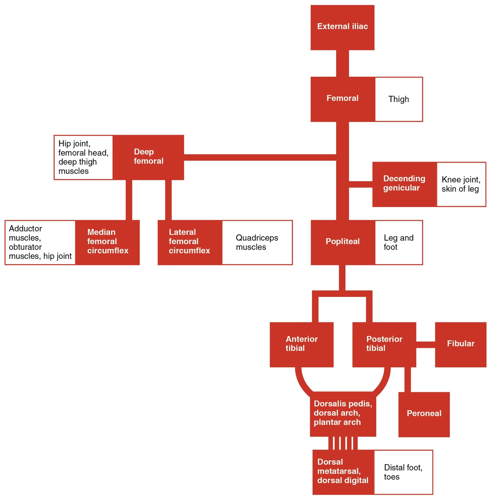
Long description
The flow chart summarizes the distribution of the systemic arteries from the external iliac artery into the lower limb.

Figure 1: The flow chart summarizes the distribution of the systemic arteries from the external iliac artery into the lower limb.
Within the femoral triangle, it gives rise to the deep femoral artery (profunda femoris), which is the major supplier of the thigh muscles. The popliteal artery lies deep to the popliteal fascia and is closely related to the knee joint, the tibial nerve, and the popliteal veins. [1]
Exam trapPopliteal Artery
Do not confuse the popliteal artery with the popliteal vein; the artery is deeper and more susceptible to injury during knee dislocations or fractures of the distal femur.
The lower limb arterial supply originates from the abdominal aorta, which bifurcates into the common iliac arteries. The external iliac artery continues as the femoral artery after passing under the inguinal ligament. The superficial femoral artery travels through the adductor canal and exits via the adductor hiatus to become the popliteal artery behind the knee. The tibioperoneal trunk further bifurcates into the posterior tibial artery and the fibular (peroneal) artery. [1]
The lower limb arterial supply originates from the abdominal aorta, which bifurcates into the common iliac arteries. The external iliac artery continues as the femoral artery after passing under the inguinal ligament. The superficial femoral artery travels through the adductor canal and exits via the adductor hiatus to become the popliteal artery behind the knee. The tibioperoneal trunk further bifurcates into the posterior tibial artery and the fibular (peroneal) artery. The anterior tibial art
The femoral artery is the direct continuation of the external iliac artery, passing deep to the inguinal ligament.
The lateral compartment of the leg includes two muscles: the fibularis longus (peroneus longus) and the fibularis brevis (peroneus brevis).
Distal to the knee, the femoral artery becomes the popliteal artery. This vessel runs through the popliteal fossa, lying deep to the popliteal fascia. It is situated between the femur and the tibia. The popliteal artery gives off several genicular branches that anastomose around the knee joint. These connections provide collateral circulation, which is vital if the main vessel is compromised. The popliteal artery then divides into the anterior and posterior tibial arteries at the lower border of the popliteus muscle.
The lower limb arterial supply originates from the abdominal aorta, which bifurcates into the common iliac arteries. The external iliac artery continues as the femoral artery after passing under the inguinal ligament. The superficial femoral artery travels through the adductor canal and exits via the adductor hiatus to become the popliteal artery behind the knee. The tibioperoneal trunk further bifurcates into the posterior tibial artery and the fibular (peroneal) artery. The anterior tibial art
The superficial femoral artery continues distally through the thigh.
| Vessel | Origin | Course | Termination |
|---|---|---|---|
| Femoral Artery | External Iliac | Through femoral triangle and adductor canal | Becomes Popliteal Artery |
| Deep Femoral Artery | Femoral Artery | Posterior thigh muscles | Perforating branches |
| Popliteal Artery | Femoral Artery | Popliteal fossa | Anterior and Posterior Tibial Arteries |
| Anterior Tibial Artery | Popliteal Artery | Anterior leg compartment | Dorsalis Pedis Artery |
| The anterior tibial artery passes anteriorly through the interosseous membrane. It runs down the anterior compartment of the leg, accompanied by the deep fibular nerve. It supplies the extensor muscles of the leg and foot. The posterior tibial artery descends posteriorly, deep to the soleus muscle. It runs alongside the tibial nerve and supplies the flexor muscles of the leg. [1] |
The lower limb arterial supply originates from the abdominal aorta, which bifurcates into the common iliac arteries. The external iliac artery continues as the femoral artery after passing under the inguinal ligament. The superficial femoral artery travels through the adductor canal and exits via the adductor hiatus to become the popliteal artery behind the knee. The anterior tibial art
The superficial femoral artery continues distally through the thigh.
DefinitionArterial Supply
The network of arteries delivering oxygenated blood to the lower limb structures, originating from the abdominal aorta and terminating in the foot arches.
The arterial system provides specific territories for each compartment. The anterior compartment of the thigh is supplied by the femoral artery branches. The anterior compartment of the leg is supplied by the anterior tibial artery. The lateral compartment of the leg is supplied by the fibular artery. The posterior [1]
| Artery | Origin | Termination |
|---|---|---|
| Femoral | External Iliac | Adductor Hiatus |
| Popliteal | Femoral | Tibioperoneal Trunk |
| Anterior Tibial | Popliteal | Dorsalis Pedis |
| Posterior Tibial | Tibioperoneal Trunk | Plantar Arch |
| Fibular | Tibioperoneal Trunk | Lateral Malleolus |
The superficial femoral artery continues distally through the thigh.
The arterial supply to the lower limb is designed to maintain perfusion even when major vessels are compromised. This resilience relies on a network of anastomoses, which are connections between arteries that allow blood to bypass an obstruction. Understanding these pathways is essential for interpreting clinical signs such as pulse deficits and for planning surgical interventions.
The central feature is the popliteal fossa, a diamond-shaped space located at the back of the knee. The boundaries of this fossa are formed by the tendons of the hamstring muscles. Specifically, the biceps femoris, semitendinosus, and semimembranosus constitute the hamstring group, which functions to flex the leg and extend the thigh. Their tendons create the superior and inferior borders of the fossa. Within this space, the popliteal artery descends, continuing from the femoral artery as it passes through the adductor hiatus. The femoral artery itself is the primary supply to the anterior thigh, while the popliteal artery serves as the conduit to the lower leg. These vessels run down the leg, providing collateral routes. The anastomoses around the knee joint connect the femoral and popliteal systems with the genicular branches, ensuring that if the femoral artery is blocked, blood can still reach the popliteal and tibial arteries via these collateral channels. The tibial arteries further anastomose around the ankle, creating a robust network that protects the foot from ischemia. [1]
DefinitionCollateral Circulation
Collateral circulation refers to the network of interconnected arteries that provide alternative routes for blood flow when a primary vessel is obstructed.
When an arterial occlusion occurs, the body’s ability to compensate depends on the maturity of these collaterals. In the lower limb, the anastomoses around the knee are particularly significant. The genicular arteries, which branch from the popliteal artery, anastomose with branches of the femoral artery above the knee and the tibial arteries below. This arrangement means that a blockage in the femoral artery may not immediately cause critical ischemia if the collateral pathways are well-developed. However, the extent of collateral development varies between individuals. [2]
| Artery Segment | Origin | Course | Clinical Significance |
|---|---|---|---|
| Femoral Artery | External Iliac | Anterior thigh | Primary supply; occlusion affects thigh and leg via collaterals. |
| Popliteal Artery | Femoral (via adductor hiatus) | Posterior knee (popliteal fossa) | Central hub for knee anastomoses; bounded by hamstring tendons. |
| Tibial Arteries | Popliteal | Anterior and posterior lower leg | Distal supply; anastomose around ankle to supply foot. |
| Muscle Group | Function | Tendinous Contribution |
|---|---|---|
| Hamstrings | Flex leg, extend thigh | Form boundaries of popliteal fossa. |
The assessment of pulses is a direct clinical application of this anatomy.
Exam trapPulse Palpation
Do not confuse the popliteal pulse with the femoral pulse; the former is located posteriorly in the popliteal fossa, bounded by the hamstring tendons, while the latter is anterior in the groin. [1]
In summary, the arterial supply of the lower limb is characterized by extensive anastomoses, particularly around the knee and ankle. The femoral and popliteal arteries form the central axis, with the tibial arteries providing distal distribution. The hamstring tendons define the popliteal fossa, housing the popliteal artery. These structures work together to ensure that blood reaches the foot, even in the presence of partial occlusions.
The arterial supply of the lower limb is generally consistent, yet significant anatomical variations occur that are critical for surgical planning and interpreting imaging. These variations primarily involve the origin and termination of the femoral artery and the configuration of the popliteal artery.
The femoral artery is the direct continuation of the external iliac artery, passing deep to the inguinal ligament.
The popliteal artery is formed by the continuation of the femoral artery through the adductor hiatus. While the standard course runs straight down the posterior thigh, variations in its bifurcation are frequent. The popliteal artery typically divides into the anterior and posterior tibial arteries at the lower border of the popliteus muscle. In some cases, the bifurcation occurs higher up in the popliteal fossa, which can complicate surgical approaches to the knee joint. Furthermore, the anterio [1]
Figure 22129ab Lower Limb Arteries Anterior Posterior
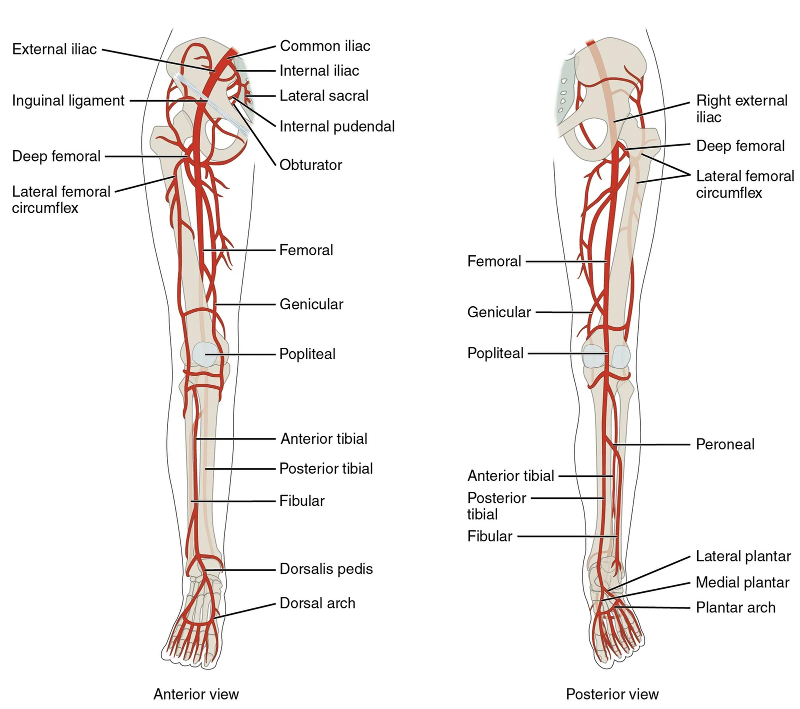
Long description
Major arteries serving the lower limb are shown in anterior and posterior views.

Figure 2: Major arteries serving the lower limb are shown in anterior and posterior views.
The peroneal artery, also known as the fibular artery, is a large branch of the posterior tibial artery. It typically arises from the posterior tibial artery in the upper part of the leg. A notable variation is the early origin of the peroneal artery, where it arises directly from the popliteal artery instead of the posterior tibial artery. Surgeons must be aware of this variation to avoid inadvertent injury during ankle surgery or when harvesting the peroneal artery for free tissue transfer. [1]
It supplies the medial aspect of the foot and the first three and a half toes. Variations in its size and anastomotic connections with the lateral plantar artery can affect the collateral circulation in the foot. In some individuals, the medial plantar artery is hypoplastic, relying more heavily on the deep plantar arch for perfusion. This has implications for patients with peripheral arterial disease, where collateral flow becomes crucial for tissue viability. [1]
Figure 3Gray555
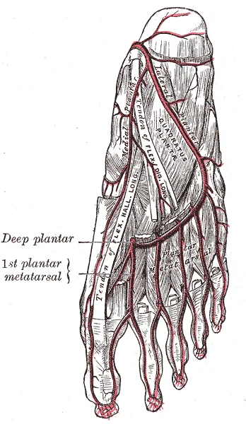
Long description
The plantar arteries. Deep view. Labelled illustration from Wikimedia Commons, used under its open licence.

Figure 3: The plantar arteries (deep view).
NoteClinical Relevance
Understanding these variations is essential for interpreting angiograms and planning vascular interventions to prevent iatrogenic injury.
| Structure | Variation | Clinical Significance |
|---|---|---|
| Femoral Artery | High origin from external iliac | Risk during pelvic surgery |
| Obturator Artery | Accessory origin from inferior epigastric | Altered hip joint supply |
| Popliteal Artery | High bifurcation | Complicates knee surgery |
| Peroneal Artery | Early origin from popliteal | Alters leg compartment supply |
The arterial supply of the lower limb is critical for distal perfusion, and its clinical assessment relies on identifying specific pulse points, access sites, and potential complications such as aneurysms or the need for ligation. Understanding the anatomical course of these vessels allows for accurate physical examination and intervention.
The femoral artery is the primary continuation of the external iliac artery as it passes beneath the inguinal ligament. It is the most accessible site for vascular access in emergency and diagnostic settings.
This location is crucial for assessing systemic perfusion. The femoral artery is also the standard access point for cardiac catheterisation and angiography.
Surgical ligation of the femoral artery is rarely performed due to the risk of distal ischaemia, but if necessary, collateral circulation via the profunda femoris artery may preserve limb viability.
The popliteal artery is the direct continuation of the femoral artery, passing through the adductor hiatus to enter the popliteal fossa. It is deep within the posterior knee, making it less accessible for pulse palpation but critical for knee joint perfusion.
Pulse Point The popliteal pulse is difficult to palpate directly due to its depth. It is best assessed by pressing the posterior knee against a firm surface with the knee slightly flexed. Absence of this pulse suggests occlusion of the femoral artery or a popliteal aneurysm.
Aneurysm Sites Popliteal artery aneurysms are the most common peripheral arterial aneurysms. They are often bilateral and can lead to distal embolisation or thrombosis, causing acute limb ischaemia. Compression of adjacent structures, such as the popliteal vein or tibial nerve, may also occur.
The anterior tibial artery arises from the popliteal artery and passes through the interosseous membrane to enter the anterior compartment of the leg. It supplies the muscles in this compartment, including the tibialis anterior, extensor hallucis longus, and extensor digitorum longus.
Pulse Point The assessment of pulses is a direct clinical application of this anatomy.
Ligation and Access Ligation of the anterior tibial artery is generally well-tolerated due to extensive collateral circulation. It is not a common site for vascular access due to its smaller calibre and deeper location in the leg compared to the femoral artery.
Understanding their compartmental location is crucial for diagnosing nerve injuries, as the superficial fibular nerve supplies them. [1]
This strong tendon inserts into the calcaneal bone of the ankle. The gastrocnemius is the most superficial and visible muscle of the calf. It lies superficial to the wide, flat soleus muscle. These muscles are large and strong and keep humans upright. [1]
Some people may have two of these muscles, whereas no plantaris is observed in about seven percent of other cadaver dissections. The plantaris tendon is a desirable substitute for the fascia lata in hernia repair, tendon transplants, and repair of ligaments. [1]
The foot also has intrinsic muscles, which originate and insert within it. These muscles primarily provide support for the foot and its arch, and contribute to movements of the toes. The principal support for the longitudinal arch of the foot is a deep fascia called plantar aponeurosis. This runs from the calcaneus bone to the toes. Inflammation of this tissue is the cause of “plantar fasciitis,” which can affect runners. [1]
The intrinsic muscles of the foot consist of two groups. The dorsal group includes only one muscle, the extensor digitorum brevis. The second group is the plantar group, which consists of four layers, starting with the most superficial. [1]
It can be detected in approximately 2–5% of hypertensive patients. In patients with coexisting hypertension and renal failure, RAS is found in 30–40% of cases. [2]
It increases the risk of heart failure by 3.5 times. The risk of stroke is increased by 3 times. The risk of death is increased by 2.6 times. [2]
In patients with coronary artery disease, the incidence of RAS is approximately 30–33%. Of these, 7–15% have a stenosis of greater than 50%. The prevalence of RAS is estimated to be as high as 40% in patients with PAD. It is as high as 50% in patients with multilevel atherosclerosis. [2]
This is generally combined with stent implantation. This approach significantly reduces the incidence of recurrent stenosis and improves long-term outcomes. It has a low complication rate of less than 5% in experienced centers. [2]
This uses a treadmill gradient of 12 degrees and a walking speed of 3.2 km/h. This allows for an objective assessment of intermittent claudication severity. It provides a starting point for ischemic preconditioning. [2]
They categorized patients by age structure and ankle-brachial index. Myocardial infarction and stroke were significantly more common in PAD patients diagnosed before the age of 60 years (10%) compared with patients whose PAD was diagnosed after the age of 70 years (6.8%). [2]
The risk of major adverse limb events and amputation was lowest in the group of patients over 70 years of age. It gradually increased in the groups of younger patients. [2]
Of the eight completed trials, only the two small ASPIRE 2 and RENAISSANCE trials showed the superiority of PTA over pharmacological treatment. This was only regarding blood pressure, with no effect on renal function, major adverse cardiovascular and cerebrovascular event rates, or survival. Other studies, including the two largest, the ASTRAL and CORAL studies, did not show an advantage of invasive procedure over pharmacotherapy in any of the parameters analyzed. [2]
It also increases the risk of mortality, cerebrovascular accidents, and episodes of acute heart failure. [2]
Students frequently confuse the anatomical boundaries and vascular supply of the lower limb compartments, leading to errors in clinical localisation. A primary error is assuming the anterior compartment muscles are primarily responsible for hip flexion. While the rectus femoris does flex the hip, the primary hip flexors originate from the pelvic girdle. The psoas major and iliacus form the iliopsoas group, which is distinct from the anterior thigh compartment muscles that primarily extend the leg at the knee. The quadriceps femoris group, comprising the rectus femoris, vastus lateralis, vastus medialis, vastus intermedius, and sartorius, is located in the anterior compartment and functions to extend the knee. Confusing the origin of these muscles can lead to incorrect diagnosis of nerve injuries, as the femoral nerve supplies the anterior compartment while the sciatic nerve supplies the posterior compartment. [1]
Another significant pitfall involves the interpretation of vascular imaging and risk stratification in elderly patients. Students often overlook the strong correlation between age and vascular stiffness. Age is an unmodifiable risk factor for vascular stiffness and atherosclerosis, independent of other atherogenic factors[1]. This means that vascular changes are not merely pathological but are part of a broader physiological decline that must be accounted for in diagnostic criteria. For instance, renal artery stenosis is predominantly an age-dependent pathology, with a prevalence of less than 1% in the general population but rising to 7% above the age of 65 years[1]. Failing to adjust expectations for vascular health based on age can lead to over-diagnosis or under-treatment. [2]
Misinterpretation of peripheral artery disease (PAD) prevalence is also common. Students may assume PAD is rare in younger populations, but the data shows a steep increase with age. The prevalence of PAD is 6% in males and 5% in females aged between 60 and 69 years, rising to 11% and 9% in those aged between 70 and 79 years, and 26% and 21% in those over 80 years of age, respectively[1]. Sykora et al. assessed the risk of MACCE in more than 22,000 outpatients with PAD according to age structure (<50, 50–59, 60–69, ≥70 years) and ABI (<1.0; 1.0–1.4; >1.4). Ignoring this gradient can result in missed diagnoses in older adults presenting with non-specific [2]
A third pitfall relates to the management of familial hypercholesterolemia (FH). Students often underestimate the severity of LDL-C elevation in FH. In most FH cases, the mean LDL-C level exceeds 190 mg/dL in adults and over 160 mg/dL in children[1]. This level of elevation is associated with a premature manifestation of atherosclerotic disease and a risk of developing coronary artery disease that is approximately 13-fold higher than in the general population[1]. Treating these patients requires aggressive intervention, as standard risk factors do not apply in the same way.
Finally, students may misinterpret the stability of atherosclerotic plaques. The presence of unstable plaque features does not necessarily indicate an acute ischemic event[1]. Necrotic lipid changes in atherosclerotic plaques, which may be ulcerated or even micro-ruptured, are not uncommon and may heal over time[1]. Therefore, imaging findings must be correlated with clinical symptoms rather than treated as immediate emergencies. Histopathological findings indicate that as the patient’s age increases, atherosclerotic lesions become larger, containing locally more inflammatory cells and MMP-9 and fewer smooth muscle cells[1]. This structural change predisposes patients to symptoms of cerebral ischemia, increasing the risk of death and stroke in patients over 75 years of age[1]. [2]
Knowledge of lower limb arterial supply and associated pathology is critical at the bedside for assessing peripheral perfusion. Clinicians use this anatomical understanding to localise occlusions based on symptom distribution. For example, pain in the calf muscles may indicate superficial femoral artery disease, while foot pain may suggest tibial or peroneal artery involvement. The assessment of pulses, such as the dorsalis pedis and posterior tibial arteries, relies on knowing the termination o [1]
In the operating theatre, precise knowledge of vascular anatomy is essential for surgical interventions. Bypass grafting procedures require identification of suitable inflow and outflow vessels. For instance, a femoropopliteal bypass requires knowledge of the profunda femoris artery and its branches. Surgeons must also be aware of the relationships between arteries and nerves to avoid iatrogenic injury. The femoral nerve lies lateral to the femoral artery, while the sciatic nerve is posterior to the hip joint. Understanding these relations prevents complications during hip or knee arthroplasty.
Imaging plays a pivotal role in diagnosing and managing vascular disease. Intravascular imaging tools such as optical coherence tomography (OCT) and intravascular ultrasound (IVUS), as well as imaging of the atherosclerotic plaque using CT angiography (CTA) and magnetic resonance angiography (MRA), allow us to detect the coexistence of atherosclerotic lesions at various stages of development in an individual patient[1]. These modalities provide detailed views of plaque morphology, helping to determine the need for intervention. For example, identifying a vulnerable plaque with a thin fibrous cap may prompt earlier surgical referral. [2]
In examinations, students are tested on their ability to identify structures and correlate anatomy with clinical signs. Questions may ask for the origin and insertion of muscles involved in lower limb movement, such as the quadriceps femoris group. The quadriceps tendon inserts into the patella and continues as the patellar ligament, which attaches to the tibial tuberosity. Understanding these attachments helps explain the mechanism of knee extension and the consequences of tendon rupture. Additionally, students must recognise the risk factors for vascular disease, such as age and familial hypercholesterolemia[1].
Clinical pearlClinical Pearl
Always correlate imaging findings with clinical symptoms; unstable plaques may be asymptomatic.[1]
Assessment of Lower Limb Ischaemia
- Patient presents with leg painAssess history and risk factors
- Question: Pulses palpable?
Yes
- Consider non-vascular causes
No
- Perform ABI measurement
- Question: ABI < 0.9?
Yes
- Refer for CTA/MRA
No
- Reassure and monitor
| Muscle | Action | Nerve Supply |
|---|---|---|
| Quadriceps Femoris | Extends leg at knee | Femoral nerve |
| Sartorius | Flexes, abducts, and laterally rotates leg at hip | Femoral nerve |
The psoas major and iliacus form the iliopsoas group, originating on the pelvic girdle. The quadriceps femoris group comprises four muscles that extend and stabilize the knee. [1] Age is an unmodifiable risk factor for vascular stiffness and atherosclerosis[1]. Renal artery stenosis prevalence rises to 7% above age 65 and 40% above age 75[1]. [2] PAD affects over 230 million adults worldwide[1]. [2] Familial hypercholesterolemia is associated with a 13-fold higher risk of CAD[1]. [2] Unstable plaque features do not necessarily indicate an acute ischemic event[1]. Imaging tools like OCT and IVUS detect atherosclerotic lesions at various stages[1].
Answer each one in your head first, then open the answer.
Which muscles make up the iliopsoas group?
Show answerHide answer
Psoas major and iliacusWhat is the prevalence of PAD in males aged 60-69?
Show answerHide answer
6%Which imaging tools detect atherosclerotic lesions at various stages?
Show answerHide answer
OCT, IVUS, CTA, MRAWhat is the risk of CAD in FH patients compared to the general population?
Show answerHide answer
13-fold higherWhat is the prevalence of RAS above age 75?
Show answerHide answer
Approximately 40%
- Betts JG, Young KA, Wise JA, et al. Appendicular Muscles of the Pelvic Girdle and Lower Limbs. OpenStax, Anatomy and Physiology 2e (CC BY-NC-SA 4.0). Accessed .
- Piechocki M, Przewłocki T, Pieniążek P, et al. A Non-Coronary, Peripheral Arterial Atherosclerotic Disease (Carotid, Renal, Lower Limb) in Elderly Patients-A Review: Part I-Epidemiology, Risk Factors, and Atherosclerosis-Related Diversities in Elderly Patients. Journal of clinical medicine 2024;13(5):1471. doi:10.3390/jcm13051471. Accessed .
Figure credits
Educational content for medical students and doctors. It is not a substitute for clinical judgement or local guidelines.