Lower limb: venous drainage
Anatomical pathways for venous return from the lower extremities to the heart.
The venous architecture of the lower limb is organised into two distinct systems: superficial and deep veins. This dual arrangement serves both circulatory and thermoregulatory functions. The deeper veins generally correspond to the complementary arteries. These deep vessels are responsible for the bulk of venous return from the muscles and bones. In contrast, the superficial veins do not normally have direct arterial counterparts. Beyond returning blood, they also make contributions to the maintenance of body temperature. When the ambient temperature is warm, more blood is diverted to the superficial veins where heat can be more easily dissipated to the environment. In colder weather, there is more constriction of the superficial veins and blood is diverted deeper where the body can retain more of the heat. [1]
The primary superficial vessels are the great saphenous vein and the small saphenous vein. [3]
The great saphenous vein terminates by piercing the cribriform fascia to join the femoral vein in the femoral triangle. The small saphenous vein typically terminates in the popliteal vein within the popliteal fossa. [1]
Connecting the superficial and deep systems are perforating veins. These vessels contain one-way valves that normally permit flow from superficial to deep veins. Valvular incompetence in these perforators allows retrograde flow, increasing hydrostatic pressure in the superficial system. This chronic hypertension leads to venous dilation and tortuosity, clinically recognised as varicose veins. In the lower limb, the saphenous veins are the primary sites for such access and pathology. [1]
As the femoral vein penetrates the body wall from the femoral portion of the lower limb, it becomes the external iliac vein, a large vein that drains blood from the leg to the common iliac vein. The external and internal iliac veins combine near the inferior portion of the sacroiliac joint to form the common iliac vein. Similar to the common iliac arteries, the common iliac veins come together at the level of L5 to form the inferior vena cava. [1]
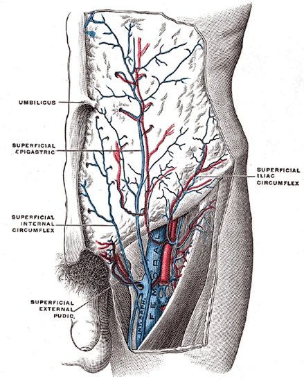
Figure 1Gray584
Long description
The femoral vein and its tributaries. (Poirier and Charpy.)

Figure 1: The femoral vein and its tributaries (Poirier and Charpy.).
Venous drainage is closely associated with lymphatic drainage. Superficial lymphatics follow the saphenous veins. The superficial inguinal nodes receive lymph from the lower limb, superficial abdominal wall, and perineum. These nodes drain into the deep inguinal nodes, which then empty into the external iliac nodes. This pathway ensures that fluid and immune cells from the lower extremity are filtered before entering the systemic circulation via the thoracic duct or right lymphatic duct.
The venous drainage of the lower limb begins in the deep tissues and superficial skin, converging to return deoxygenated blood to the heart. Systemic venous circulation from beneath the diaphragm, which includes the abdominal and pelvic regions and the lower limbs, enters the inferior vena cava. This contrasts with blood from above the diaphragm, which generally flows into the superior vena cava. [1]
The superficial venous system consists of two main trunks: the great saphenous vein and the small saphenous vein. The great saphenous vein is a prominent surface vessel located on the medial surface of the leg and thigh. It originates from the dorsal venous arch of the foot and ascends anterior to the medial malleolus. It travels up the medial leg, posterior to the medial border of the tibia, and continues along the medial thigh.
It drains blood from the superficial regions of the lower leg and foot. It originates from the lateral side of the dorsal venous arch and ascends posterior to the lateral malleolus. It travels up the posterior calf, lying between the two heads of the gastrocnemius muscle. It flows into the popliteal vein. [1]
The deep venous system accompanies the major arteries of the lower limb. The venae comitantes, or paired veins, run alongside the posterior tibial and peroneal arteries in the leg. These merge to form the popliteal vein behind the knee. As the popliteal vein passes behind the knee in the popliteal region, it becomes the femoral vein. The femoral vein ascends through the adductor canal and into the femoral triangle. It passes under the inguinal ligament to become the external iliac vein. The external iliac vein then joins the internal iliac vein to form the common iliac vein. The right and left common iliac veins unite to form the inferior vena cava. [1]
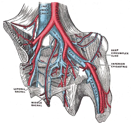
Figure 2Gray586
Long description
The iliac veins. (Poirier and Charpy.). Labelled illustration from Wikimedia Commons, used under its open licence.

Figure 2: The iliac veins (Poirier and Charpy.).
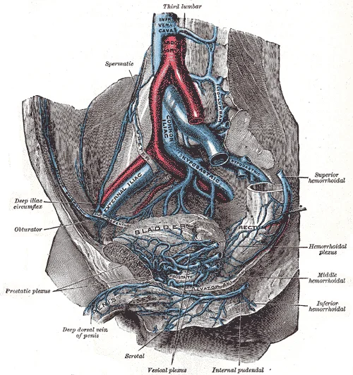
Figure 3Gray585
Long description
The veins of the right half of the male pelvis. (Spalteholz).

Figure 3: The veins of the right half of the male pelvis (Spalteholz).
Veins in the lower limb are equipped with valves to prevent backflow. Many veins of the body, particularly those of the limbs, contain valves that assist the unidirectional flow of blood toward the heart. This is critical because blood flow becomes sluggish in the extremities, as a result of the lower pressure and the effects of gravity. In comparison to arteries, venules and veins withstand a much lower pressure from the blood that flows through them. Their walls are considerably thinner and their lumens are correspondingly larger in diameter, allowing more blood to flow with less vessel resistance. [3]
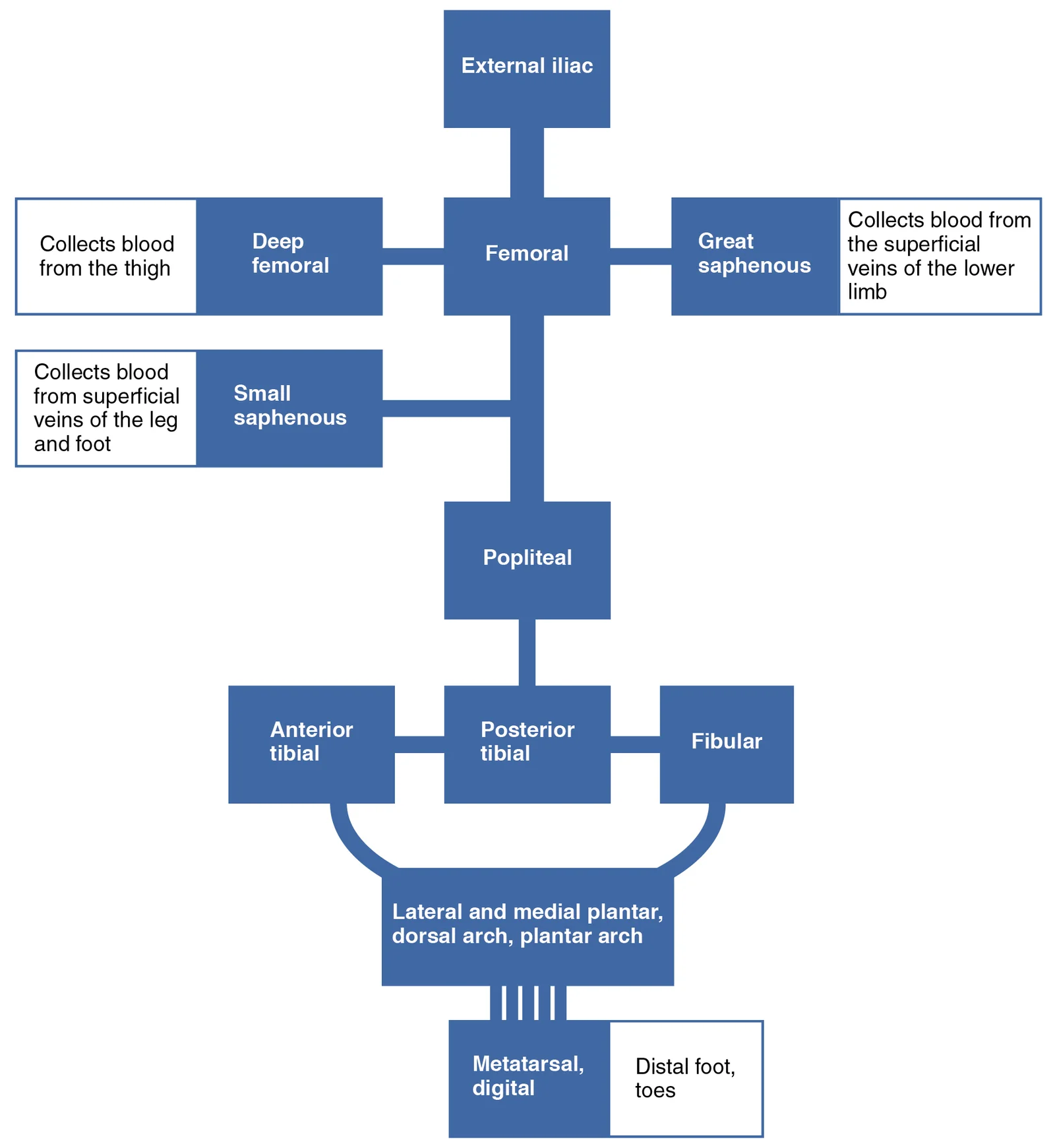
Figure 42137 Lower Limb Veins Chart
Long description
The flow chart summarizes venous flow from the lower limb.

Figure 4: The flow chart summarizes venous flow from the lower limb.
DefinitionVenous Return
The process by which blood flows from the lower limbs back to the right atrium of the heart, aided by valves and muscle pumps.
| Vein | Origin | Termination |
|---|---|---|
| Great Saphenous | Dorsal venous arch | Femoral vein |
| Small Saphenous | Dorsal venous arch | Popliteal vein |
| Femoral | Popliteal vein | External iliac vein |
| Popliteal | Deep veins of leg | Femoral vein |
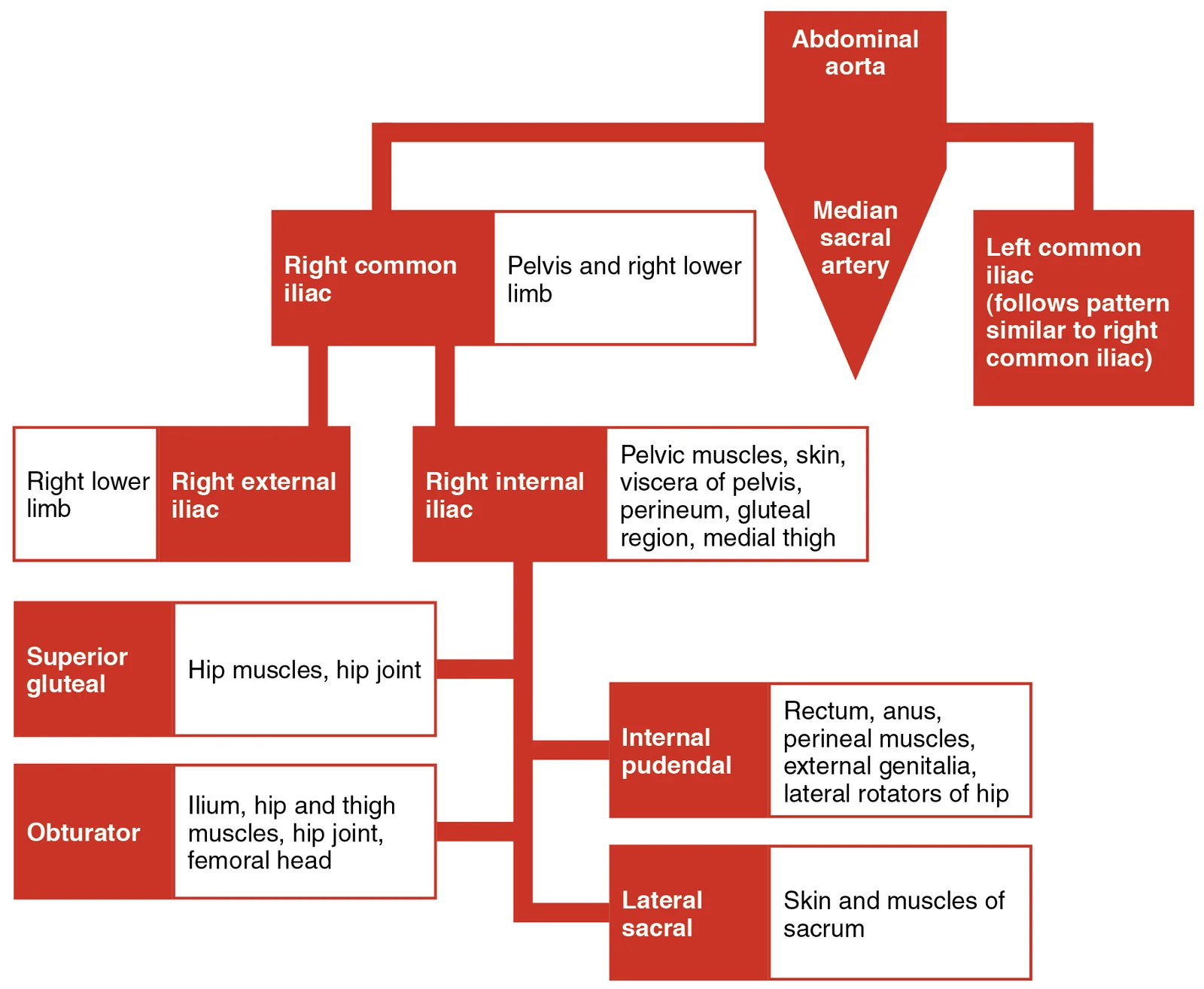
Figure 52126 Iliac Artery Branches Chart
Long description
The flow chart summarizes the distribution of the major branches of the common iliac arteries into the pelvis and lower limbs. The left side follows a similar pattern to the right.

Figure 5: The flow chart summarizes the distribution of the major branches of the common iliac arteries into the pelvis and lower limbs.
The venous drainage of the lower limb is a complex network that returns deoxygenated blood to the heart. It is divided into two main systems: the superficial veins, which lie in the subcutaneous tissue, and the deep veins, which accompany the major arteries. These systems are connected by perforating veins that allow blood to flow from the superficial to the deep system. Understanding this anatomy is critical for managing conditions such as deep vein thrombosis and varicose veins.
The superficial system consists primarily of two large veins: the great saphenous vein and the small saphenous vein. The great saphenous vein is a prominent surface vessel located on the medial surface of the leg and thigh. It originates from the dorsal venous arch of the foot, passes anterior to the medial malleolus, and ascends along the medial side of the leg and thigh. It terminates by draining into the femoral vein at the saphenofemoral junction, located in the groin. The small saphenous vein originates from the lateral side of the dorsal [1]
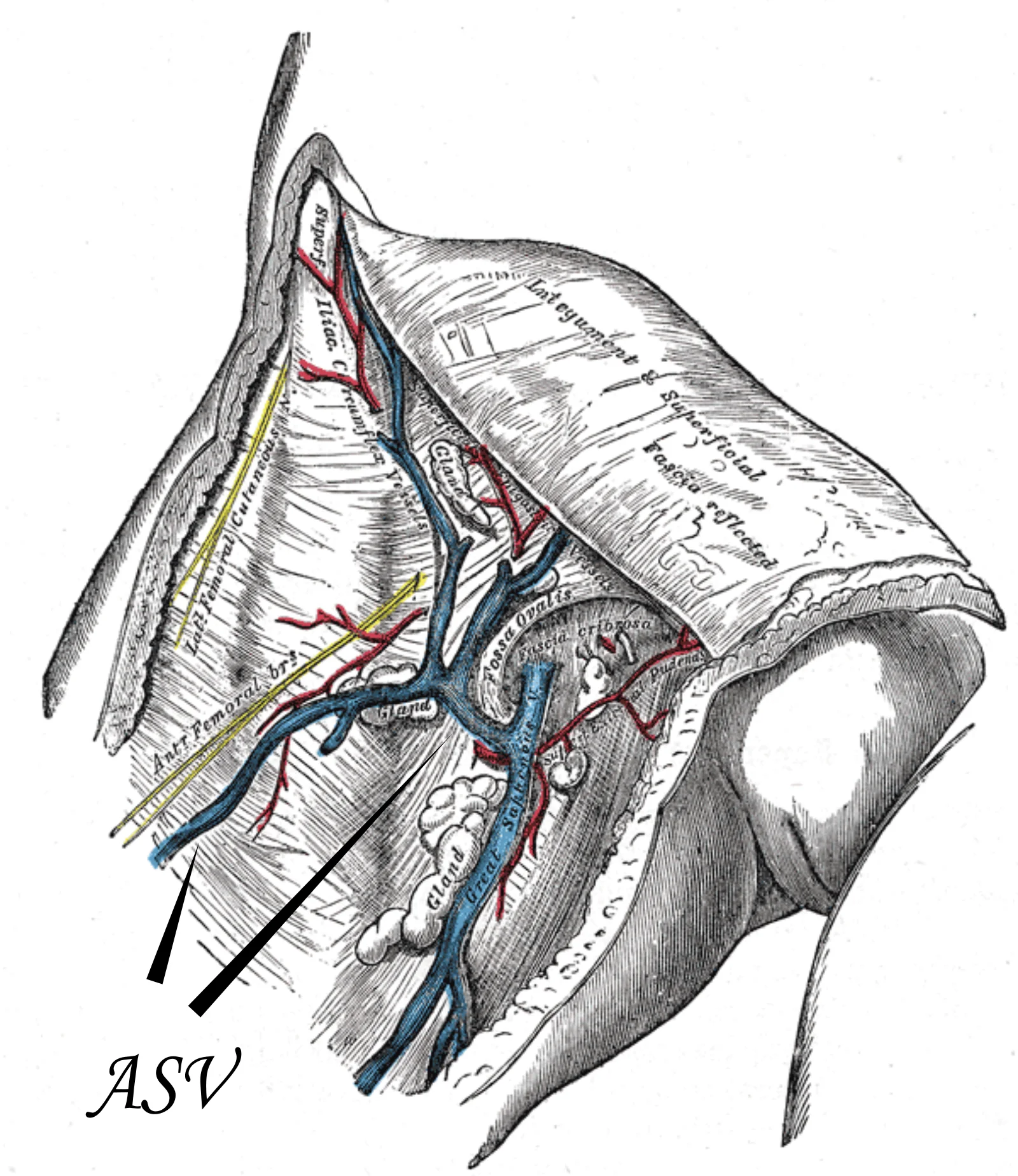
Figure 6Gray580
Long description
The great saphenous vein and its tributaries at the fossa ovalis.

Figure 6: The great saphenous vein and its tributaries at the fossa ovalis.
The deep veins run in the fascial compartments alongside the major arteries. In the foot, the deep veins are paired with the dorsalis pedis and posterior tibial arteries. In the leg, the deep veins are paired with the anterior tibial, posterior tibial, and peroneal arteries. In the thigh, the deep veins are paired with the femoral artery. The deep veins are generally larger than the superficial veins and have thicker walls. They are responsible for the majority of venous return from the lower limb.
Perforating veins are short, valve-bearing vessels that connect the superficial veins to the deep veins. They are named after the deep vein into which they drain. For example, the perforating veins of the calf drain into the posterior tibial and peroneal veins. The valves in the perforating veins ensure that blood flows only from the superficial to the deep system. If these valves fail, blood can flow in the reverse direction, leading to venous hypertension in the superficial system and the development of varicose veins.
| Vein | Origin | Course | Termination |
|---|---|---|---|
| Great Saphenous | Dorsal venous arch | Anterior to medial malleolus, medial leg/thigh | Femoral vein |
| Small Saphenous | Lateral dorsal venous arch | Posterior to lateral malleolus, posterior calf | Popliteal vein |
| Femoral Vein | Popliteal vein | Thigh | External iliac vein |
| Popliteal Vein | Anterior and posterior tibial veins | Popliteal fossa | Femoral vein |
The deep veins are more susceptible to thrombosis due to their larger diameter and the slower flow of blood during periods of immobility. Both systems are important in the pathophysiology of venous disease.
Systemic veins return deoxygenated blood to the right atrium of the heart. Since this blood has already passed through systemic capillaries, it is relatively low in oxygen concentration. In many cases, veins draining organs and regions of the body share the same name as the arteries that supplied these regions, and the two vessels often parallel one another. This arrangement is described as a “complementary” pattern. However, there is a great deal more variability in the venous circulation than normally occurs in the arteries. For the sake of brevity and clarity, this text will discuss only the most commonly encountered patterns. Keep this variation in mind when you move from the classroom to clinical practice. [1]
In addition, many veins, particularly in the lower limbs, contain valves formed by sections of thickened endothelium that are reinforced with connective tissue, extending into the lumen. These valves prevent backflow, ensuring that blood moves toward the heart against gravity. [3]
NoteClinical Variation
Venous anatomy varies significantly; always consider individual differences when interpreting imaging or performing procedures.
| Vein | Location | Termination |
|---|---|---|
| Great saphenous | Medial leg and thigh | Femoral vein |
| Small saphenous | Posterior calf | Popliteal vein |
| Femoral | Anterior thigh | External iliac vein |
| Popliteal | Popliteal fossa | Femoral vein |
The venous system of the lower limb is characterised by a complex network of connections that ensure robust blood return to the heart. Unlike the arterial system, which operates under high pressure with thick-walled vessels, the venous system is a low-pressure environment. Veins possess larger lumens and thinner walls, often appearing flattened in cross-section. This structural difference necessitates efficient mechanisms to maintain flow, particularly against gravity. Anastomoses are particularly common in veins, serving as critical pathways to maintain blood flow when a primary vessel is blocked or narrowed. [3] [1]
Think of the venous network like a river system with many tributaries and channels, several of which interconnect. This analogy helps in understanding how blood can be rerouted. Just as a street changes name as it passes through an intersection, a vein can change names as it passes an anatomical landmark. For instance, vessels may transition from superficial to deep systems, or connect between different regional groups. These connections are vital for collateral circulation. [1]
DefinitionAnastomosis
A connection between two blood vessels that allows blood to flow from one to the other, often providing alternative routes if the primary path is obstructed.
In the lower limb, these anastomoses occur between superficial and deep veins, as well as between veins on opposite sides of the body. The superficial veins, such as the great saphenous and small saphenous veins, run close to the skin surface. The deep veins accompany the major arteries. Communication between these two systems occurs via perforating veins. These perforators allow blood to move from the superficial system into the deep system, which has higher pressure gradients and more efficient muscle pump mechanisms.
NoteClinical Relevance
Failure of valves in perforating veins can lead to venous hypertension and varicose ulcers, highlighting the importance of these anastomotic pathways.
When tracing blood flow, we move from the capillaries into the smallest veins and follow the direction of blood flow into larger veins and back to the heart. This path is facilitated by the numerous anastomoses. For example, anastomoses exist between the medial and lateral plantar veins, and between the dorsal venous arch and the deep veins of the foot. These connections ensure that even if one route is compromised, blood can still reach the deep system for return to the inferior vena cava. [1]
The inferior vena cava drains most of the veins inferior to the diaphragm. Therefore, the efficiency of lower limb venous drainage is crucial for overall circulatory homeostasis. The presence of numerous anastomoses means that the venous system is resilient. However, it also means that pathology in one area can affect others. Understanding these connections is essential for diagnosing and treating venous disorders. [1]
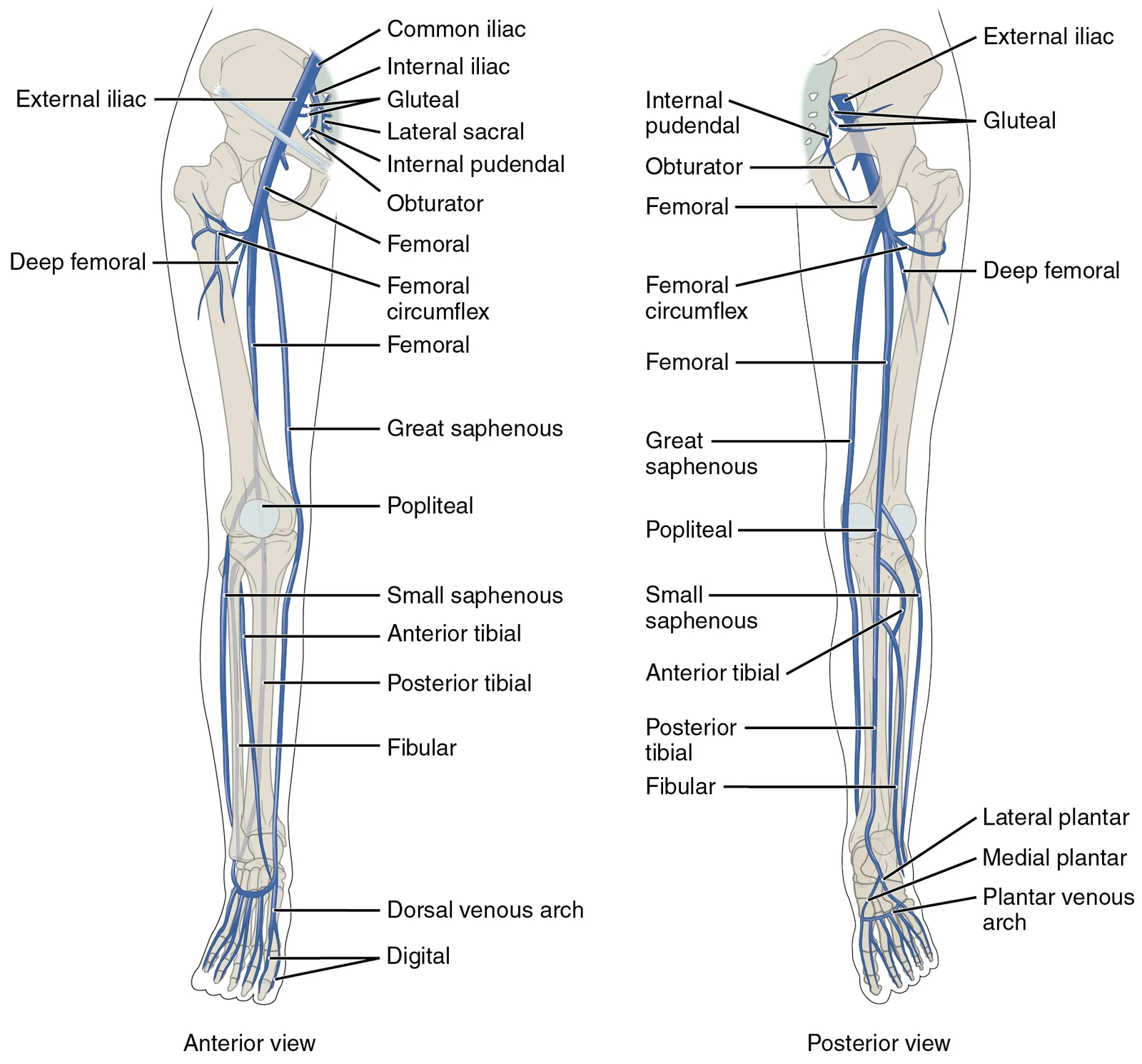
Figure 72136ab Lower Limb Veins Anterior Posterior
Long description
Illustration from Anatomy & Physiology, Connexions Web site. http://cnx.org/content/col11496/1.6/, Jun 19, 2013.

Figure 7: Illustration from Anatomy & Physiology, Connexions Web site (http://cnx.org/content/col11496/1.6/, jun 19, 2013).
| Structure | Type | Function |
|---|---|---|
| Perforating Veins | Superficial to Deep | Allow blood flow from superficial to deep system |
| Great Saphenous Vein | Superficial | Drains medial leg and thigh |
| Small Saphenous Vein | Superficial | Drains posterior calf |
| Popliteal Vein | Deep | Formed by union of tibial veins |
The venous system of the lower limb is a complex network that drains deoxygenated blood from the foot, leg, and thigh back to the heart. While standard anatomical texts describe a consistent pattern of superficial and deep veins, individual variations in circulation patterns are not uncommon. Understanding these variations is critical for clinical practice, particularly when performing venous access or interpreting imaging studies. The primary distinction in lower limb venous anatomy lies between the superficial veins, which lie in the subcutaneous tissue, and the deep veins, which accompany the major arteries and are surrounded by muscle. [1]
The two major superficial veins of the lower limb are the great saphenous vein and the small saphenous vein. The great saphenous vein is a prominent surface vessel located on the medial surface of the leg and thigh. It originates from the medial marginal vein of the foot and the anterior venous arch. It ascends along the medial side of the leg and thigh. Its termination is at the femoral vein, where it pierces the femoral sheath at the saphenous opening in the fascia lata. The small saphenous vein originates from the lateral marginal vein of t
The deep veins of the lower limb are paired veins that accompany the major arteries. In the foot, the deep venous system is formed by the plantar venous arch. In the leg, the deep veins are the anterior tibial veins and the posterior tibial veins. The anterior tibial veins accompany the anterior tibial artery, while the posterior tibial veins accompany the posterior tibial artery. These veins join to form the popliteal vein at the apex of the popliteal fossa. The femoral vein is the direct continuation of the popliteal vein and runs alongside the femoral artery in the femoral triangle. It terminates at the inguinal ligament, where it becomes the external iliac vein. The deep venous system is responsible for the majority of venous return from the lower limb.
Perforating veins, also known as communicating veins, connect the superficial venous system to the deep venous system. They are found throughout the leg and thigh. The most clinically significant perforating veins are located in the medial calf and the thigh. These veins contain valves that ensure unidirectional flow from the superficial to the deep system. Failure of these valves can lead to venous hypertension and the development of varicose veins. The Cockett perforating veins are a specific group of perforating veins in the medial calf that are frequently implicated in venous insufficiency.
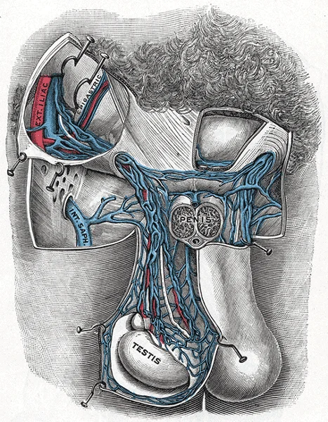
Figure 8Gray590
Long description
Spermatic veins. (Testut.). Labelled illustration from Wikimedia Commons, used under its open licence.

Figure 8: Spermatic veins (Testut.).
Variations in the venous anatomy can have significant clinical implications. For example, the great saphenous vein may have multiple tributaries or may terminate in an atypical location. The small saphenous vein may drain into the femoral vein rather than the popliteal vein in some individuals. These variations must be considered when planning surgical procedures, such as vein grafting for coronary artery bypass, or when performing ultrasound-guided venous access. A thorough understanding of the standard anatomy and its potential variations is essential for safe and effective clinical practice.
NoteAnatomical Variability
Individual variations in circulation patterns are not uncommon, so clinicians must always verify venous anatomy using imaging before invasive procedures.
| Vein | Origin | Course | Termination |
|---|---|---|---|
| Great Saphenous | Medial marginal vein of foot | Medial leg and thigh | Femoral vein |
| Small Saphenous | Lateral marginal vein of foot | Posterior calf | Popliteal vein |
The venous drainage of the lower limb is clinically significant for understanding fluid dynamics, access routes, and pathological states. The superficial venous system, primarily the great and small saphenous veins, is often accessed for venous cutdowns or cannulation. This location is superficial and easily palpable, making it a preferred site when deep veins
Aneurysms are focal dilations of blood vessels. While arterial aneurysms are more common in the lower limb (e.g., popliteal or femoral), venous aneurysms are rare but can occur in the saphenous veins. They present as soft, compressible masses that may increase in size with dependency or Valsalva manoeuvre. Ligation of superficial veins, such as in varicose vein surgery, requires precise identification to prevent damage to accompanying nerves. The saphenous nerve accompanies the great saphenous v
Pulse points are typically associated with arteries, not veins, as venous pressure is low and pulsations are rarely palpable unless there is right heart failure causing jugular venous distension. However, the femoral vein lies medial to the femoral artery in the femoral triangle. During resuscitation, if arterial access is difficult, venous access can be established here, although it is not a pulse point. The anatomical relationship is critical: the order from lateral to medial is Nerve, Artery, [3]
Clinical pearlSaphenous Landmark
The great saphenous vein passes anterior to the medial malleolus, making it a reliable landmark for venous access. [3]
| Vein | Origin | Course | Termination |
|---|---|---|---|
| Great Saphenous | Medial marginal vein | Anterior to medial malleolus, medial leg/thigh | Femoral vein |
| Small Saphenous | Lateral marginal vein | Posterior to lateral malleolus, posterior calf | Popliteal vein |
Deep veins accompany the arteries. Risk factors include stasis, endothelial injury, and hypercoagulability. Compression of the femoral vein by a thrombus can lead to swelling and pain. Ultrasound is the primary imaging modality for diagnosing DVT, assessing compressibility of the vein.
Exam trapNAVEL Mnemonic
Remember the order in the femoral triangle: Nerve, Artery, Vein, Lymphatics, Empty space. This prevents accidental arterial puncture during venous cannulation.
The venous drainage of the lower limb is divided into superficial and deep systems, which communicate via perforating veins. A key anatomical principle is that many arteries and veins share the same names, parallel one another throughout the body, and are very similar on the right and left sides of the body[1]. However, some superficial veins, such as the great saphenous vein in the femoral region, have no arterial counterpart[1].
DefinitionSuperficial Veins
The superficial system lies within the subcutaneous tissue and does not accompany arteries closely. It includes the great and small saphenous veins.
The great saphenous vein is a prominent superficial vein. It originates from the dorsal venous arch of the foot and ascends anterior to the medial malleolus. It travels up the medial aspect of the leg and thigh, finally draining into the femoral vein in the femoral triangle. This vein is a common site for varicosities and is frequently harvested for coronary artery bypass grafting.
Exam trapCommon Trap
Students often confuse the course of the great saphenous vein with the small saphenous vein. Remember: Great = Medial; Small = Lateral/Posterior.
It ascends posterior to the lateral malleolus and travels up the posterior aspect of the leg. It typically terminates by draining into the popliteal vein behind the knee. [1][3]
Clinical pearlPerforating Veins
Perforating veins connect the superficial and deep systems. They contain valves that normally allow flow from superficial to deep veins, preventing backflow. Incompetence in these valves leads to venous hypertension and varicose veins.
The deep venous system consists of paired veins that accompany the major arteries of the lower limb. These include the anterior and posterior tibial veins, the fibular (peroneal) veins, and the popliteal vein. The deep veins run in pairs (venae comitantes) and drain blood from the muscles and bones.
DefinitionDeep Veins
Deep veins are named after their accompanying arteries. They are located deep to the deep fascia and are crucial for returning blood from the muscle pumps of the calf.
It receives the great saphenous vein and the deep veins of the thigh. It passes under the inguinal ligament to become the external iliac vein.
NoteClinical Note
Deep vein thrombosis (DVT) most commonly occurs in the deep veins of the lower limb, particularly in the calf and popliteal veins. Risk factors include immobility, surgery, and hypercoagulable states.
| Vein | Origin | Course | Termination |
|---|---|---|---|
| Great Saphenous | Dorsal venous arch (medial) | Anterior to medial malleolus, medial leg/thigh | Femoral vein |
| Small Saphenous | Dorsal venous arch (lateral) | Posterior to lateral malleolus, posterior leg | Popliteal vein |
Clinical pearlValve Distribution
Veins in the lower limb have numerous valves to combat gravity. Valve failure is the primary cause of varicose veins.
Understanding the distinction between superficial and deep systems is vital for clinical procedures such as venous cannulation and sclerotherapy. The superficial veins are more accessible but carry a higher risk of thrombophlebitis if damaged.
Exam trapAnatomical Variation
While the main pathways are consistent, there can be variations in the termination of the small saphenous vein. It may drain into the great saphenous vein or directly into the femoral vein in some individuals.
The lymphatic drainage of the lower limb generally follows the venous drainage. Superficial lymphatics follow the great and small saphenous veins to the inguinal and popliteal lymph nodes, respectively. Deep lymphatics follow the deep veins to the external iliac and common iliac nodes.
DefinitionLymphatic Drainage
Lymph from the lower limb drains into the inguinal nodes superficially and the iliac nodes deeply. Infection in the foot can lead to inguinal lymphadenopathy.
In summary, the venous drainage of the lower limb is a complex network of superficial and deep veins that work together to return blood to the heart. The superficial system includes the great and small saphenous veins, while the deep system includes the tibial, fibular, popliteal, and femoral veins. Perforating veins connect these systems and are critical for maintaining unidirectional blood flow.
NoteKey Takeaway
Always remember the rule: "Great is Medial, Small is Lateral/Posterior." This mnemonic helps in identifying the course and termination of the major superficial veins.
Students frequently confuse the directional flow of blood within the systemic circulation, particularly regarding the distinction between arteries and veins. A fundamental error is assuming that all arteries carry oxygenated blood. While this is true for the systemic circuit, the pulmonary arteries carry deoxygenated blood to the lungs. Conversely, pulmonary veins carry oxygenated blood back to the heart. This distinction is critical when interpreting imaging or understanding gas exchange. The abdominal aorta remains to the left of the vertebral column and is embedded in adipose tissue behind the peritoneal cavity. Anoth [1]
A third pitfall relates to the structural composition of blood vessels. Students may overlook that veins are larger blood vessels that return blood to the heart, formed after blood exits capillaries and combines into venules. The misconception that veins are merely "smaller arteries" ignores their role in the final stage of the circulatory pathway. Furthermore, when studying the lower limb, students often confuse the muscle compartments. The anterior compartment of the thigh contains the quadriceps femoris group, which comprises four muscles that extend and stabilize the knee. The rectus femoris is on the anterior aspect, the vastus lateralis on the lateral aspect, the vastus medialis on the medial aspect, and the vastus intermedius is deep to the rectus femoris. Mistaking the location of these muscles can lead to incorrect identification of neurovascular bundles, as the femoral artery and vein lie anterior to the hip joint and medial to the femoral nerve, closely associated with these anterior compartment muscles. [2]
The fourth error involves the termination of major vessels. The aorta arises from the left ventricle and eventually descends to the abdominal region. At the base of the aorta is the aortic semilunar valve that prevents backflow of blood into the left ventricle while the heart is relaxing. Students sometimes confuse the aortic semilunar valve with the pulmonary semilunar valve, which is located at the base of the pulmonary trunk and prevents backflow from the pulmonary trunk[1]. Understanding that the left ventricle ejects blood into the aorta through the aortic semilunar valve is essential for distinguishing systemic from pulmonary circulation[1]. [4]
The fifth pitfall is ignoring the complex swirling pattern of cardiac muscle. The myocardium is built upon a framework of collagenous fibers, plus the blood vessels that supply the myocardium and the nerve fibers that help regulate the heart[1]. The muscle pattern is elegant and complex, as the muscle cells swirl and spiral around the chambers of the heart[1]. They form a figure 8 pattern around the atria and around the bases of the great vessels[1]. Deeper ventricular muscles also form a figure 8 around the two ventricles and proceed toward the apex[1]. This complex swirling pattern allows the heart to pump blood more effectively than a simple linear pattern would[1]. Failing to appreciate this structural efficiency can lead to misunderstandings about cardiac output and ventricular function.
Exam trapDirectional Flow
Remember: Arteries carry blood away from the heart, while veins return blood to the heart, regardless of oxygenation status. [3]
DefinitionAbdominal Aorta Position
The abdominal aorta remains to the left of the vertebral column and is embedded in adipose tissue behind the peritoneal cavity.
Students frequently confuse the directional flow of blood within the systemic circulation, particularly regarding the distinction between arteries and veins. A fundamental error is assuming that all arteries carry oxygenated blood. While this is true for the systemic circuit, the pulmonary arteries carry deoxygenated blood to the lungs. Conversely, pulmonary veins carry oxygenated blood back to the heart. This distinction is critical when interpreting imaging or understanding gas exchange. The abdominal aorta formally ends at approximately the level of vertebra L4, where it bifurcates to form the common iliac arteries. Anoth [1]
In the operating theatre, precise knowledge of vascular anatomy prevents iatrogenic injury. Surgeons must be aware that the abdominal aorta is embedded in adipose tissue behind the peritoneal cavity. This retroperitoneal position influences surgical approaches to the aorta and its branches. Additionally, understanding the muscle compartments of the thigh is vital. The quadriceps femoris group, which comprises four muscles that extend and stabilize the knee, is located in the anterior compartment. The rectus femoris is on the anterior aspect of the thigh, the vastus lateralis is on the lateral aspect of the thigh, the vastus medialis is on the medial aspect of the thigh, and the vastus intermedius is between the vastus lateralis and vastus medialis and deep to the rectus femoris. Surgeons performing knee or hip procedures must navigate these muscles carefully to avoid damaging the underlying neurovascular structures. [2]
On imaging, radiologists use these anatomical relationships to identify pathology. The distinction between the aortic semilunar valve and the pulmonary semilunar valve is critical in echocardiography. The aortic semilunar valve prevents backflow of blood into the left ventricle while the heart is relaxing. The pulmonary semilunar valve prevents backflow from the pulmonary trunk[1]. Recognizing these valves helps in diagnosing valvular heart disease. Furthermore, understanding that the left ventricle is the major pumping chamber for the systemic circuit, ejecting blood into the aorta through the aortic semilunar valve, aids in assessing cardiac function[1].
In examinations, students are tested on their ability to describe structures in a fixed order: origin, course, relations, branches, and termination. For example, describing the abdominal aorta requires stating that it crosses the diaphragm at the aortic hiatus, remains to the left of the vertebral column, and ends at L4. Similarly, describing the quadriceps femoris group involves listing the four muscles and their actions. The rectus femoris is on the anterior aspect of the thigh, the vastus lateralis is on the lateral aspect of the thigh, the vastus medialis is on the medial aspect of the thigh, and the vastus intermedius is between the vastus lateralis and vastus medialis and deep to the rectus femoris. This structured approach ensures clarity and completeness in answers.
NoteSurgical Landmark
The bifurcation of the aorta at L4 is a key landmark for abdominal surgery and imaging interpretation.
| Muscle | Location | Action |
|---|---|---|
| Rectus Femoris | Anterior aspect of the thigh | Extends and stabilizes the knee |
| Vastus Lateralis | Lateral aspect of the thigh | Extends and stabilizes the knee |
| Vastus Medialis | Medial aspect of the thigh | Extends and stabilizes the knee |
| Vastus Intermedius | Deep to rectus femoris | Extends and stabilizes the knee |
- Arteries carry blood away from the heart, while veins return blood to the heart.
- The abdominal aorta remains to the left of the vertebral column and is embedded in adipose tissue behind the peritoneal cavity.
- The aorta bifurcates at the level of the fourth lumbar vertebra into the two common iliac arteries. [1]
- The abdominal aorta formally ends at approximately the level of vertebra L4, where it bifurcates to form the common iliac arteries.
- The quadriceps femoris group comprises four muscles that extend and stabilize the knee.
- The rectus femoris is on the anterior aspect of the thigh, the vastus lateralis is on the lateral aspect of the thigh, the vastus medialis is on the medial aspect of the thigh, and the vastus intermedius is between the vastus lateralis and vastus medialis and deep to the rectus femoris.
- The aortic semilunar valve prevents backflow of blood into the left ventricle while the heart is relaxing.
- The pulmonary semilunar valve prevents backflow from the pulmonary trunk[1].
Answer each one in your head first, then open the answer.
What is the primary function of arteries according to basic circulatory principles?
Show answerHide answer
Arteries carry blood away from the heartWhere does the abdominal aorta formally end and what does it form?
Show answerHide answer
At approximately the level of vertebra L4, where it bifurcates to form the common iliac arteriesWhich four muscles comprise the quadriceps femoris group?
Show answerHide answer
Rectus femoris, vastus lateralis, vastus medialis, and vastus intermediusWhat prevents backflow of blood into the left ventricle while the heart is relaxing?
Show answerHide answer
The aortic semilunar valveWhere is the abdominal aorta positioned relative to the vertebral column?
Show answerHide answer
To the left of the vertebral column and embedded in adipose tissue behind the peritoneal cavity
- Betts JG, Young KA, Wise JA, et al. Circulatory Pathways. OpenStax, Anatomy and Physiology 2e (CC BY-NC-SA 4.0). Accessed .
- Betts JG, Young KA, Wise JA, et al. Appendicular Muscles of the Pelvic Girdle and Lower Limbs. OpenStax, Anatomy and Physiology 2e (CC BY-NC-SA 4.0). Accessed .
- Betts JG, Young KA, Wise JA, et al. Structure and Function of Blood Vessels. OpenStax, Anatomy and Physiology 2e (CC BY-NC-SA 4.0). Accessed .
- Betts JG, Young KA, Wise JA, et al. Heart Anatomy. OpenStax, Anatomy and Physiology 2e (CC BY-NC-SA 4.0). Accessed .
Figure credits
- Figure 1: Henry Vandyke Carter, via Wikimedia Commons, Public domain, Public domain.
- Figure 2: Henry Vandyke Carter, via Wikimedia Commons, Public domain, Public domain.
- Figure 3: Henry Vandyke Carter, via Wikimedia Commons, Public domain, Public domain.
- Figure 4: OpenStax College, via Wikimedia Commons, CC BY 3.0, CC BY 3.0. resized.
- Figure 5: OpenStax College, via Wikimedia Commons, CC BY 3.0, CC BY 3.0. resized.
- Figure 6: Henry Vandyke Carter, via Wikimedia Commons, Public domain, Public domain.
- Figure 7: OpenStax College, via Wikimedia Commons, CC BY 3.0, CC BY 3.0. resized.
- Figure 8: Henry Vandyke Carter, via Wikimedia Commons, Public domain, Public domain.
Educational content for medical students and doctors. It is not a substitute for clinical judgement or local guidelines.