Lumbosacral plexus
Network of nerves from lumbar and sacral spinal roots supplying the lower limb and pelvis.
The lumbosacral plexus is a complex network of nerve bundles interconnecting spinal nerve roots and peripheral nerves of the lower extremities. It is formed by the convergence of the lumbar plexus and the sacral plexus. The lumbar plexus arises from all the lumbar spinal nerves and gives rise to nerves innervating the pelvic region and the anterior leg[1]. The sacral plexus comes from the lower lumbar nerves L4 and L5 and the sacral nerves S1 to S4[1]. [2]
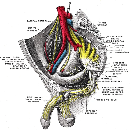
Figure 1Gray837
Long description
Sacral plexus of the right side. (Testut).

Figure 1: Sacral plexus of the right side (Testut).
DefinitionPlexus
A plexus, in a general sense, is a network of fibers or vessels, applying here to nervous tissue connecting spinal roots to peripheral targets.
The most significant systemic nerve to come from this plexus is the sciatic nerve, which is a combination of the tibial nerve and the fibular nerve[1]. The sciatic nerve extends across the hip joint and is most commonly associated with the condition sciatica, which is the result of compression or irritation of the nerve or any of the spinal nerves giving rise to it[1].
NoteClinical Context
Traumatic injury of the LSP is relatively uncommon because LSP is less mobile than the brachial plexus, and protected by pelvic bone, spine and abdominal wall. [2]
The iliohypogastric nerve and the lateral femoral cutaneous nerve are key branches supplying the abdominal wall and thigh. The iliohypogastric nerve provides sensory innervation to the suprapubic region. The lateral femoral cutaneous nerve supplies sensation to the lateral aspect of the thigh.
The sciatic nerve is the largest nerve in the body. It divides into the common fibular (peroneal) nerve and the tibial nerve. The common fibular nerve wraps around the neck of the fibula and is susceptible to injury. The tibial nerve runs down the posterior compartment of the leg.
The superior gluteal nerve innervates the gluteus medius, gluteus minimus, and tensor fasciae latae. The inferior gluteal nerve (L5-S2) supplies the gluteus maximus. Lesions here affect hip extension and abduction.
The pudendal nerve (S2-S4) provides sensory and motor innervation to the perineum. It passes through the greater sciatic foramen and re-enters the pelvis via the lesser sciatic foramen.
Ankle sprains often involve the lateral ligaments. The neurovascular pairing in the lower limb follows specific anatomical corridors. For instance, the posterior tibial neurovascular bundle passes posterior to the medial malleolus.
Exam trapAnatomical Landmark
Do not confuse the anterior tibial artery, which passes anterior to the ankle, with the posterior tibial artery, which is located posterior to the medial malleolus.
While the brachial plexus supplies the upper limb, understanding the lumbosacral plexus requires distinguishing it from the cervical and brachial plexuses. The axilla contains the brachial plexus cords, not the lumbosacral plexus. The lateral thorax is supplied by intercostal nerves.
The axillary nerve wraps around the surgical neck of the humerus. This is a brachial plexus structure, not lumbosacral. Injury here causes deltoid paralysis.
The radial nerve runs in the radial groove of the midshaft of the humerus. This is also part of the brachial plexus. Fractures here can cause wrist drop.
Figure 2Gray822
Long description
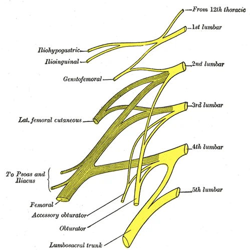
Plan of lumbar plexus. Labelled illustration from Wikimedia Commons, used under its open licence.

Figure 2: Plan of lumbar plexus.
The lumbosacral plexus represents a complex reorganisation of axons from spinal nerves to innervate the lower limb and pelvic structures. Spinal nerves are mixed nerves containing both sensory and motor fibres. There are thirty-one pairs of spinal nerves, including five pairs of lumbar nerves designated L1 to L5 and five pairs of sacral nerves designated S1 to S5. These nerves emerge from the vertebral column through the intervertebral foramen at their respective levels. The lumbosacral plexus is formed by the anterior rami of these spinal nerves, which reorganise into networks of nerve fibres with no associated cell bodies. [1]
The lumbar plexus arises from all the lumbar spinal nerves. Its branches include the iliohypogastric, ilioinguinal, genitofemoral, lateral femoral cutaneous, obturator, and femoral nerves. The iliohypogastric and ilioinguinal nerves course over the psoas muscle and quadratus lumborum muscles[1].
The sacral plexus is formed by the lumbosacral trunk (L4 and L5) and the anterior rami of S1 to S4[1]. Its major branches include the sciatic, common fibular, tibial, superior and inferior gluteal, and pudendal nerves. The sciatic nerve is the largest nerve in the body, formed by the common fibular and tibial components. It exits the pelvis via the greater sciatic foramen. The superior and inferior gluteal nerves innervate the gluteal muscles. The pudendal nerve arises from S2 to S4 an [2]
The obturator nerve is located medially, near the pelvic brim. The femoral nerve lies lateral to the psoas major.
The lumbosacral plexus provides motor and sensory innervation to the lower extremity. The nerves are not straight continuations of the spinal nerves but are reorganised to follow different courses. This organisation allows for efficient innervation of the complex structures of the lower limb and pelvis. Understanding the formation and branching of these nerves is essential for localising lesions and understanding clinical presentations.
Figure 3Gray827
Long description
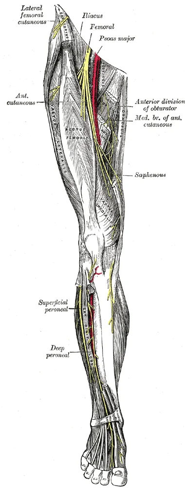
Nerves of the right lower extremity. Front view.

Figure 3: Nerves of the right lower extremity (front view).
DefinitionPlexus
A network of intersecting nerves with no associated cell bodies, formed by the reorganisation of axons from spinal nerves.
| Nerve | Roots | Key Branches/Relations |
|---|---|---|
| Iliohypogastric | L1 | Courses over psoas and quadratus lumborum[1] |
| Ilioinguinal | L1 | Courses over psoas and quadratus lumborum[1] |
| Genitofemoral | L1-L2 | Emerges from anterior surface of psoas major |
| Lateral Femoral Cutaneous | L2-L3 | Pierces iliacus fascia |
| Obturator | L2-L4 | Passes through obturator foramen |
| Femoral | L2-L4 | Largest branch; lies lateral to psoas major |
| Nerve | Roots | Key Branches/Relations |
|---|---|---|
| Sciatic | L4-S3 | Formed by common fibular and tibial components |
| Common Fibular | L4-S2 | Part of sciatic nerve |
| Tibial | L4-S3 | Part of sciatic nerve |
| Superior Gluteal | L4-S1 | Innervates gluteal muscles |
| Inferior Gluteal | L5-S2 | Innervates gluteal muscles |
| Pudendal | S2-S4 | Supplies the perineum |
The lumbosacral plexus (LSP) is a complex network of nerves derived from the anterior rami of the lower spinal nerves. It is not a single linear structure but a branching system located in the retroperitoneal and pelvic spaces. Understanding its course requires visualizing its origin, path, and termination in relation to surrounding musculoskeletal landmarks.
The lumbar plexus arises from all the lumbar spinal nerves. These nerves emerge from the psoas major muscle. The sacral plexus forms from L4 to S4. It lies on the posterior wall of the pelvis, anterior to the piriformis muscle. The lumbosacral trunk connects the two plexuses.
The course of the nerves is defined by their origin from the spinal roots. The roots combine to form trunks, divisions, and cords. These structures travel through specific anatomical compartments. The lumbar plexus is embedded within the psoas major muscle. The nerves emerge from the muscle borders to innervate the anterior and medial thigh. The sacral plexus is located on the pelvic wall. It is covered by the parietal pelvic fascia. The nerves exit the pelvis to reach the lower limb. The sciatic nerve travels down the posterior thigh. It divides into the tibial and common fibular nerves. This division usually occurs in the popliteal fossa. The popliteal fossa is a diamond-shaped space behind the knee. The sciatic nerve lies deep to the biceps femoris tendon. It is medial to the biceps femoris and lateral to the semimembranosus. The tibial nerve is the most superficial structure in the mid-popliteal fossa. The common fibular nerve runs along the lateral border. It winds around the neck of the fibula. This course is vulnerable to injury.
Figure 4Gray824
Long description
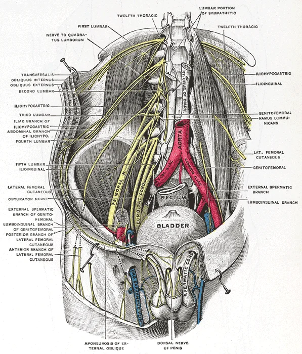
Deep and superficial dissection of the lumbar plexus. (Testut.)

Figure 4: Deep and superficial dissection of the lumbar plexus (Testut.).
The relations of the plexus are vital for diagnosis. The plexus is related to the lumbar vertebrae and sacrum. It is anterior to the quadratus lumborum and piriformis muscles. It is medial to the iliacus muscle. The ureter and genital branch of the genitofemoral nerve are related to the lumbar plexus. The internal iliac vessels are related to the sacral plexus. These relations explain the symptoms of space-occupying lesions. Tumours in the retroperitoneum can compress the plexus. This causes pain and weakness in the lower limb. The specific pattern of weakness depends on the affected nerve. For example, compression of the femoral nerve causes quadriceps weakness. Compression of the sciatic nerve causes foot drop. Understanding these relations allows for accurate localization of pathology.
| Muscle | Origin | Insertion | Action | Nerve Supply |
|---|---|---|---|---|
| Psoas major | Transverse processes of T12-L5 | Lesser trochanter | Hip flexion | Lumbar plexus |
| Iliacus | Iliac fossa | Lesser trochanter | Hip flexion | Femoral nerve |
| Piriformis | Sacrum | Greater trochanter | Hip external rotation | Sacral plexus |
The termination of the plexus is in the peripheral nerves of the lower limb. The femoral nerve terminates in the anterior compartment of the thigh. The sciatic nerve terminates in the leg and foot. The obturator nerve terminates in the medial compartment. Each nerve has a specific distribution. This distribution is tested in clinical examinations. The sensory supply follows the dermatomal pattern. The motor supply follows the myotomal pattern. This organization is consistent and predictable. It forms the basis of neurological assessment.
Figure 5Gray823
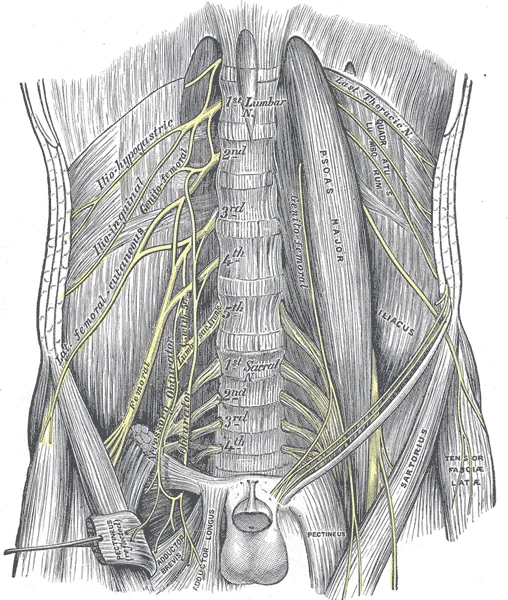
Long description
The lumbar plexus and its branches. Labelled illustration from Wikimedia Commons, used under its open licence.

Figure 5: The lumbar plexus and its branches.
Figure 61321 Spinal Nerve Plexuses
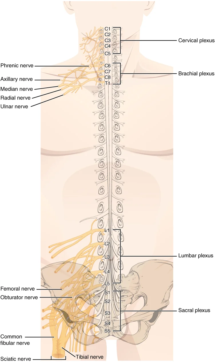
Long description
There are four main nerve plexuses in the human body. The cervical plexus supplies nerves to the posterior head and neck, as well as to the diaphragm. The brachial plexus supplies nerves to the arm. The lumbar plexus supplies nerves to the anterior leg. The sacral plexus supplies nerves to the poste

Figure 6: There are four main nerve plexuses in the human body.
These nerves emerge from the spinal cord and divide into roots, trunks, divisions, cords, and finally terminal branches. The plexus is not a single entity but two interconnected networks: the lumbar plexus and the sacral plexus. Understanding their branches requires tracing the path from the spinal nerve to the specific muscle or skin area it innervates. [1]
The lumbar plexus is situated within the substance of the psoas major muscle. The sacral plexus lies on the posterior wall of the pelvis, anterior to the piriformis muscle. The two plexuses are connected by the lumbosacral trunk, which passes over the sacroiliac joint. The femoral nerve emerges from the lateral border of the psoas major. The obturator nerve emerges from the medial border. The sciatic nerve is the largest branch, emerging from the greater sciatic foramen below the piriformis. The pudendal nerve exits the pelvis through the greater sciatic foramen and re-enters through the lesser sciatic foramen.
Figure 7Gray829
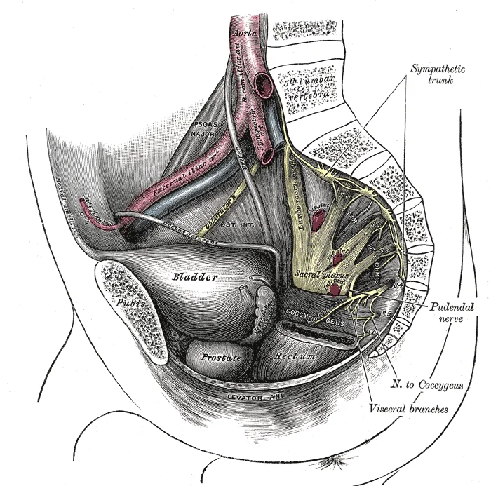
Long description
Dissection of side wall of pelvis showing sacral and pudendal plexuses. (Testut.)

Figure 7: Dissection of side wall of pelvis showing sacral and pudendal plexuses (Testut.).
Figure 8Gray828
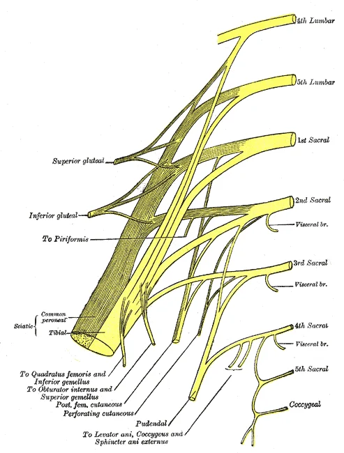
Long description
Plan of sacral and pudendal plexuses. Labelled illustration from Wikimedia Commons, used under its open licence.

Figure 8: Plan of sacral and pudendal plexuses.
| Nerve | Origin | Motor Supply | Sensory Supply |
|---|---|---|---|
| Femoral Nerve | L2-L4 | Iliopsoas, Sartorius, Quadriceps | Anterior thigh, medial leg |
| Obturator Nerve | L2-L4 | Adductor muscles | Medial thigh |
| Sciatic Nerve | L4-S3 | Hamstrings, muscles of leg and foot | Posterior thigh, leg, foot |
| Pudendal Nerve | S2-S4 | Perineal muscles | Perineum, external genitalia |
The femoral nerve is the largest branch of the lumbar plexus. It supplies the anterior compartment of the thigh. The primary muscles innervated are the quadriceps femoris, which extends the knee. The sartorius and pectineus are also supplied. Sensory innervation covers the anterior thigh and the medial aspect of the leg via the saphenous nerve.
The obturator nerve arises from the medial aspect of the lumbar plexus. It passes through the pelvis and enters the thigh via the obturator canal. It supplies the medial compartment of the thigh, including the adductor longus, adductor brevis, and adductor magnus. It provides sensory innervation to the medial thigh.
The sacral plexus gives rise to the sciatic nerve. This nerve is formed by the anterior rami of L4 through S3. It exits the pelvis via the greater sciatic foramen. It divides into the tibial and common fibular nerves. The tibial nerve supplies the posterior compartment of the leg and the sole of the foot. The common fibular nerve supplies the lateral and anterior compartments of the leg.
The pudendal nerve arises from S2 to S4. It supplies the perineum and external genitalia. It innervates the external urethral and anal sphincters. Sensory innervation includes the skin of the perineum and the external genitalia.
DefinitionDermatome
A dermatome is an area of skin supplied by a single spinal nerve.
DefinitionMyotome
A myotome is a group of muscles primarily innervated by a single spinal nerve.
Lesions of these nerves result in specific motor and sensory deficits. A femoral nerve lesion causes weakness in knee extension and sensory loss in the anterior thigh. An obturator nerve lesion leads to weakness in thigh adduction. A sciatic nerve lesion results in weakness in knee flexion and foot movement. A pudendal nerve lesion causes perineal sensory loss and sphincter dysfunction.
| Nerve | Motor Deficit | Sensory Deficit |
|---|---|---|
| Femoral | Weak knee extension | Anterior thigh, medial leg |
| Obturator | Weak thigh adduction | Medial thigh |
| Sciatic | Weak knee flexion, foot drop | Posterior leg, foot |
| Pudendal | Sphincter dysfunction | Perineum, genitalia |
The lumbosacral plexus is susceptible to pathology occurring both within and outside the spinal canal. Extraspinal causes are diverse, including entrapment neuropathies, traumatic injuries, infectious or inflammatory diseases, primary or secondary tumours, and vascular lesions. Timely detection of these lesions is facilitated by magnetic resonance neurography (MRN). [2]
The lumbosacral plexus nerves emerge from the psoas muscle belly.
Routine MRN protocols include 2D and isotropic 3D sequences with T1- and T2-weighted contrast. Axial T1-weighted images without fat suppression and fluid-sensitive T2-weighted images are obtained with high resolution (matrix size more than 256 in both directions) to depict the fascicular pattern of the nerves. Coronal T1-weighted and short tau inversion recovery (STIR), and proton density-weighted Spectral Attenuated Inversion Recovery (SPAIR) images show lesions along the long axis of the nerves are composed of more than just nervous tissue. [1][2]
Uniform fat suppression is critical in assessing the signal intensity of nerves. Frequency-selective fat suppression techniques are vulnerable to B0 and B1 inhomogeneity in large FOV imaging. STIR imaging offers robust fat suppression. However, TE should be lowered around 30–50 ms to maintain good SNR. 3D FSE combined with STIR is a promising technique for LSP evaluation due to its high CNR between nerve and soft tissues and capability of multiplanar reconstruction. 3D STIR FSE depicts lesions along the long axis of the nerves and provides anatomic information that helps optimize preoperative planning. [2]
The role of intravenous contrast in MRN is variably reported in the literature. Intravenous contrast may provide additional benefit in pathologic conditions, which may violate the integrity of the blood-nerve barrier. Such conditions include neural and perineural mass lesions, postoperative adhesion, radiation injury, inflammation, polyneuropathy conditions including lymphoma, amyloidosis, demyelinating neuropathies, and hereditary neuropathies. [2]
Metal artifacts degrade image quality and mimic abnormalities of peripheral nerves, in particular increased T2 signal on fat-suppressed T2-weighted images. 1.5T is preferred to 3T scanner for reducing metallic artifact if metal is present in the area of clinical concern. However, the optimal quality for nerve imaging at 3T outweighs the image degradation. Conventional artifacts reduction strategies should be implemented by decreasing TE, increasing receiver bandwidth, placing frequency-encoding gradient along the axis of metallic instrument, using high resolution matrix and thin slice thickness. [2]
| Feature | Description |
|---|---|
| Pathology Location | Within or outside spinal canal |
| Extraspinal Causes | Entrapment, trauma, infection, inflammation, tumour, vascular |
| Key Imaging Sequence | 2D/3D T1/T2 with high resolution matrix >256 |
| Fat Suppression | STIR preferred for robust suppression and metal artifact reduction, |
| Contrast Indication | Blood-nerve barrier violation (tumour, inflammation, neuropathy) |
| Metal Artifact Strategy | 1.5T preferred; FSE 3D over GRE 3D |
Clinical pearlImaging Pearl
STIR sequences are the most viable option for effective fat suppression under the influence of metallic artifacts. [2]
NoteClinical Note
Intravenous contrast may provide additional benefit in pathologic conditions, which may violate the integrity of the blood-nerve barrier.
The clinical examination of the lumbosacral plexus requires a systematic approach to localise lesions within the peripheral nervous system. Understanding the structural organisation of these nerves is fundamental. Bundles of axons in the peripheral nervous system are referred to as nerves. These structures differ from their central counterparts, known as tracts, by their composition and investment. Nerves are not solely nervous tissue; they contain connective tissues and blood vessels that supply. Individual axons are surrounded by loose connective tissue called the endoneurium. [1]
DefinitionNerve Structure
Nerves consist of axons bundled into fascicles, surrounded by epineurium, perineurium, and endoneurium, with blood vessels providing nourishment.
Clinical assessment begins with identifying signs of radiculopathy, which often mimic plexus pathology. Magnetic resonance neurography (MRN) is a valuable tool for displaying the distribution of peripheral nerve disease and guiding perineural injections. It is particularly useful for assessing extraspinal causes of sciatica. The mainstay imaging sequence is three-dimensional short-tau inversion recovery turbo spin-echo combined with vascular suppression. This technique allows for high-resolution [2]
NoteImaging Modality
MRN is useful for displaying peripheral nerve disease distribution and guiding injections. Three-dimensional short-tau inversion recovery turbo spin-echo with vascular suppression is the mainstay technique.
Diffusion-weighted imaging (DWI) effectively improves nerve conspicuity by suppressing background tissues and vessels. For the lumbosacral plexus, axial diffusion tensor imaging is obtained with a b-value of 600–900 and 15–20 directions. This provides quantitative values such as the apparent diffusion coefficient and fractional anisotropy. There is a significant correlation between abnormal intraneural T2 signal on MRN and active radiculopathy verified on electromyography. However, positive MRN [2]
Exam trapImaging Limitations
Contrast-enhanced imaging does not add value in trauma or entrapment neuropathy cases. Most patients undergo MRN in subacute stages where contrast does not change diagnostic interpretation. There is no difference in nerve visualization with or without contrast-enhancement. [2]
Nerve injuries are classified by their anatomical level. Spinal nerves of the thoracic region, T2 through T11, are not part of the plexuses. They emerge and give rise to the intercostal nerves found between the ribs. These nerves articulate with the vertebrae surrounding the spinal nerve. In contrast, the lumbosacral plexus involves complex branching patterns. MRN led to an alternative diagnosis in 18 out of 23 patients with clinically suspected chronic cauda equina syndrome. This highlights the diagnostic certainty MRN provides in complex cases. [2] [1]
| Feature | Description | Clinical Utility |
|---|---|---|
| STIR Sequence | Three-dimensional short-tau inversion recovery with vascular suppression | Mainstay for displaying nerve disease distribution |
| DWI/DTI | b-value 600–900, 15–20 directions | Provides ADC and FA values; improves conspicuity |
| Contrast | Not routinely used | No added value in trauma or entrapment neuropathy |
Diagnostic Approach to Sciatica
- Patient presents with sciaticaAssess for radiculopathy vs plexus lesion
- Question: Routine spine MRI normal?
No
- Treat identified spinal pathology
Yes
- Perform abbreviated MRN (coronal STIR)
Clinical pearlDiagnostic Insight
MRN led to an alternative diagnosis in 18 out of 23 patients with clinically suspected chronic cauda equina syndrome. Abbreviated MRN can discover abnormalities missed by routine spine MRI.
The lumbosacral plexus spans a large area, making implementation of MRN challenging. However, it plays an important role in diagnostic certainty and treatment decision-making. While most studies are retrospective, the growing body of evidence supports its utility. Future work should focus on clinical validation and developing practical guidelines. [2]
The lumbosacral plexus is formed by the anterior rami of the lower thoracic, lumbar, and sacral spinal nerves. Understanding its anatomy requires distinguishing between the lumbar plexus, located within the psoas major muscle, and the sacral plexus, situated on the posterior pelvic wall.
The lumbar plexus arises from all the lumbar spinal nerves. These nerves emerge from the vertebral column and enter the substance of the psoas major muscle. Within this muscle, the rami communicate to form the plexus. The plexus is located in the posterior part of the abdomen, embedded within the psoas major. [1]
DefinitionLumbar Plexus Origin
The lumbar plexus arises from all the lumbar spinal nerves.
The sacral plexus is formed by the lumbosacral trunk (L4-L5) and the anterior rami of S1 to S4. The lumbosacral trunk descends over the sacral ala to join the sacral nerves. The plexus lies on the posterior wall of the pelvis, anterior to the piriformis muscle. It is covered by the pelvic parietal peritoneum anteriorly and the sacrum posteriorly.
DefinitionSacral Plexus Origin
The sacral plexus is formed by the lumbosacral trunk and anterior rami of S1 to S4.
The femoral nerve (L2-L4) is a major branch of the lumbar plexus. It passes under the inguinal ligament to enter the femoral triangle. It supplies the anterior compartment of the thigh. Injury to the femoral nerve results in weakness of knee extension and loss of the patellar reflex.
The sciatic nerve (L4-S3) is the largest nerve in the body. It exits the pelvis through the greater sciatic foramen, below the piriformis muscle. Compression or injury can lead to foot drop and sensory loss in the leg and foot.
Exam trapSciatic Nerve Path
The sciatic nerve typically passes inferior to the piriformis muscle, not through it.
The following table summarises the key muscles innervated by the lumbar plexus.
| Muscle | Nerve Supply | Action |
|---|---|---|
| Iliopsoas | Lumbar plexus (direct branches) | Hip flexion |
| Sartorius | Femoral nerve | Hip flexion, knee flexion |
| Quadriceps Femoris | Femoral nerve | Knee extension |
The posterior compartment of the thigh is innervated by the sciatic nerve.
| Muscle | Nerve Supply | Action |
|---|---|---|
| Biceps Femoris | Sciatic nerve | Knee flexion, hip extension |
| Semitendinosus | Sciatic nerve | Knee flexion, hip extension |
| Semimembranosus | Sciatic nerve | Knee flexion, hip extension |
Trauma to the lower back or pelvis can damage the lumbosacral plexus. Obstetric injuries during childbirth may affect the sacral plexus. Diabetic amyotrophy is a condition that can cause lumbosacral plexopathy. Symptoms include pain, weakness, and sensory deficits in the distribution of the affected nerves.
NoteDiabetic Amyotrophy
This condition typically presents with asymmetric pain and weakness in the proximal lower limbs.
The lumbosacral plexus also has connections with the autonomic nervous system. Parasympathetic input to the pelvic organs arises from sacral spinal nerves S2 to S4. These nerves contribute to the formation of the pelvic plexus.
DefinitionParasympathetic Input
Sacral spinal nerves S2 to S4 provide parasympathetic input to the pelvic organs.
Students frequently confuse the anatomical boundaries of the lumbosacral plexus, leading to errors in localisation and clinical correlation. A primary error is assuming the plexus is confined strictly to the pelvic cavity. In reality, the plexus originates from the anterior rami of the lumbar and sacral spinal nerves, which emerge from the vertebral column before descending into the retroperitoneal and pelvic spaces. The lumbar plexus forms within the substance of the psoas major muscle, while the sacral plexus lies on the posterior pelvic wall. Misidentifying the origin as purely pelvic ignores the lumbar contribution from L1 to L4.
Another frequent mistake involves the relationship between the plexus and the vertebral column. Students often overlook that the plexus is anterior to the sacrum and piriformis muscle. The sacral plexus is formed by the lumbosacral trunk (L4–L5) and the anterior rami of S1–S4. It lies on the posterior wall of the pelvis, anterior to the piriformis muscle, and is covered by the pelvic fascia. Confusing this with the lumbar plexus, which is embedded in the psoas major, leads to incorrect surgical approaches and imaging interpretations.
A third pitfall concerns the interpretation of magnetic resonance neurography (MRN) sequences. Students may not appreciate the importance of the field of view (FOV) and coil selection. Dedicated high-resolution multichannel phased-array surface coils are essential to enhance the signal over a larger field-of-view without sacrificing the signal-to-noise ratio (SNR). Using a standard spine coil without combining it with multi-channel body matrix coils can result in poor image quality for the lower plexus. Furthermore, the optimal FOV for LSP imaging should include the lower lumbar spines, pelvis, and proximal thighs. [2]
Metal artifacts degrade image quality and mimic abnormalities of peripheral nerves, in particular increased T2 signal on fat-suppressed T2-weighted images. 1.5T is preferred to 3T scanner for reducing metallic artifact if metal is present in the area of clinical concern. However, the optimal quality for nerve imaging at 3T outweighs the image degradation. Conventional artifacts reduction strategies should be implemented by decreasing TE, increasing receiver bandwidth, placing frequency-encoding gradient along the axis of metallic instrument, using high resolution matrix and thin slice thickness. Instead of GRE-based 3D sequences, FSE-based 3D sequence should be chosen. [2]
Hyperintense vascular structures accompanying nerves can hinder a clear view of peripheral nerves on fluid-sensitive sequences. Students often mistake these vascular signals for nerve pathology or inflammation. Techniques such as PSIF (reversed fast imaging with steady-state precession) can reduce hyperintense vessels by dropping the signal in flowing blood. However, this sequence is vulnerable to local field inhomogeneity with a large FOV, limiting its utility for the entire lumbosacral plexus.
The use of gadolinium contrast is another area of confusion. While gadolinium enhancement is preferable in inflammatory or tumorous involvement of the LSP, its routine use is controversial. Intravenous gadolinium with STIR can clearly visualise the LSP by suppressing vascular signal, but it is unclear whether this improves diagnostic performance over suboptimal vascular suppression without contrast. Students should not assume contrast is always required for a diagnostic study. [2]
Finally, diffusion tensor imaging (DTI) is technically demanding for peripheral nerves. Although DTI is well-established in the brain, it requires high SNR and high resolution in an affordable scan time for clinical practice. Students should not rely on DTI as a routine part of the LSP protocol without understanding its technical limitations and the need for further evidence-based validation.
Knowledge of the lumbosacral plexus is critical at the bedside for localising neurological deficits. When a patient presents with weakness or sensory loss in the lower limb, the clinician must determine if the lesion is radicular, plexus-related, or peripheral nerve-specific. For example, weakness in hip flexion and knee extension suggests involvement of the femoral nerve, which arises from the lumbar plexus (L2–L4). Sensory loss over the medial thigh and leg further supports this localisation. Understanding the specific roots (L2–L4) helps differentiate between a high lumbar disc herniation and a plexus lesion.
In the operating theatre, anatomical knowledge is vital for surgical approaches to the pelvis and retroperitoneum. Surgeons performing procedures such as retroperitoneal lymph node dissection or pelvic tumour resection must identify the plexus to avoid iatrogenic injury. The sacral plexus lies on the posterior pelvic wall, anterior to the piriformis muscle. Injury to the sciatic nerve, a major branch of the sacral plexus, can result in foot drop and sensory loss in the lower leg and foot. Preoperative planning involves mapping the relationship between the tumour and the plexus.
On imaging, MRN is the gold standard for evaluating the lumbosacral plexus. Optimal imaging requires a coronal FOV of 300–320 mm centered at L5 to cover the LSP from the L1 root to the pelvic floor. This ensures visualization of the pudendal nerves, which innervate the pelvic floor. For evaluating the sciatic nerves, lowering the center of the FOV to S1–S2 root levels is more advantageous. Including both sides in the FOV facilitates side-to-side comparison of nerve morphology and signal intensity. [2]
Axial slice thickness should be increased up to 5–6 mm with a 2–4 mm interslice gap to achieve adequate coverage in a reasonable scan time. High in-plane resolution is maintained to detect subtle nerve abnormalities. After screening a large area, imaging on a specific area of interest using a small FOV can be performed to obtain high-resolution MRN images. This targeted approach improves diagnostic accuracy for focal lesions. [2]
In examinations, students are often tested on the components and branches of the plexus. The lumbar plexus gives rise to the iliohypogastric, ilioinguinal, genitofemoral, lateral femoral cutaneous, and femoral nerves. The sacral plexus gives rise to the sciatic, superior gluteal, inferior gluteal, pudendal, and posterior femoral cutaneous nerves. Memorising the origin, course, and termination of these nerves is essential. For instance, the sciatic nerve is formed by the anterior divisions of L4–S3 and exits the pelvis through the greater sciatic foramen below the piriformis muscle.
Lumbosacral Plexus Imaging Protocol
- StartPatient with suspected LSP pathology
- Question: Select Coil
Yes
- Use dedicated high-resolution multichannel phased-array surface coils
No
- Combine spine array coil with multi-channel body matrix coils
- Question: Set FOV
General LSP
- Coronal FOV 300–320 mm centered at L5
Sciatic Nerve
- Lower FOV center to S1–S2 root levels
- Question: Sequence Parameters
Non-fat-suppressed T2
- Effective TE > 90 ms
Fat-suppressed T2
- Effective TE 60–80 ms
- Complete ScanInclude both sides for comparison; consider contrast if tumour/inflammation suspected
| Nerve | Origin | Key Relations | Clinical Significance |
|---|---|---|---|
| Femoral | L2–L4 | Within psoas major, passes under inguinal ligament | Weakness in knee extension, sensory loss in anterior thigh |
| Sciatic | L4–S3 | Exits pelvis below piriformis | Foot drop, sensory loss in leg and foot |
| Pudendal | S2–S4 | Exits pelvis via greater sciatic foramen, re-enters via lesser sciatic foramen | Pelvic floor dysfunction, perineal sensory loss |
| Superior Gluteal | L4–S1 | Exits pelvis above piriformis | Weakness in hip abduction, Trendelenburg gait |
The lumbosacral plexus originates from the anterior rami of lumbar and sacral spinal nerves. The lumbar plexus forms within the psoas major muscle, while the sacral plexus lies on the posterior pelvic wall. Dedicated high-resolution multichannel phased-array surface coils are essential for high-quality MRN. Optimal FOV for LSP imaging includes the lower lumbar spines, pelvis, and proximal thighs. Effective TE should be >90 ms for non-fat-suppressed T2 and 60–80 ms for fat-suppressed T2. [2] Gadolinium contrast is preferable in inflammatory or tumorous involvement but is controversial for routine use. DTI is technically demanding and not yet standard for routine LSP clinical practice. Side-to-side comparison is facilitated by including both sides in the FOV.
Answer each one in your head first, then open the answer.
What is the optimal FOV for general LSP imaging?
Show answerHide answer
300–320 mm centered at L5What is the effective TE for non-fat-suppressed 3D FSE T2-weighted imaging?
Show answerHide answer
More than 90 msWhich coils are essential for enhancing signal in LSP imaging?
Show answerHide answer
Dedicated high-resolution multichannel phased-array surface coilsWhere does the sacral plexus lie?
Show answerHide answer
On the posterior pelvic wall, anterior to the piriformis muscleWhat is the advantage of lowering the FOV center to S1–S2?
Show answerHide answer
More advantageous in evaluating the sciatic nerves
- Betts JG, Young KA, Wise JA, et al. The Peripheral Nervous System. OpenStax, Anatomy and Physiology 2e (CC BY-NC-SA 4.0). Accessed .
- Kim SG, Jung JY. Role of MR Neurography for Evaluation of the Lumbosacral Plexus: A Scoping Review. Journal of the Korean Society of Radiology 2022;83(6):1273-1285. doi:10.3348/jksr.2022.0001. Accessed .
Figure credits
- Figure 1: Henry Vandyke Carter, via Wikimedia Commons, Public domain, Public domain.
- Figure 2: Henry Vandyke Carter, via Wikimedia Commons, Public domain, Public domain.
- Figure 3: Henry Vandyke Carter, via Wikimedia Commons, Public domain, Public domain.
- Figure 4: Henry Vandyke Carter, via Wikimedia Commons, Public domain, Public domain.
- Figure 5: Henry Vandyke Carter, via Wikimedia Commons, Public domain, Public domain.
- Figure 6: OpenStax, via Wikimedia Commons, CC BY 4.0, CC BY 4.0.
- Figure 7: Henry Vandyke Carter, via Wikimedia Commons, Public domain, Public domain.
- Figure 8: Henry Vandyke Carter, via Wikimedia Commons, Public domain, Public domain.
Educational content for medical students and doctors. It is not a substitute for clinical judgement or local guidelines.