Meninges
The meninges are the three protective layers enclosing the central nervous system, defining spaces for CSF and vessels.
Figure 1Gray567
Long description
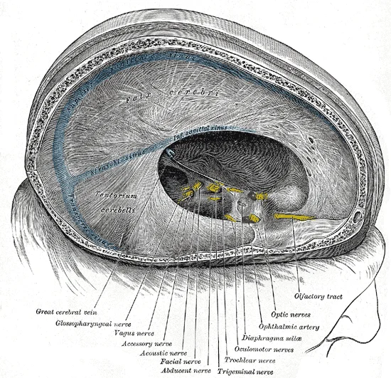
Dura mater and its processes exposed by removing part of the right half of the skull, and the brain.

Figure 1: Dura mater and its processes exposed by removing part of the right half of the skull, and the brain.
Figure 2Gray765
Long description
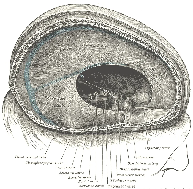
Dura mater and its processes exposed by removing part of the right half of the skull and the brain.

Figure 2: Dura mater and its processes exposed by removing part of the right half of the skull and the brain.
The central nervous system components are encased within three distinct layers known as the meninges[1]. The outermost layer is the dura mater, which lies in close contact with the periosteum of the skull[1]. The middle layer is the arachnoid mater, described as spiderweb-like, and it is loosely adherent to the brain[1]. The innermost layer is the pia mater, which is closely adherent to the brain and follows the contours of the sulci and gyri on the cerebral cortex[1]. [2]
These layers define three spaces. The epidural space exists between the skull and the dura mater[1]. The subdural space is found between the dura and arachnoid mater[1]. The subarachnoid space lies between the arachnoid and pia mater[1]. The epidural and subdural spaces are potential spaces[1]. The subarachnoid space contains cerebrospinal fluid (CSF), major vascular structures, and the subarachnoid cisterns[1].
Figure 3Gray768
Long description
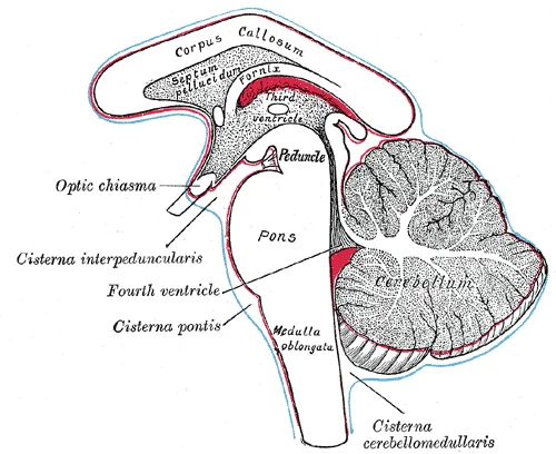
Diagram showing the positions of the three principal subarachnoid cisternæ.

Figure 3: Diagram showing the positions of the three principal subarachnoid cisternæ.
The subarachnoid cisterns are clinically and surgically significant because they transmit important neurovascular structures, including cranial nerves and intracranial vessels[1]. Although often described as distinct compartments, the cisterns are in free communication with each other and with the rest of the subarachnoid space[1]. They are not anatomically separate entities[1].
On imaging, cisternal anatomy is key to diagnosing various pathologies. The medullary cistern encases the medulla oblongata and communicates with the cisterna magna via the cerebello-medullary fissure. This cistern contains the glossopharyngeal nerve (CN IX), vagus nerve (CN X), and accessory nerve (CN XI). It also contains the PICA, lateral medullary vein, pontomedullary vein, and transverse medullary vein. Recognizing these [1]
DefinitionMeninges
The three protective layers (dura, arachnoid, pia) enclosing the CNS, defining the epidural, subdural, and subarachnoid spaces.
| Layer | Position | Adherence |
|---|---|---|
| Dura mater | Outermost | Close to periosteum |
| Arachnoid mater | Middle | Loose adherence |
| Pia mater | Innermost | Close to brain surface |
The innervation of the meninges involves sensory supply from the trigeminal nerve for supratentorial structures and upper cervical nerves for infratentorial structures. This differential innervation explains why pathology in different regions causes distinct headache patterns.
Meningitis involves inflammation of these layers, leading to meningeal signs such as neck stiffness. The anatomy of lumbar puncture targets the subarachnoid space in the lumbar region, below the termination of the spinal cord, to access CSF without damaging neural tissue.
Herniation syndromes occur when increased intracranial pressure forces brain tissue across rigid dural folds or through foramina. For instance, uncal herniation involves the uncus moving over the tentorial edge, compressing the oculomotor nerve and cerebral peduncle. [2]
Exam trapPotential Spaces
The epidural and subdural spaces are potential, meaning they only open under pathological conditions like bleeding or fluid accumulation.
The meninges are the protective membranes enveloping the brain and spinal cord. They consist of three distinct layers, arranged from superficial to deep: the dura mater, the arachnoid mater, and the pia mater. Understanding their gross anatomical relationships is fundamental to neurosurgical navigation and radiological interpretation. [1]
The outermost layer, the dura mater, appears as a thick, dense fibrous membrane adhering tightly to the inner surface of the skull. Beneath the dura lies the potential subdural space, which is normally collapsed. The middle layer, the arachnoid mater, is a delicate, avascular web-like membrane. It does not dip into the cerebral sulci but bridges across them. Between the arachnoid and the pia mater lies the subarachnoid space, which is wide and contains cerebrospinal fluid. The innermost layer, the pia mater, is a thin, highly vascular membrane that adheres directly to the surface of the brain, dipping into every sulcus and fissure.
Figure 4Gray769
Long description
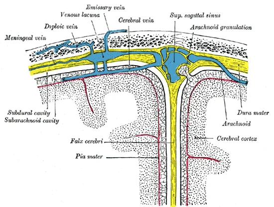
Diagrammatic representation of a section across the top of the skull, showing the membranes of the brain, etc. (Modified from Testut.)

Figure 4: Diagrammatic representation of a section across the top of the skull, showing the membranes of the brain, etc (Modified from Testut.).
DefinitionDura Mater
The tough, outermost meningeal layer derived from the endosteal and meningeal layers, providing structural support and containing venous sinuses.
The dura mater is the strongest and thickest of the three meningeal layers. It consists of two layers: the periosteal layer, which lines the inner surface of the skull bones, and the meningeal layer, which forms the true dura. These two layers separate in certain regions to form dural venous sinuses, which drain blood from the brain. The dura mater is innervated by the trigeminal nerve in the cranial cavity and by spinal nerves in the vertebral canal.
DefinitionArachnoid Mater
The intermediate, web-like meningeal layer that lies beneath the dura mater and above the pia mater, separated from the dura by the subdural space.
The outermost bright line represents the skull. Immediately internal to the bone is the dura mater. In cases of trauma, blood may accumulate between the skull and dura, creating an epidural haematoma.
DefinitionPia Mater
The delicate, innermost meningeal layer that adheres directly to the surface of the brain and spinal cord, following all contours and dipping into sulci.
The pia mater is a thin, highly vascular membrane that closely adheres to the surface of the brain and spinal cord. It follows every contour of the underlying neural tissue, dipping into the sulci and fissures. The pia mater provides a protective barrier and supplies blood vessels to the underlying brain tissue. It is continuous with the ependymal lining of the ventricles at the foramina of Luschka and Magendie.
The subarachnoid space lies between the arachnoid mater and the pia mater. It is filled with cerebrospinal fluid and contains the major arteries and veins of the brain. The space is wider at the base of the brain, forming the subarachnoid cisterns. These cisterns are important landmarks for neurosurgeons and radiologists.
The meninges consist of three distinct layers: the dura mater, arachnoid mater, and pia mater. In cross-section, these layers define the subarachnoid space, which is filled with cerebrospinal fluid (CSF) and contains major cerebral vessels. The arachnoid mater bridges across the surface of the brain, creating enclosed CSF-filled spaces known as cisterns. These cisterns are critical neuroanatomical landmarks where nerves and arteries traverse the cranial cavity. Understanding their internal structure requires examining specific named cisterns, their boundaries, and their contents.
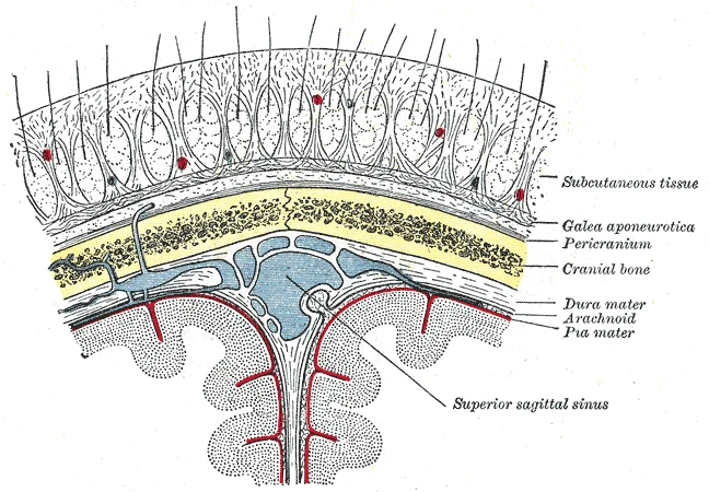
Figure 5Gray1196
Long description
Layers of the scalp, skull and meninges, including the superior sagittal sinus

Figure 5: Layers of the scalp, skull and meninges, including the superior sagittal sinus.
Anterior to this is the cistern of the lamina terminalis, a tent-shaped structure in the midline of the basal telencephalon. Posteriorly, the pericallosal cistern extends under the falx cerebri between the cerebral hemispheres and above the corpus callosum.
The lamina terminalis forms the cistern of lamina terminalis’s posterior wall. [1]
The pericallosal cistern encases the pericallosal artery. It also houses the origins of the frontopolar, fronto-orbital, and callosomarginal arteries. Anterior cerebral and orbital veins are also present. [1]
The cisterna magna is the biggest of all subarachnoid cisterns. It is located in the midline under the cerebellum and behind the medulla. The PICA membrane divides it into the vallecular cistern (medial) and PICA cistern (lateral). The fifth segment of the PICA lies in the vallecular cistern, while the third and fourth segments lie in the PICA cistern. Other structures include the vertebral arteries, inferior vermian vein, and medial posterior medullary vein. [1]
The olfactory cisterns are situated in the olfactory sulcus, delimited laterally by the posterior orbital gyri and medially by the rectus gyri. They contain the olfactory bulb, tracts, nerves, and orbitofrontal artery. [2]
DefinitionSubarachnoid cistern
An enclosed subarachnoid space containing CSF and neurovascular structures.
The meninges are not merely passive coverings but active interfaces connecting the central nervous system to the peripheral vascular and lymphatic networks. Understanding these connections requires tracing the afferent sensory supply and the efferent venous drainage, which dictate both pain perception and the spread of pathology.
Sensory innervation of the meninges is segmental and follows a predictable pattern based on the anatomical region. The dura mater of the anterior and middle cranial fossae receives afferent fibres from the ophthalmic division of the trigeminal nerve (CN V1). [1]
In contrast, the dura of the posterior cranial fossa is innervated by the upper cervical spinal nerves (C1–C3) and the vagus nerve (CN X). Pain arising from the meninges is thus strictly dural or vascular in origin. [1]
Clinical pearlReferred Pain
Pain from the posterior fossa dura is referred to the C2–C3 dermatomes, manifesting as occipital or suboccipital headache.
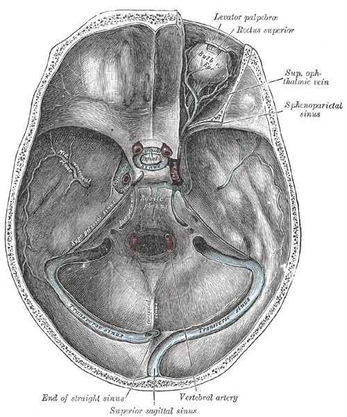
The venous drainage of the meninges is complex, involving both extracranial and intracranial pathways that bypass the blood-brain barrier. The dural venous sinuses collect blood from the brain and drain into the internal jugular veins. [1]
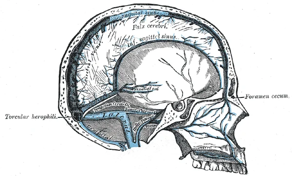
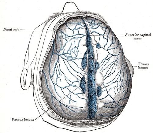
The superior sagittal sinus runs along the attached margin of the falx cerebri and drains into the confluence of sinuses. It receives arachnoid granulations, which allow cerebrospinal fluid to enter the venous system. The inferior sagittal sinus runs in the free edge of the falx cerebri and joins the great cerebral vein to form the straight sinus. [1]
Figure 6Gray568
Long description
Sagittal section of the skull, showing the sinuses of the dura.

Figure 6: Sagittal section of the skull, showing the sinuses of the dura.
Figure 7Gray566
Long description
Superior sagittal sinus laid open after remova of the skull cap. The chordæ Willisii are clearly seen. The venous lacunæ are also well shown; from two of them probes are passed into the superior sagittal sinus. (Poirier and Charpy.)

Figure 7: Superior sagittal sinus laid open after remova of the skull cap.
| Sinus | Location | Drainage Destination |
|---|---|---|
| Superior Sagittal Sinus | Attached margin of falx cerebri | Confluence of sinuses |
| Inferior Sagittal Sinus | Free edge of falx cerebri | Great cerebral vein |
| Straight Sinus | Junction of falx cerebri and tentorium | Confluence of sinuses |
| The suprasellar cistern is situated superiorly to sella turcica. [1] |
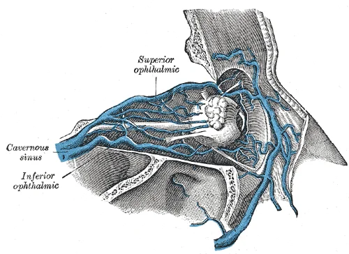
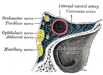
The cavernous sinus is a critical connection point, receiving blood from the ophthalmic veins and the superficial middle cerebral vein. It contains the internal carotid artery and cranial nerves III, IV, V1, V2, and VI within its lateral wall. Infection or thrombosis here can spread rapidly to the contralateral side via intercavernous sinuses. [1]
Figure 8Gray572
Long description
Veins of orbit. (Poirier and Charpy.). Labelled illustration from Wikimedia Commons, used under its open licence.

Figure 8: Veins of orbit (Poirier and Charpy.).
Figure 9Gray571
Long description
Oblique section through the cavernous sinus.

Figure 9: Oblique section through the cavernous sinus.
Figure 10Gray1198
Long description
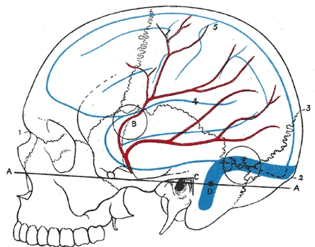
Relations of the brain and middle meningeal artery to the surface of the skull. 1. Nasion. 2. Inion. 3. Lambda. 4. Lateral cerebral fissure. 5. Central sulcus. AA. Reid’s base line. B. Point for trephining the anterior branch of the middle meningeal artery. C. Suprameatal triangle. D. Sigmoid bend o

Figure 10: Relations of the brain and middle meningeal artery to the surface of the skull (1).
The vascular architecture of the meninges is distinct from the brain parenchyma, reflecting the protective and structural roles of these coverings. Understanding this supply requires distinguishing between the three layers: the dura mater, the arachnoid mater, and the pia mater. Each layer receives its blood supply from different sources, primarily branches of the external carotid artery and the internal carotid artery, with significant anastomoses ensuring robust perfusion.
The dura mater is the tough, outermost meningeal layer. It is supplied mainly by meningeal branches of the external carotid artery and the internal carotid artery. The middle meningeal artery is the largest and most clinically significant vessel. It arises from the maxillary artery, a branch of the external carotid artery. It enters the cranial cavity through the foramen spinosum in the sphenoid bone. It runs between the dura mater and the inner surface of the skull. It divides into anterior and posterior branches. The anterior branch runs forward and upward, supplying the frontal and parietal lobes. The posterior branch runs backward, supplying the occipital and parietal regions. The anterior branch is more superficial and lies closer to the pterion, a thin area of the skull where the frontal, parietal, temporal, and sphenoid bones meet. This makes it vulnerable to rupture in temporal bone fractures. Rupture of this artery leads to an epidural haematoma, a neurosurgical emergency.
Other meningeal branches include the anterior meningeal arteries, which arise from the anterior ethmoidal artery (a branch of the ophthalmic artery, itself from the internal carotid). These supply the frontal lobes. The posterior meningeal arteries arise from the vertebral artery or the ascending pharyngeal artery. They supply the posterior fossa dura. The venous drainage of the dura follows the arteries, emptying into the dural venous sinuses. These sinuses are located between the periosteal and meningeal layers of the dura. They drain into the internal jugular vein. [1] [2]
The arachnoid mater is the middle, web-like layer. It is avascular. It receives its nourishment by diffusion from the cerebrospinal fluid and the pia mater. The pia mater is the delicate, innermost layer that adheres directly to the brain surface. It is highly vascular. It is supplied by small penetrating branches from the cerebral arteries. These branches follow the blood vessels into the brain tissue. The pia mater forms the outer covering of the brain and enters the sulci and fissures. It also forms the tela choroidea in the ventricles, where it contributes to the production of cerebrospinal fluid by the choroid plexus.
The subarachnoid space lies between the arachnoid and pia mater. It contains cerebrospinal fluid and large blood vessels that branch as they penetrate the brain. These vessels are anchored by arachnoid trabeculae. The blood supply to the brain parenchyma itself is separate, arising from the internal carotid and vertebral systems, but the meningeal supply is crucial for the integrity of the protective coverings. [2]
The meninges serve as the essential protective and supportive framework for the central nervous system, ensuring the brain and spinal cord remain suspended, cushioned, and metabolically stable within the cranial and vertebral cavities. Their primary role is mechanical protection, absorbing shock from external impacts to prevent direct trauma to the delicate neural tissue. This is achieved through the layered arrangement of the dura mater, arachnoid mater, and pia mater, each contributing distinct structural and physiological properties.
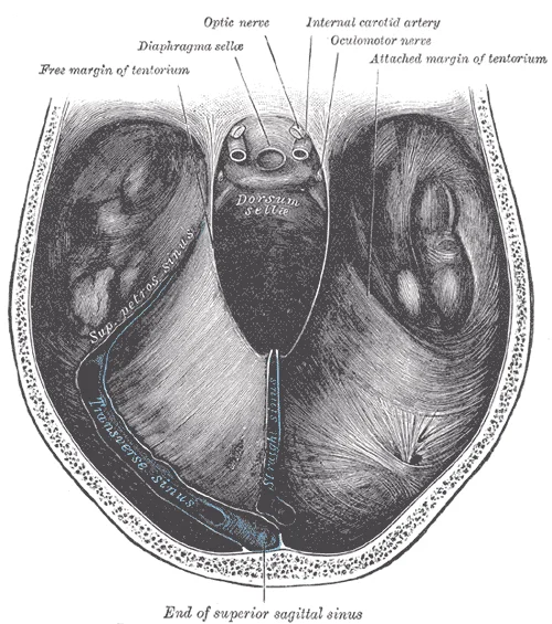
The outermost layer, the dura mater, is a thick, durable membrane adhering tightly to the inner surface of the skull. It forms rigid folds, such as the falx cerebri and tentorium cerebelli, which separate major brain compartments and provide anchor points for venous sinuses.
Figure 11Gray766
Long description
Tentorium cerebelli seen from above. Labelled illustration from Wikimedia Commons, used under its open licence.

Figure 11: Tentorium cerebelli seen from above.
Figure 12Gray570
Long description
The sinuses at the base of the skull. Labelled illustration from Wikimedia Commons, used under its open licence.

Figure 12: The sinuses at the base of the skull.
Figure 13Gray569
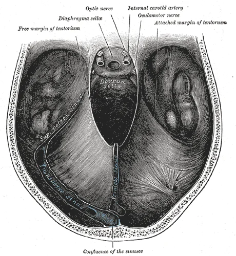
Long description
Tentorium cerebelli from above. Labelled illustration from Wikimedia Commons, used under its open licence.

Figure 13: Tentorium cerebelli from above.
DefinitionMeningeal Protection
The meninges protect the central nervous system through mechanical cushioning via CSF, structural support via dural folds, and metabolic stability via vascular adherence.
Beyond physical protection, the meninges are critical for homeostasis. The arachnoid villi, projections of the arachnoid mater into the dural venous sinuses, facilitate the reabsorption of CSF into the venous circulation, maintaining intracranial pressure within normal limits. This process is vital for preventing hydrocephalus and ensuring adequate perfusion of neural tissue. The pia mater’s close association with cerebral blood vessels allows for efficient nutrient exchange and waste removal, supporting the high metabolic demands of the brain.
| Layer | Location | Key Feature | Primary Function |
|---|---|---|---|
| Dura Mater | Outermost, against skull | Rigid folds (falx, tentorium) | Structural support, venous sinus housing |
| Arachnoid Mater | Middle, web-like | Arachnoid villi | CSF reabsorption, potential subdural space |
| Pia Mater | Innermost, on brain surface | Vascular adherence | Nutrient exchange, contour following |
Disruption of these functions can lead to severe neurological consequences. For instance, a tear in the middle meningeal artery, often due to a temporal bone fracture, can cause an epidural haematoma, compressing the brain against the rigid skull. Similarly, impaired CSF reabsorption can lead to increased intracranial pressure, damaging neural tissue. Understanding the precise anatomy and function of each meningeal layer is therefore fundamental to diagnosing and managing neurological emergencies. The meninges are not merely passive coverings but active participants in maintaining the delicate balance required for normal brain function.
The meninges form a protective envelope for the central nervous system, but their anatomical relationships dictate the clinical presentation of lesions. Understanding the layers—dura, arachnoid, and pia—is essential for interpreting imaging and symptoms. The dura mater is the tough outer layer, while the arachnoid mater and pia mater form the leptomeninges. Between these layers lie specific spaces that are critical in pathology.
The outermost bright line represents the skull. Immediately internal to the bone is the dura mater. In cases of trauma, blood may accumulate between the skull and dura, creating an epidural haematoma.
DefinitionSubarachnoid Cisterns
Named spaces within the subarachnoid space that facilitate CSF dynamics and provide pathways for surgical access.
The cistern of the lamina terminalis is a tent-shaped cistern situated in the midline of the basal telencephalon. [1]
| Space | Boundaries | Common Cause |
|---|---|---|
| Epidural | Skull and Dura | Arterial tear (Middle Meningeal Artery) |
| Subdural | Dura and Arachnoid | Venous tear (Bridging Veins) |
| Subarachnoid | Arachnoid and Pia | Aneurysm rupture or Trauma |
Meningitis involves inflammation of these membranes. Bacterial meningitis affects the subarachnoid space, leading to purulent exudate. The subarachnoid trabeculae can bridge between the arterial, venous, and brain elements in various conformations. In subarachnoid haemorrhage, blood fills these cisterns, visible on CT. Correlation with neuroimaging allows precise identification of these compartments and enhances understanding of their role in pathological conditions such as subarachnoid hemorrhage, tumors, and hydrocephalus. The pia mater adheres firmly to the cortex and deepens the sulci giving the SAS an irregular geometry. This complex solid-fluid relationship between the brain and the CSF is vital for mechanical support. [2]
Exam trapClinical Pearl
Do not confuse the potential subdural space with the real subarachnoid space. Subdural haematomas are venous; epidural are arterial.
Knowledge of the limits and contents of these compartments has made micro neurosurgery possible. [2]
The arachnoid mater is an avascular, web-like membrane that bridges the subarachnoid space, separating the pia mater from the dura mater. Its name derives from the Greek word for spider, reflecting its delicate, cobweb-like appearance under microscopy. This structure is crucial for cushioning the brain and facilitating cerebrospinal fluid (CSF) circulation.
DefinitionSubarachnoid Cisterns
Large expansions of the subarachnoid space where CSF accumulates, containing major cerebral arteries and cranial nerves.
Cisterns are not anatomical compartments with walls but rather potential spaces defined by the adherence of arachnoid membranes to the brain surface. They serve as critical pathways for neurovascular structures.
Specifically, it houses the A1 and proximal A2 segments of the anterior cerebral artery (ACA), the anterior communicating artery (ACoA), and the recurrent artery of Heubner. Surgical access to this area requires careful navigation to avoid damaging these vital vessels. [1]
The Sylvian cistern is divided into three compartments by named arachnoid membranes and imaginary lines. It contains the superficial Sylvian veins and the most proximal part of the cortical segment (M4) of the middle cerebral artery (MCA). [2]
This plane is an imaginary line passing through the deepest point of opposition between the frontoparietal and temporal opercula. It contains the opercular segment (M3) of the MCA lying on the intermediate Sylvian membrane. [2]
Other structures here include a segment of the internal carotid artery from the anterior choroidal artery to its terminal division. The origin of the basal veins of Rosenthal and the lenticulostriate arteries from M1 are also found along the Sylvian cistern. [2]
The interpeduncular cistern contains the bifurcation of the basilar artery and the peduncular segments of the posterior cerebral arteries. It also houses the thalamo-perforating arteries, peduncular segments of the superior cerebellar arteries, and perforating branches of the posterior cerebral arteries. The posterior communicating arteries connect with the midpoint of the peduncular segments of the posterior cerebral arteries in this cistern. [2]
It extends from the upper edge of the atlas to the pyramid of the cerebellar vermis. The posterior inferior cerebellar artery (PICA) membranes divide this cistern into three compartments: one medial and two lateral. [2]
However, detailed descriptions of the subarachnoid space (SAS) were not available until Axel Key and Gustaf Retizius published their work in 1875. They were the first to use the term “cistern” to describe these CSF-filled spaces. [2]
Exam trapSurgical Approach
This modification lessens the need for brain retraction compared to standard approaches. [2]
| Cistern | Key Contents | Defining Boundaries/Membranes |
|---|---|---|
| Lamina Terminalis | A1/A2 ACA, ACoA, Heubner’s artery | Lamina terminalis |
| Sylvian (Superficial) | Superficial Sylvian veins, M4 segment | Outer arachnoid membrane |
| Sylvian (Deep) | M3 segment, intermediate Sylvian membrane | Lateral Sylvian membrane |
| Interpeduncular | Basilar bifurcation, PCA peduncular segments | Interpeduncular fossa |
| Posterior Cerebellomedullary | PICA, medulla, cerebellar vermis | PICA membranes |
Understanding these anatomical relationships is essential for interpreting neuroimaging and planning surgical interventions. Misidentification of these compartments can lead to catastrophic vascular injury.
Students frequently misinterpret the nature of the subarachnoid space (SAS) and its cisterns, leading to errors in both anatomical understanding and clinical application. A primary misconception is viewing the cisterns as static, empty spaces. In reality, they are dynamic compartments filled with cerebrospinal fluid (CSF) that serve as critical pathways for neurovascular structures. A common error is assuming that normal radiological studies always provide clear visualization of these structures. Radiological evaluation of the cisterns is limited in normal studies, however, in the presence of accumulations or pathologies, the radiological visualization of cisterns is enhanced. Students must remember that absence of visible pathology on a standard scan does not imply the cisterns are irrelevant or absent; they are simply not distended. [1]
Another frequent pitfall involves the confusion between different cisternal boundaries and their specific contents. For instance, the oculomotor cistern is often misunderstood regarding its termination. It is a paired, small CSF-filled dural cuff that surrounds the oculomotor nerve (CN III) as it enters the cavernous sinus. It is worth noting that although the CN III enters the orbital apex after penetrating the cavernous sinus, the oculomotor cistern tapers and terminates at the anterior clinoid process. Students often incorrectly assume it extends further into the orbit or fails to recognize its surgical utility as an avascular space for manipulating CN III during cavernous sinus surgery. [1]
Misidentifying the contents of the interpeduncular cistern is another significant error. This cistern is not merely a void but a conical space containing major arterial branches. It is made by the convergence of the subarachnoid space of the supratentorial and infratentorial regions and is limited anteriorly by the sella turcica, pituitary stalk, and the optic chiasm. The division of basilar artery, peduncular segments of the PCA and SCA, thalamogeniculate arteries, and medial and lateral posterior choroidal arteries are present in it. Confusing these vessels with those in the prepontine cistern can lead to catastrophic surgical errors. [1]
Students also struggle with the compartmentalization of the crural cistern. It is not a single uniform space but is divided into superior and inferior compartments by the intracrural membrane that travels anteromedially from the uncus to the cerebral peduncle. The anterior choroid artery courses above the intracrural membrane, while the posterior communicating artery courses below it. Failing to recognize this membrane and the relative positions of these arteries is a common cause of intraoperat [2]
Furthermore, there is a tendency to over-rely on standard MRI sequences for detailed cisternal anatomy. Routine 3D-CISS sequence MRI was studied for a patient with trigeminal neuralgia. 3D-CISS is a gradient echo MRI sequence that is useful in evaluating a variety of pathological states where regular MRI does not provide appropriate anatomical information. 3D-CISS sequences are highly useful in evaluating cranial nerves, CSF rhinorrhea, and cisternal spaces due to the detection of subtle CSF intensity lesions that may be missed on routine sequences. The superiority and higher sensitivity of the 3D-CISS sequence are attributable to the accentuation of T2 values between CSF and other structures. Ignoring this modality when routine MRI is inconclusive is a pitfall. [1]
Finally, students often underestimate the impact of anatomical variations on surgical planning. Deep knowledge of the SAS and its contents is mandatory and key to success when performing intracranial surgery. Imaging studies such as CT cisternography and T2 MRI sequence are widely available and are a great aid when planning the surgical approach; however, they may be subject to pitfalls such as anatomic variations and lack of significant enlargement of SAS that allows visualization of the structures. Assuming standard anatomy without accounting for variations can lead to failed surgical corridors. [2]
Knowledge of meningeal cisterns is indispensable at the bedside, in the operating theatre, on imaging, and in examinations. At the bedside, understanding cisternal anatomy is crucial for interpreting signs of raised intracranial pressure or meningeal irritation. For example, in cases of suspected CSF rhinorrhea, clinical presentation of watery nasal discharge requires specific imaging protocols. CT cisternography was performed for a 35-year-old female who presented with watery nasal discharge to evaluate for suspected CSF rhinorrhea. CT cisternography is a standard imaging modality that is used to diagnose the occult site of CSF leak in cases of CSF rhinorrhea. This involves a pre-contrast plain CT to identify areas of defects in the cribriform plate, roofs of ethmoidal cells, or skull base. Three to 10 mL of iodinated nonionic contrast agent is then administered into the thecal sac, following which the patient is placed in Trendelenburg position for opacification of cisterns, and CT scan is performed with thin slices in coronal and axial planes in both prone and supine positions. Post-contrast and pre-contrast images are then compared to identify CSF and contrast leak areas. [1]
In the operating theatre, cisterns serve as natural corridors for neurosurgical access. Subarachnoid cisterns act as natural corridors, and deep knowledge of neuroanatomy is essential to safely navigate throughout them. Anatomic variants must be considered when planning the surgical approach for the wide range of pathologies that can affect the SAS. For instance, the oculomotor cistern is a surgically relevant space as it is avascular and can be used to expose and manipulate CN III during cavernous sinus surgery. Similarly, the interpeduncular cistern provides access to the basilar artery tip and surrounding structures. The Liliequist membrane and the mammillary bodies are the anterosuperior limit, while the top basilar membrane that separates the prepontine from the interpeduncular cistern forms the inferior limit. Surgeons must navigate these boundaries carefully to avoid damaging the division of basilar artery, peduncular segments of the PCA and SCA, thalamogeniculate arteries, and medial and lateral posterior choroidal arteries. [1] [2]
On imaging, cisternal anatomy is key to diagnosing various pathologies. The medullary cistern encases the medulla oblongata and communicates with the cisterna magna via the cerebello-medullary fissure. This cistern contains the glossopharyngeal nerve (CN IX), vagus nerve (CN X), and accessory nerve (CN XI). It also contains the PICA, lateral medullary vein, pontomedullary vein, and transverse medullary vein. Recognizing these
In examinations, students are tested on the precise boundaries and contents of cisterns. The crural cistern is divided into superior and inferior compartments by the intracrural membrane that travels anteromedially from the uncus to the cerebral peduncle. The anterior choroid artery courses above the intracrural membrane, while the posterior communicating artery courses below it. Questions often focus on the relationship between these arteries and the membrane. Additionally, the classical hypothesis dictates that the CSF is actively secreted by the choroid plexuses to flow in a unidirectional way from the ventricles to end up in the SAS after passing through the foramens of Magendie and Luschka and then reabsorbed by the arachnoid granulations. While novel evidence suggests different mechanisms, the classical hypothesis remains the basis for defining hydrocephalus and treatment strategies. Understanding this flow is essential for answering questions on CSF dynamics. [2] [1]
Diagnostic Approach to CSF Leak
- Patient presents with watery nasal dischargeSuspect CSF rhinorrhea
- Question: Perform pre-contrast plain CT
Identify bone defect
- Proceed to CT cisternography
No defect seen
- Still proceed to CT cisternography
- Administer 3-10 mL iodinated nonionic contrast into thecal sacPlace patient in Trendelenburg position
- Perform thin-slice CT in coronal/axial planes (prone/supine)Compare post-contrast and pre-contrast images
- Identify CSF and contrast leak areasDiagnose occult site of CSF leak
- Radiological visualization of cisterns is enhanced only in the presence of accumulations or pathologies.
- The oculomotor cistern tapers and terminates at the anterior clinoid process.
- The interpeduncular cistern contains the division of basilar artery and peduncular segments of the PCA and SCA.
- The crural cistern is divided by the intracrural membrane, with the anterior choroid artery above and posterior communicating artery below.
- 3D-CISS MRI is superior for detecting subtle CSF intensity lesions in cisternal spaces.
- CT cisternography uses 3 to 10 mL of iodinated nonionic contrast for diagnosing CSF rhinorrhea.
- The medullary cistern contains CN IX, X, and XI, along with PICA and associated veins.
- Deep knowledge of SAS contents is mandatory for successful intracranial surgery.
Answer each one in your head first, then open the answer.
Where does the oculomotor cistern terminate?
Show answerHide answer
Anterior clinoid processWhich membrane divides the crural cistern into superior and inferior compartments?
Show answerHide answer
Intracrural membraneWhat artery courses above the intracrural membrane?
Show answerHide answer
Anterior choroid arteryWhat is the volume of contrast used in CT cisternography for CSF rhinorrhea?
Show answerHide answer
3 to 10 mLWhich cistern contains the glossopharyngeal, vagus, and accessory nerves?
Show answerHide answer
Medullary cistern
- Rai S, Srivastava S, Kamath M, et al. Delineation of Subarachnoid Cisterns Using CT Cisternography, CT Brain Positive and Negative Contrast, and a Three Dimensional MRI Sequence: A Pictorial Review. Cureus 2022;14(4):e23741. doi:10.7759/cureus.23741. Accessed .
- Pérez-Carrillo N, Baldoncini M, Contreras Salazar A, et al. Subarachnoid cisterns as surgical corridors: Integrating microsurgical anatomy and neuroimaging for intracranial navigation. Surgical neurology international 2026;17:283. doi:10.25259/sni_362_2026. Accessed .
Figure credits
- Figure 1: Henry Vandyke Carter, via Wikimedia Commons, Public domain, Public domain.
- Figure 2: Henry Vandyke Carter, via Wikimedia Commons, Public domain, Public domain.
- Figure 3: Henry Vandyke Carter, via Wikimedia Commons, Public domain, Public domain.
- Figure 4: Henry Vandyke Carter, via Wikimedia Commons, Public domain, Public domain.
- Figure 5: Henry Vandyke Carter, via Wikimedia Commons, Public domain, Public domain.
- Figure 6: Henry Vandyke Carter, via Wikimedia Commons, Public domain, Public domain.
- Figure 7: Henry Vandyke Carter, via Wikimedia Commons, Public domain, Public domain.
- Figure 8: Henry Vandyke Carter, via Wikimedia Commons, Public domain, Public domain.
- Figure 9: Henry Vandyke Carter, via Wikimedia Commons, Public domain, Public domain.
- Figure 10: Henry Vandyke Carter, via Wikimedia Commons, Public domain, Public domain.
- Figure 11: Henry Vandyke Carter, via Wikimedia Commons, Public domain, Public domain.
- Figure 12: Henry Vandyke Carter, via Wikimedia Commons, Public domain, Public domain.
- Figure 13: Henry Vandyke Carter, via Wikimedia Commons, Public domain, Public domain.
Educational content for medical students and doctors. It is not a substitute for clinical judgement or local guidelines.