Cerebral circulation
Cerebral circulation supplies blood to the brain via arteries and drains through venous sinuses.
The cerebral circulation is supplied by two main arterial systems: the internal carotid arteries and the vertebral arteries. The internal carotid arteries enter the cranium through the carotid canal in the temporal bone, while the vertebral arteries pass through the transverse foramina of the cervical vertebrae and enter the cranium through the foramen magnum. These vessels contribute to the unique circle of Willis. [1]
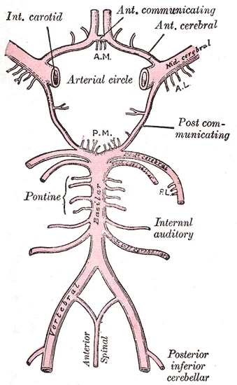
The circle of Willis is a critical anastomotic ring at the base of the brain that connects the internal carotid and vertebral arterial systems. It consists of the anterior communicating artery, bilateral anterior cerebral arteries, bilateral internal carotid arteries, bilateral posterior communicating arteries, and bilateral posterior cerebral arteries. Variations in the circle of Willis are common, with studies showing that complete anatomical variations occur in approximately 50% of individual [1]
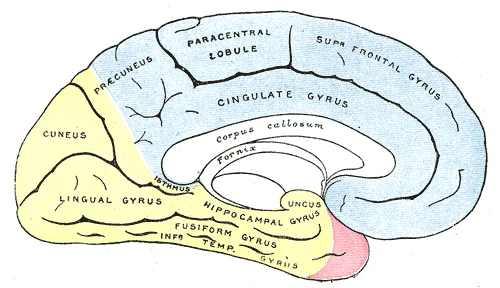
The cerebral arteries have distinct cortical distributions. The anterior cerebral artery supplies the medial surface of the frontal and parietal lobes, including the motor and sensory areas for the lower extremity. The middle cerebral artery supplies the lateral surface of the frontal, parietal, and temporal lobes, including the motor and sensory areas for the face and upper extremity. The posterior cerebral artery supplies the occipital lobe and parts of the temporal lobe, including the visual cortex. [1]
Figure 1Gray518
Long description
Medial surface of cerebral hemisphere, showing areas supplied by cerebral arteries.

Figure 1: Medial surface of cerebral hemisphere, showing areas supplied by cerebral arteries.
Watershed zones are border areas between major arterial territories that are vulnerable to hypoperfusion. These include the anterior border zone between the anterior and middle cerebral arteries, and the posterior border zone between the middle and posterior cerebral arteries. These zones are particularly susceptible to ischemic damage in conditions of reduced blood flow.
Venous drainage occurs through a series of dural sinuses and veins. The superior sagittal sinus runs in the groove of the longitudinal fissure and drains to the confluence of sinuses, along with the occipital sinuses and straight sinus. The transverse sinuses connect to the sigmoid sinuses, which then drain into the jugular veins. The arachnoid granulations are outpocketings of the arachnoid membrane into the dural sinuses that allow CSF to be reabsorbed into the blood. [1] [2]
Aneurysms most commonly occur at arterial bifurcations, with the most common sites being the anterior communicating artery, the bifurcation of the middle cerebral artery, and the top of the basilar artery. These locations are particularly susceptible to aneurysm formation due to hemodynamic stress.
Stroke syndromes vary depending on the affected vascular territory. Anterior circulation strokes affect the internal carotid artery territory and may present with contralateral weakness, sensory loss, aphasia, or neglect. Middle cerebral artery strokes often cause contralateral hemiparesis with greater involvement of the face and arm, sensory loss, and possibly aphasia or neglect. Posterior circulation strokes affect the vertebral and basilar artery territories and may present with vertigo, diplopia, dysarthria, dysphagia, or ataxia.
Cerebral perfusion is maintained through autoregulation, which keeps blood flow relatively constant despite changes in blood pressure. This is particularly important in the brain, which cannot store nutrients and requires a constant supply of oxygen and glucose.
The homunculus is a representation of the body on the cerebral cortex, showing the relative amount of cortical tissue dedicated to different body parts. The motor homunculus has a disproportionately large representation for the hands, face, and tongue, reflecting the fine motor control required for these areas.
Watershed zones, also known as border zones, are areas between major arterial territories that receive dual blood supply. These zones are particularly vulnerable to hypoperfusion, as they represent the terminal zones of the cerebral arterial circulation. There are two main types of watershed zones: cortical and subcortical.
Cortical watershed zones are located at the border between the anterior cerebral artery (ACA) and middle cerebral artery (MCA) territories, and between the MCA and posterior cerebral artery (PCA) territories. The ACA-MCA watershed zone is located in the frontal parasagittal region, while the MCA-PCA watershed zone is located in the parieto-occipital region. These zones are particularly susceptible to ischemic damage in conditions of reduced blood flow, such as hypotension or carotid stenosis.
Subcortical watershed zones are located in the deep white matter at the border between the territories of the deep penetrating branches of the cerebral arteries. These zones include the area between the territories of the lenticulostriate arteries (from the MCA) and the thalamoperforating arteries (from the PCA). Subcortical watershed infarcts typically appear as linear or band-like lesions in the deep white matter.
Watershed infarcts can occur in various clinical settings, including systemic hypotension, cardiac arrest, and carotid occlusive disease. The pattern of infarction depends on the severity and duration of the hypoperfusion. Complete watershed infarcts involve the entire border zone, while partial watershed infarcts involve only part of the border zone.
The circle of Willis is a polygonal anastomotic ring at the base of the brain that connects the internal carotid and vertebral arterial systems. It is formed by the anterior communicating artery, bilateral anterior cerebral arteries, bilateral internal carotid arteries, bilateral posterior communicating arteries, and bilateral posterior cerebral arteries. This arrangement allows for collateral circulation between the anterior and posterior circulations, as well as between the left and right sides of the brain.
The circle of Willis has several important functions. It provides collateral pathways that can maintain perfusion of the brain even if one of the major arteries is narrowed or blocked. This is particularly important in conditions such as carotid stenosis or vertebral artery occlusion. The circle of Willis also equalizes blood pressure between the two sides of the brain and between the anterior and posterior circulations. [1]
Variations in the anatomy of the circle of Willis are common. Studies have shown that complete anatomical variations occur in approximately 50% of individuals. Common variations include hypoplasia or absence of the anterior communicating artery, hypoplasia or absence of one or both posterior communicating arteries, and fenestration of the basilar artery. These variations can affect the efficiency of collateral circulation and may influence the risk of stroke.
The circle of Willis is also a common site for aneurysms, particularly at the junction of the anterior communicating artery with the anterior cerebral arteries, at the bifurcation of the middle cerebral artery, and at the junction of the posterior communicating artery with the posterior cerebral artery. These locations are particularly susceptible to aneurysm formation due to hemodynamic stress and the presence of elastic tissue in the arterial wall.
The cerebral circulation is characterized by a specialized arrangement of blood vessels that supply the central nervous system (CNS). This privileged blood supply is essential due to the critical function of CNS tissue and the presence of the blood-brain barrier, which strictly controls what can move from the general circulation into the brain and spinal cord.
The brain is protected by three meningeal layers: the dura mater, arachnoid mater, and pia mater. The dura mater is a thick, fibrous layer that forms a strong protective sheath over the entire brain and spinal cord, anchored to the inner surface of the cranium and vertebral cavity. The arachnoid mater is a thin fibrous membrane that forms a loose sac around the CNS, with a filamentous mesh called the arachnoid trabeculae beneath it. The pia mater is a thin fibrous membrane that follows the convolutions of the cerebral cortex, extending into every sulcus and groove. Its name comes from the Latin for "tender mother," reflecting its delicate covering of the brain.
The ventricular system consists of four cavities within the brain, all derived from the original hollow space within the neural tube. The lateral ventricles are located deep within the cerebrum and connect to the third ventricle via the interventricular foramina. The third ventricle lies between the left and right sides of the diencephalon and opens into the cerebral aqueduct, which passes through the midbrain. The aqueduct opens into the fourth ventricle, situated between the cerebellum and the pons and upper medulla. The floor of the fourth ventricle is formed by the dorsal surface of the pons and upper medulla, and it narrows into the central canal of the spinal cord.
Choroid plexuses, which appear as soft, fuzzy structures, are found in all four ventricles and produce cerebrospinal fluid (CSF). CSF flows from the lateral ventricles to the third ventricle, then through the cerebral aqueduct to the fourth ventricle, with additional CSF produced at each location. Approximately 500 milliliters of CSF are produced daily, continuously pulsing through the ventricular system. From the fourth ventricle, most CSF exits through the median and lateral apertures into the subarachnoid space, while a small amount continues down the central canal of the spinal cord. [1]
In cases of severe intracranial hypertension with impending cerebral herniation, immediate treatment is required, which may include decompressive craniectomy, ventriculoperitoneal shunt, or hematoma evacuation. Short-term dehydration therapy with mannitol or furosemide may be initiated to gain time for surgery in patients with intracranial hypertension, progressive visual loss, or cerebral herniation, though excessive dehydration is prohibited as it can lead to blood concentration and aggravation of cerebral venous sinus thrombosis. Acetazolamide may be considered in certain patients to reduce intracranial pressure by decreasing CSF secretion.
The cerebral circulation is supplied by two main arterial systems: the internal carotid system and the vertebrobasilar system. The internal carotid arteries originate from the common carotid arteries, which branch directly from the aorta. These arteries ascend through the neck and enter the cranial cavity through the carotid canal in the temporal bone. The internal carotid artery terminates by dividing into the anterior cerebral artery and middle cerebral artery, forming the anterior part of the circle of Willis.
The vertebrobasilar system begins with the vertebral arteries, which arise from the subclavian arteries. Each vertebral artery ascends through the transverse foramina of the cervical vertebrae, enters the cranial cavity through the foramen magnum, and unites with its counterpart to form the basilar artery at the pontomedullary junction. The basilar artery gives rise to branches to the brain stem and cerebellum.
The circle of Willis is a crucial anastomotic structure that provides constant perfusion to the brain even if one vessel is compromised. It is formed by the anterior communicating artery connecting the two anterior cerebral arteries, the posterior communicating arteries connecting the internal carotid arteries with the posterior cerebral arteries, and the basilar artery connecting the vertebral arteries.
The cerebral arteries can be categorized into cortical branches and central (penetrating) branches. The cortical branches supply the cerebral cortex, while the central branches penetrate deep into the brain to supply structures such as the basal ganglia, internal capsule, and thalamus. These penetrating arteries are particularly vulnerable to hypertension, which can lead to lacunar infarcts.
The circle of Willis is clearly demonstrated with the anterior and posterior communicating arteries completing the anastomotic ring.
The cerebral circulation receives its primary blood supply from the internal carotid arteries and the vertebral arteries, which anastomose to form the Circle of Willis. This anastomotic circle provides collateral circulation in cases of occlusion. The internal carotid arteries contribute to the anterior circulation, while the vertebral arteries merge into the basilar artery and contribute to the posterior circulation. [1]
The anterior cerebral artery arises from the internal carotid artery and supplies the medial surface of the frontal and parietal lobes. It is connected to its counterpart on the opposite side via the anterior communicating artery. The middle cerebral artery, also arising from the internal carotid artery, supplies the lateral surface of the cerebral hemisphere. It is connected to the posterior cerebral artery via the posterior communicating artery. & [1]
The vertebral arteries ascend through the foramina transversaria of the cervical vertebrae and enter the cranial cavity through the foramen magnum. They unite at the pontomedullary junction to form the basilar artery. The basilar artery gives rise to branches to the brain stem and cerebellum. [1]
The Circle of Willis is a complete anastomotic ring located at the base of the brain, surrounding the optic chiasm and the infundibulum of the pituitary gland. It consists of the anterior communicating artery, bilateral anterior cerebral arteries, bilateral internal carotid arteries, bilateral posterior communicating arteries, and bilateral posterior cerebral arteries. This arrangement allows for collateral blood flow if one of the major vessels becomes occluded. [1]
Figure 2Gray519
Long description
Diagram of the arterial circulation at the base of the brain. A.L. Antero-lateral. A.M. Antero-medial. P.L. Postero-lateral. P.M. Posteromedial ganglionic branches.

Figure 2: Diagram of the arterial circulation at the base of the brain (a.l).
The central nervous system requires a continuous and uninterrupted supply of oxygenated blood, as any interruption can be devastating[1]. To ensure this perfusion, the cardiovascular system employs specific regulatory reflexes and multiple specialized routes for blood entry[1]. The primary arterial supply is divided into two major systems: the carotid system and the vertebrobasilar system. These systems converge at the base of the brain to form the circle of Willis, an anastomotic ring that provides collateral circulation. This arrangement maximizes the brain's ability to maintain perfusion even if one of the major feeding arteries is compromised[1].
The posterior cerebral arteries, derived from the vertebrobasilar system, connect to the posterior communicating arteries, completing the circle. The middle cerebral arteries, the largest branches of the internal carotids, extend laterally into the Sylvian fissure. The blood-brain barrier is represented by the tight junctions between endothelial cells of the capillaries within the nervous tissue, distinguishing the vascular supply from the neural parenchyma. [2] [1]
The carotid system supplies the anterior and middle portions of the brain. The internal carotid arteries ascend through the neck and enter the skull via the carotid canal. They give off the ophthalmic artery and then bifurcate into the anterior and middle cerebral arteries. The vertebrobasilar system supplies the posterior brain, including the brainstem and cerebellum. The vertebral arteries ascend through the transverse foramina of the cervical vertebrae and join to form the basilar artery at the pontomedullary junction. The basilar artery bifurcates into the posterior cerebral arteries.
The circle of Willis is a polygonal anastomosis at the base of the brain. It connects the anterior circulation (carotid) with the posterior circulation (vertebrobasilar). The key components are the anterior communicating artery, the anterior cerebral arteries, the internal carotid arteries, the posterior communicating arteries, and the posterior cerebral arteries. This structure allows for collateral flow, ensuring that if one artery is stenosed, blood can be redirected from the other system to maintain perfusion to the affected territory[1].
Venous drainage from the brain occurs through a system of dural venous sinuses. The dura mater contains infoldings that fit into large crevasses of the brain, such as the falx cerebri and the tentorium cerebelli[1]. These dural reflections surround and support the venous sinuses[1]. Blood from the cerebral veins empties into these sinuses, which eventually drain into the internal jugular veins. The superior sagittal sinus runs along the superior margin of the falx cerebri, while the straight sinus runs along the junction of the falx cerebri and tentorium cerebelli.
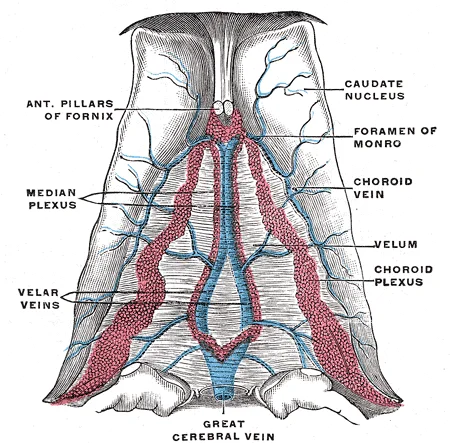
Figure 3Gray565
Long description
Velum interpositum. (Poirier and Charpy.)

Figure 3: Velum interpositum (Poirier and Charpy.).
Aneurysms are focal dilations of blood vessels, often occurring at bifurcations within the circle of Willis. Rupture of an aneurysm leads to subarachnoid haemorrhage. Stroke syndromes result from occlusion of specific arteries. Anterior cerebral artery occlusion affects the medial aspect of the frontal and parietal lobes, leading to contralateral leg weakness. Middle cerebral artery occlusion affects the lateral convexity, causing contralateral face and arm weakness and aphasia if the dominant hemisphere is involved. Posterior cerebral artery occlusion affects the occipital lobe, leading to visual field defects.
The blood-brain barrier is a physiological barrier that limits the exchange of materials between the blood and the interstitial fluid of the nervous tissue[1]. It is enforced by glial cells and tight junctions between endothelial cells of the capillaries[1]. This barrier protects the central nervous tissue from potentially harmful substances in the blood. Metabolic wastes are collected in the cerebrospinal fluid, which circulates through the central nervous system and is eventually reabsorbed into the blood via the arachnoid granulations[1]. The choroid plexus, located within the ventricles, filters blood to produce cerebrospinal fluid, which is essentially water, small molecules, and electrolytes[1].
The cerebral arteries have a specific cortical distribution that corresponds to different functional areas. The anterior cerebral artery (ACA) arises from the internal carotid artery and supplies the medial surface of the frontal and parietal lobes. It provides branches to the corpus callosum, cingulate gyrus, and medial aspects of the frontal and parietal lobes. The ACA also supplies the anterior limb of the internal capsule and parts of the basal ganglia. [1]
The middle cerebral artery (MCA) is the largest cerebral artery and arises from the internal carotid artery. It supplies the lateral surface of the frontal, parietal, and temporal lobes, including the primary motor and sensory cortices for the face and upper extremity. The MCA also supplies the insula, basal ganglia, and internal capsule. The MCA is divided into four main segments: M1 (horizontal segment), M2 (insular segment), M3 (opercular segment), and M4 (cortical segment). [2] [1]
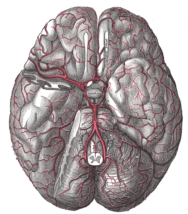
Figure 4Gray516
Long description
The arteries of the base of the brain. The tempora pole of the cerebrum and a portion of the cerebellar hemisphere have been removed on the right side.

Figure 4: The arteries of the base of the brain.
The posterior cerebral artery (PCA) arises from the basilar artery and supplies the occipital lobe and parts of the temporal lobe. It provides branches to the visual cortex, including the primary visual area (Brodmann area 17) and association visual areas. The PCA also supplies parts of the thalamus, midbrain, and hippocampus.
The cortical distribution of these arteries creates distinct vascular territories that are important for understanding stroke syndromes. The ACA territory includes the medial frontal and parietal lobes, the MCA territory includes the lateral frontal, parietal, and temporal lobes, and the PCA territory includes the occipital lobe and inferior temporal lobe.
The cerebral circulation serves as the vital lifeline for the central nervous system, ensuring that neural tissue receives the continuous supply of oxygen and glucose required for electrical signalling and metabolic homeostasis. Without this perfusion, neuronal function deteriorates rapidly, leading to ischaemia and cell death. The system is not merely a passive pipe network but a dynamic interface that also facilitates waste removal and chemical communication between the blood and the brain parenchyma.
Between these two meningeal layers lies the subarachnoid space.
The circulation supports the high metabolic demand of the brain, which consumes approximately 20% of the body's total oxygen despite representing only 2% of body weight. This demand is met by a dense capillary network that ensures short diffusion distances for gases and nutrients. The blood-brain barrier, formed by tight junctions between endothelial cells, regulates this exchange, protecting the neural tissue from toxins and fluctuations in plasma composition while allowing essential substrates to pass. [2]
Beyond metabolic support, the cerebral circulation plays a role in diagnostic medicine. Similar to clinical blood work, a sample of CSF can be withdrawn to find chemical evidence of neuropathology or metabolic traces of the biochemical functions of nervous tissue. This procedure, known as lumbar puncture, accesses the subarachnoid space where the CSF circulates. The analysis of this fluid allows clinicians to detect infections, haemorrhages, and demyelinating diseases. The presence of specific proteins, cells, or glucose levels in the CSF provides indirect information about the state of the cerebral circulation and the integrity of the blood-brain barrier. [2] [1]
The efficiency of this system relies on autoregulation, a mechanism that maintains constant blood flow despite changes in systemic blood pressure. This ensures that the brain is protected from hypoperfusion during hypotension and from hyperperfusion and oedema during hypertension. The vascular tone is adjusted by local metabolic factors, including carbon dioxide tension and pH, which act on the smooth muscle of the arterioles. This precise control is essential for maintaining the stable internal environment required for neuronal signalling.
DefinitionCerebral Circulation
The vascular network supplying the brain and spinal cord, responsible for delivering oxygen and nutrients while removing metabolic waste, and interacting with the CSF in the subarachnoid space.
The integration of blood flow and CSF dynamics is fundamental to brain health. The pulsatile nature of arterial flow influences CSF movement, aiding in the clearance of metabolic byproducts through the glymphatic system. This interplay ensures that the brain remains functional and protected from both mechanical injury and chemical imbalance. Understanding these functions is essential for diagnosing and managing conditions that affect cerebral perfusion or meningeal integrity.
| Lesion Site | Clinical Presentation | Key Features |
|---|---|---|
| Middle cerebral artery (MCA) territory | Contralateral hemiparesis, sensory loss, homonymous hemianopia, aphasia (if dominant hemisphere) | Affects face and arm more than leg; Wernicke's aphasia (receptive) with left MCA; Broca's aphasia (expressive) with left MCA |
| Anterior cerebral artery (ACA) territory | Contralateral leg weakness, sensory loss, grasp reflex, urinary incontinence | May have abulia (lack of initiative); sparing of face and arm |
| Posterior cerebral artery (PCA) territory | Contralateral homonymous hemianopia, visual agnosia, cortical blindness | May involve memory if hippocampal branches affected |
| Internal carotid artery (ICA) occlusion | Ipsilateral eye and forehead pain, contralateral hemiparesis, Horner's syndrome | May have "crossed syndromes" with brainstem signs |
| Basilar artery occlusion | "Locked-in syndrome", coma, bilateral motor/sensory loss, pupils may be spared | May have "top of the basilar" syndrome with thalamic or midbrain signs |
| Venous sinus thrombosis | Headache, seizures, focal neurological deficits, papilledema | May have hemorrhagic infarction on imaging; can present with isolated intracranial hypertension |
Cerebral venous sinus thrombosis (CVST) is a rare but serious condition where thrombosis occurs in the dural venous sinuses or cerebral veins. The clinical presentation is often variable and may include headache, seizures, focal neurological deficits, and papilledema. [2]
The diagnostic evaluation of suspected CVST should include CT/CTV and MRI/MRV as initial imaging modalities. SWI and T2*-GRE imaging can improve diagnosis, particularly in cases of isolated cortical venous thrombosis or during the acute stage. If initial imaging is inconclusive or if endovascular therapy is being considered, digital subtraction angiography (DSA) is recommended, though its limitations in detecting isolated cortical venous thrombosis should be considered. [2]
The main treatment for CVST is anticoagulation therapy with either intravenous unfractionated heparin (UFH) or subcutaneous low-molecular weight heparin (LMWH), which should be initiated immediately. After the acute stage, oral anticoagulants such as warfarin should be used for at least 3-6 months to prevent recurrence. Recent evidence suggests that dabigatran may be as safe and effective as warfarin for preventing recurrent venous thrombotic events in patients with CVST. [2]
Screening for potential prothrombotic factors is recommended in patients with undetermined aetiology, recurrent CVST, or a family history of venous thrombosis. These factors include antithrombin III, protein C or S deficiency, factor V Leiden, prothrombin mutation, anticardiolipin antibody, and lupus anticoagulant. To prevent recurrence, it is important to eliminate the root cause by identifying and treating prothrombotic conditions such as protein C/S deficiency, antiphospholipid syndrome, and factor V Leiden mutation. [2]
For patients undergoing sinus stenting, the long-term antithrombotic treatment is not clearly established. Based on clinical practice in arterial stenting, it is reasonable to use dual antiplatelet therapy for the first 3 months followed by single antiplatelet therapy for maintenance. [2]
The telencephalon represents the most anterior region of the original neural tube, yet its expansion is strictly constrained by the physical boundaries of the cranial cavity. As the cerebrum develops, it cannot grow past the limit of the frontal bone of the skull, forcing a specific morphological adaptation. Consequently, the cerebrum takes on a C-shaped formation that traverses the frontal, parietal, occipital, and finally temporal regions. This curvature is not merely superficial; the space within the telencephalon is stretched into this same C-shape to accommodate the growing tissue. [1]
A common trap is assuming the ventricles are simple spherical cavities. In reality, the two ventricles are situated on the left and right sides, reflecting the bilateral nature of the telencephalon. Historically, these structures were referred to as the first and second ventricles, a nomenclature that highlights their paired symmetry. Students must remember that the lateral ventricles are not isolated; they communicate directly with the diencephalon. The interventricular foramina serve as the critical connection between the frontal region of the lateral ventricles and the third ventricle. [1]
Another high-yield fact concerns the spatial relationships within the skull. The telencephalon is limited by the space within the skull, meaning any pathology causing mass effect must be understood in the context of this rigid boundary. The C-shaped formation is essential for understanding the trajectory of neurosurgical approaches and the spread of cerebrospinal fluid. The frontal region of the lateral ventricles is anterior, while the temporal regions form the inferior limb of the 'C'.
Clinical pearlVentricular Anatomy
The interventricular foramina connect the frontal region of the lateral ventricles with the third ventricle, maintaining CSF continuity. [1]
Exam trapDevelopmental Shape
The cerebrum takes on a C-shaped formation, not a spherical one, due to constraints within the skull.
DefinitionTelencephalon Limits
The telencephalon cannot grow past the limit of the frontal bone of the skull.
The historical naming convention of first and second ventricles is still relevant for interpreting older literature and understanding the bilateral symmetry of the lateral ventricles. The space within the telencephalon is stretched into this same C-shape, which dictates the orientation of the choroid plexus and the ependymal lining. Understanding this geometry is vital for interpreting MRI scans, where the body of the lateral ventricle follows the parietal region, and the inferior horn extends into the temporal lobe.
| Structure | Connection | Anatomical Region |
|---|---|---|
| Interventricular foramina | Lateral to third ventricle | Frontal region |
| Lateral ventricles | Paired cavities | Left and right sides |
| Telencephalon space | Stretched C-shape | Frontal to temporal |
Finally, remember that the C-shaped formation involves the frontal, parietal, occipital, and finally temporal regions, creating a continuous pathway for neural and vascular structures. This comprehensive curvature ensures that the telencephalon fits efficiently into the available intracranial space.
Students frequently misinterpret the role of D-dimer in cerebral venous sinus thrombosis (CVST). An elevated D-dimer level supports the diagnosis, but a normal D-dimer level should not exclude CVST, especially in patients presented with isolated headache or chronic symptoms. You must not rely on this single biomarker to rule out the condition. [2]
Another frequent error is the immediate initiation of anticoagulation without considering the clinical context. Anticoagulation therapy should be initiated in patients with CVST immediately. However, students often fear haemorrhage. CVST with minor intracranial haemorrhage or intracranial hypertension is not an absolute contradiction to anticoagulation therapy. The thrombotic risk generally outweighs the bleeding risk in this specific pathology. [2]
Students also confuse the imaging modalities. CT/CTV and MRI/MRV are recommended in the initial imaging evaluation of patients with suspected CVST. While CT is fast, MRI/MRV is able to diagnose most of CVST, and is recommended as the best non-invasive imaging in the follow-up of CVST. Furthermore, contrast-enhanced (CE) MRV is more reliable than time-of-flight MRV. Do not assume a non-contrast CT is sufficient for definitive diagnosis. [2]
A significant trap involves the use of prophylactic antiepileptic drugs. Prophylactic use of antiepileptic drugs in patients with CVST is not beneficial. Antiepileptic therapy should be initiated immediately only in patients with a first seizure and parenchymal lesion. In patients with a first seizure without parenchymal lesion, early initiation of antiepileptic drugs may be beneficial. Do not prescribe them routinely. [2]
Steroid use is another common misconception. Routine use of steroid in CVST is not beneficial, unless it is indicated for other underlying diseases. Students often administer steroids for cerebral oedema, but this is not supported for CVST specifically. [2]
Similarly, antiplatelet agents are often misused. Current evidence does not support the routine use of antiplatelet drugs or de-fibrin therapy in CVST, unless it is indicated for other underlying diseases. Stick to anticoagulation as the primary medical therapy. [2]
In pregnancy, students may hesitate to treat due to fetal concerns. LMWH is recommended in women with CVST during pregnancy. The safety of LMWH is superior to UFH. Subcutaneous injection of LMWH is recommended throughout the pregnancy (0.4–0.6 mL, twice a day). Do not withhold treatment. [2]
Finally, regarding oral contraceptives, it is recommended to reduce or avoid the use of oral contraceptives, especially in female patients with a previous history of CVST. Pregnancy is not contraindicated in female patients with a history of CVST, but patients should be informed of the high risk of recurrent CVST or loss of pregnancy. [2]
At the bedside, vigilance should be maintained for CVST. The possibility of CVST should be considered in patients presented with headache, papilloedema or increased intracranial pressure with unknown cause. The screening for CVST is reasonable in patients presented with unexplained seizures (including eclampsia), focal brain injury, different levels of unconsciousness, cognitive disorders, psychosis or dural arteriovenous fistula. You must maintain a high index of suspicion for these diverse presentations. [2]
In the operating theatre or procedural setting, if patients with severe CVST require intervention, mechanical thrombectomy can be considered in patients with severe CVST after adequate anticoagulation has failed and there is a need to prevent or treat brain herniation. Current evidence is not sufficient to recommend the application of intrasinus thrombolysis or mechanical thrombectomy in severe CVST. These are rescue therapies, not first-line treatments. [2]
On imaging, the initial evaluation relies on specific protocols. CT/CTV and MRI/MRV are recommended in the initial imaging evaluation of patients with suspected CVST. If initial imaging is negative but symptoms persist, a follow-up CTV/MRV is recommended in patients with CVST with persistent or evolving symptoms despite the initial imaging results. This ensures no evolving thrombus is missed. [2]
In examinations, you must know the pharmacological details. The therapeutic dosage of LMWH is 0.4–0.6 mL, injected subcutaneously twice a day. If UFH is used, the initial treatment should at least double the activated partial thromboplastin time, lasting for 1–4 weeks. The safety and efficacy of LMWH is slightly superior to UFH. LMWH is recommended for the acute management of CVST. [2]
For infectious causes, it is recommended to seek and treat the potential causes. Administration of appropriate and adequate antibiotics should be initiated immediately in patients with infection and CVST, as well as surgical removal of sources of infection. It is recommended to use antibiotics in patients with CVST and infection for 3–8 weeks. [2]
For risk factor modification, the risk factors and causes of CVST are complicated and diverse. It is recommended to screen for the risk factors contributed to prothrombotic conditions and exclude the possibility of infections associated with CVST. [2]
CVST Diagnostic and Management Pathway
- Suspect CVSTHeadache, papilloedema, seizures, focal deficit
- Question: Initial Imaging?
Yes
- CT/CTV or MRI/MRVCE MRV preferred for reliability
No
- Perform ImagingCT/CTV and MRI/MRV recommended
| Intervention | Recommendation | Evidence Level |
|---|---|---|
| Anticoagulation | Initiate immediately in all CVST patients | Class I, Level B |
| LMWH Dosage | 0.4–0.6 mL subcutaneously twice daily | Class IIa, Level B [2] |
| UFH Target | Double aPTT for 1–4 weeks | Class IIb, Level B [2] |
| Steroids | Not beneficial unless other indication | Class III, Level B |
| Antiplatelets | Not beneficial unless other indication | Class III, Level C |
| Prophylactic AEDs | Not beneficial | Class III, Level C |
Vigilance should be maintained for CVST in patients with unexplained neurological symptoms. An elevated D-dimer level supports diagnosis, but a normal level does not exclude CVST. CT/CTV and MRI/MRV are recommended for initial imaging evaluation. Anticoagulation therapy should be initiated in patients with CVST immediately. LMWH is recommended for acute management with a dosage of 0.4–0.6 mL twice daily. Routine use of steroids and antiplatelet drugs is not beneficial in CVST,. Prophylactic antiepileptic drugs are not beneficial in CVST patients. Antibiotics should be used for 3–8 weeks if infection is the cause.
Answer each one in your head first, then open the answer.
What is the recommended initial imaging for suspected CVST?
Show answerHide answer
CT/CTV and MRI/MRVShould anticoagulation be withheld if minor intracranial haemorrhage is present?
Show answerHide answer
No, it is not an absolute contradictionWhat is the therapeutic dosage of LMWH for CVST?
Show answerHide answer
0.4–0.6 mL, injected subcutaneously twice a dayIs routine steroid use beneficial in CVST?
Show answerHide answer
No, routine use is not beneficialWhen should mechanical thrombectomy be considered?
Show answerHide answer
After adequate anticoagulation has failed and there is a need to prevent or treat brain herniation
- Betts JG, Young KA, Wise JA, et al. Circulation and the Central Nervous System. OpenStax, Anatomy and Physiology 2e (CC BY-NC-SA 4.0). Accessed .
- Fan Y, Yu J, Chen H, et al. Chinese Stroke Association guidelines for clinical management of cerebrovascular disorders: executive summary and 2019 update of clinical management of cerebral venous sinus thrombosis. Stroke and vascular neurology 2020;5(2):152-158. doi:10.1136/svn-2020-000358. Accessed .
Figure credits
- Figure 1: Henry Vandyke Carter, via Wikimedia Commons, Public domain, Public domain.
- Figure 2: Henry Vandyke Carter, via Wikimedia Commons, Public domain, Public domain.
- Figure 3: Henry Vandyke Carter, via Wikimedia Commons, Public domain, Public domain.
- Figure 4: Henry Vandyke Carter, via Wikimedia Commons, Public domain, Public domain.
Educational content for medical students and doctors. It is not a substitute for clinical judgement or local guidelines.