Autonomic nervous system
The autonomic nervous system regulates involuntary functions controlling cardiac and smooth muscle, and glands.
Figure 1Gray799
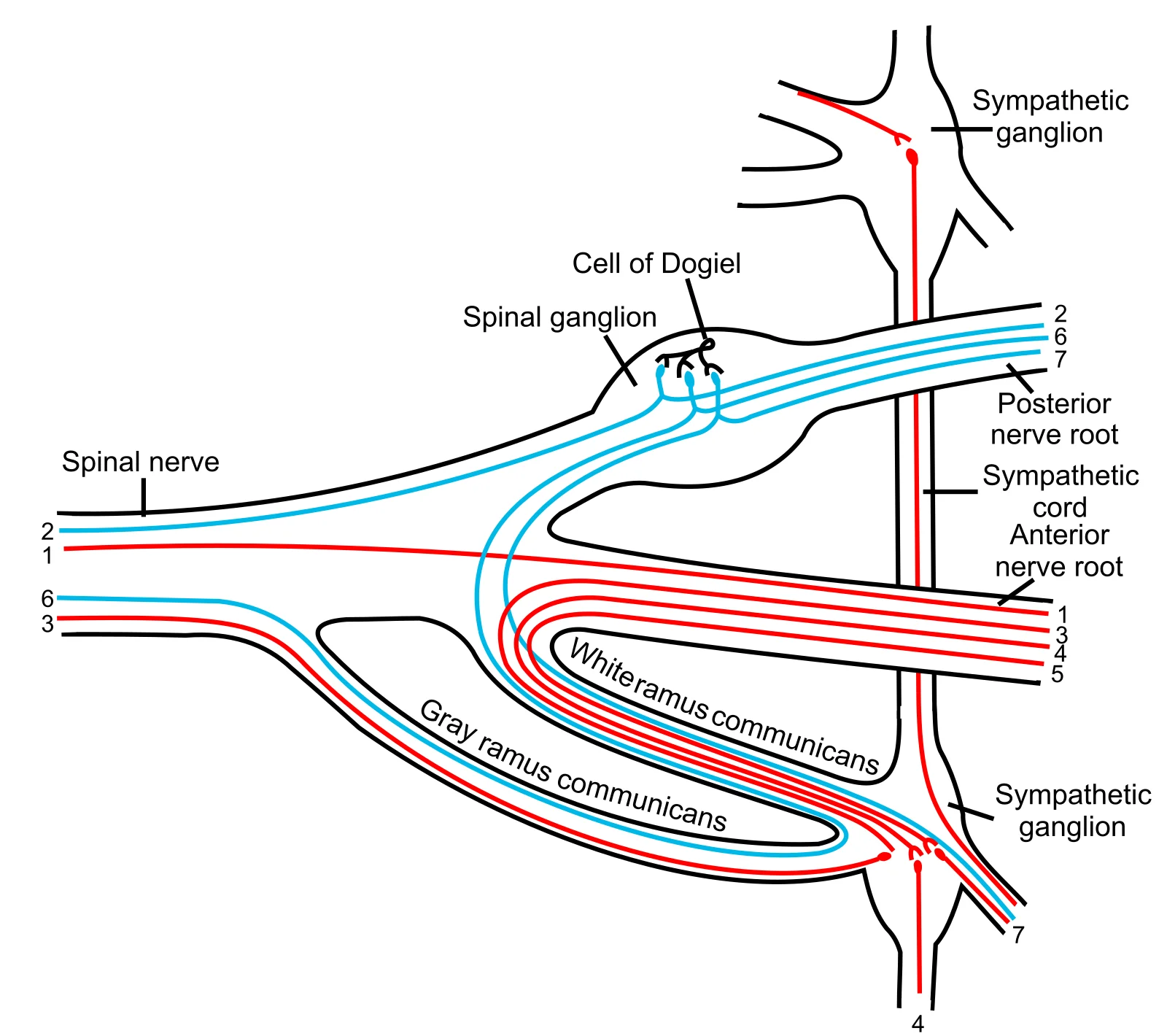
Long description
Scheme showing structure of a typical spinal nerve. 1. Somatic efferent. 2. Somatic afferent. 3,4,5. Sympathetic efferent. 6,7. Autonomic nervous system (ANS) afferent.

Figure 1: Scheme showing structure of a typical spinal nerve (1). The autonomic nervous system is a functional division of the nervous system that controls cardiac and smooth muscle, as well as glandular tissue, in contrast to the somatic nervous system which causes skeletal muscle contraction[1]. It regulates many internal organs through a balance of two divisions: the sympathetic division, associated with the fight-or-flight response, and the parasympathetic division, responsible for rest and digest functions[1]. Homeostasis is maintained
The hypothalamus serves as the major integration center for autonomic functions, coordinating responses to maintain internal balance. Brainstem autonomic centers include the nucleus tractus solitarius, which receives visceral afferent information and helps regulate cardiovascular, respiratory, and digestive functions. Descending autonomic pathways originate from these central regions and project to preganglionic neurons in the brainstem and spinal cord. [4]
Visceral afferents carry sensory information from internal organs to the central nervous system. These afferents terminate primarily in the nucleus tractus solitarius in the medulla oblongata, where the information is processed and integrated to produce appropriate autonomic responses. Unlike somatic sensations, visceral sensations typically do not reach conscious perception unless they are particularly intense, such as cardiac pain. [2]
The autonomic nervous system operates through visceral reflexes that maintain balance in organ systems. These reflexes have a two-neuron efferent pathway, with a central neuron projecting from the spinal cord or brainstem to synapse on a ganglionic neuron that then projects to the effector. The sympathetic system increases heart rate and dilates pupils, while the parasympathetic system decreases heart rate and constricts pupils. At rest, heart rate is normally under parasympathetic tone, while blood vessels maintain slight constriction through sympathetic tone. [2]
The autonomic nervous system (ANS) is the division of the peripheral nervous system responsible for regulating involuntary physiological processes. It operates largely below the level of conscious awareness to maintain homeostasis. The system is anatomically and functionally divided into two primary branches: the sympathetic division and the parasympathetic division. These divisions often exert opposing effects on target organs, a concept known as dual innervation[1]. The balance betwe [2]
The sympathetic division is referred to as the thoracolumbar system because its preganglionic neurons originate in the lateral horn of the thoracic and upper lumbar spinal cord. A central neuron in the lateral horn of any of these spinal regions projects to ganglia adjacent to the vertebral column through the ventral spinal roots. The majority of sympathetic ganglia belong to a network of sympathetic chain ganglia that runs alongside the vertebral column. There are typically 23 ganglia in the chain on either side of the spinal column. Three correspond to the cervical region, 12 are in the thoracic region, four are in the lumbar region, and four correspond to the sacral region. Superior to the chain ganglia are three paravertebral ganglia in the cervical region. Other autonomic ganglia related to the sympathetic chain are the prevertebral ganglia, which are located anterior to the vertebral column. [1] [2][3]
Figure 2Gray844
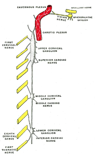
Long description
Diagram of the cervical sympathetic. (Testut.)

Figure 2: Diagram of the cervical sympathetic (Testut.).
Figure 3Gray842
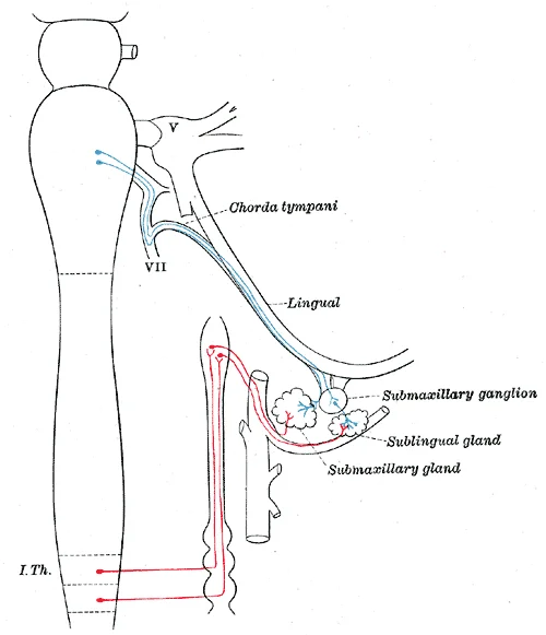
Long description
Sympathetic connections of the submaxillary and superior cervical ganglia.

Figure 3: Sympathetic connections of the submaxillary and superior cervical ganglia.
Figure 4Gray841
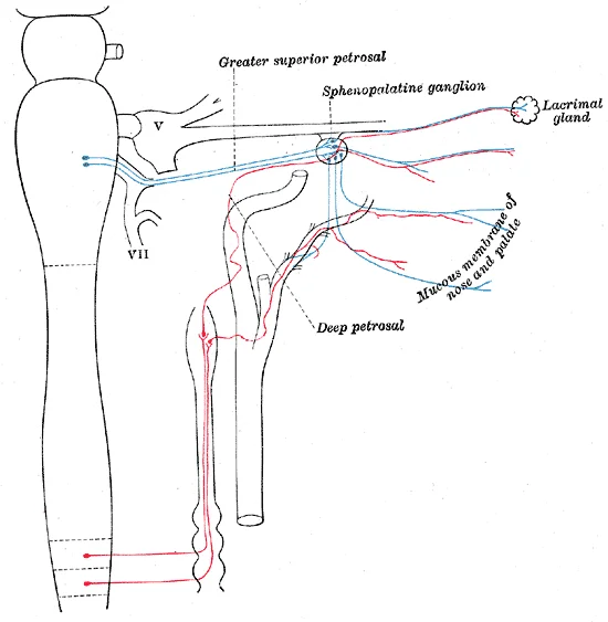
Long description
Sympathetic connections of the sphenopalatine and superior cervical ganglia.

Figure 4: Sympathetic connections of the sphenopalatine and superior cervical ganglia.
Figure 5Gray840
Long description
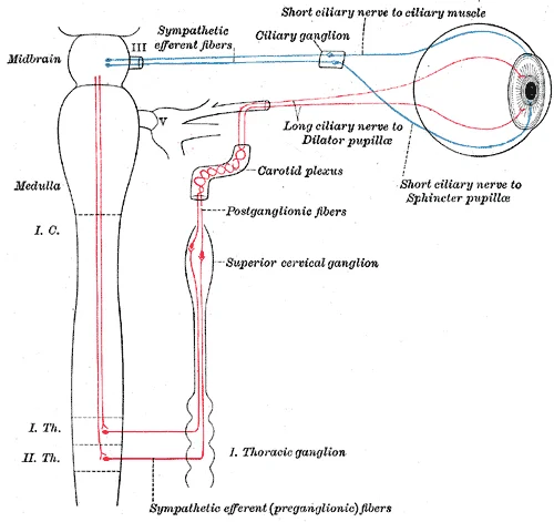
Sympathetic connections of the ciliary and superior cervical ganglia.

Figure 5: Sympathetic connections of the ciliary and superior cervical ganglia.
In contrast, the parasympathetic system is referred to as the craniosacral system because its preganglionic neurons are located in nuclei of the brain stem and the lateral horn of the sacral spinal cord. Three of the cranial nerves contain autonomic fibers: the oculomotor, facial, and glossopharyngeal nerves. The oculomotor fibers initiate pupillary constriction, whereas the facial and glossopharyngeal fibers both initiate salivation. The vagus nerve primarily targets autonomic ganglia in the thoracic and upper abdominal cavities. [1] [3]
The hypothalamus serves as the executive region in charge of the autonomic nervous system[1]. It coordinates homeostatic functions through the autonomic and endocrine systems[1]. When the body responds to a threat, the sympathetic system causes divergent effects as many different effector organs are activated together for a common purpose. To coordinate all these responses, the connections in the sympathetic system diverge from a limited region of the central nervous system to a wide array of ganglia that project to the many effector organs simultaneously. [2][4]
DefinitionAutonomic Tone
The resting level of activity in an organ system, determined by the balance of sympathetic and parasympathetic input[1].
| Feature | Sympathetic Division | Parasympathetic Division |
|---|---|---|
| Origin | Thoracic and upper lumbar spinal cord | Brain stem nuclei and sacral spinal cord |
| Ganglia Location | Paravertebral and prevertebral ganglia | Terminal ganglia near or within organs |
| Functional Role | Fight or flight; divergent activation | Rest and digest; homeostatic balance[1] |
The autonomic nervous system is organised into two main divisions, each with distinct anatomical origins and internal structures within the central nervous system. Understanding these origins requires examining the spinal cord and brainstem in cross-section to identify the specific nuclei that house preganglionic neurons.
In the spinal cord, the gray matter appears as a bulbous capital “H” in cross-section. This gray matter is subdivided into horns. The lateral horn is only found in the thoracic, upper lumbar, and sacral regions. It contains cell bodies of motor neurons of the autonomic nervous system. Specifically, the thoracolumbar spinal cord houses the lateral horn neurons for the sympathetic division. The sacral spinal cord is at the level of the upper lumbar vertebral bones. Neurons from the lateral horn of the sacral spinal cord serve as preganglionic neurons for the parasympathetic division. [4] [1]
In the brainstem, the parasympathetic output is based in specific nuclei. These nuclei project to terminal ganglia located close to or within the wall of target effectors. The preganglionic fibers from these brainstem nuclei release acetylcholine onto nicotinic receptors to excite the ganglionic neurons. [1]
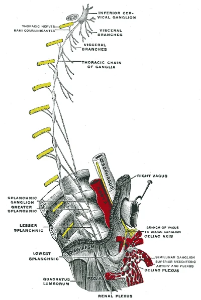
The sympathetic division originates from the lateral horn in the thoracolumbar spinal cord. An axon that leaves a central neuron of the lateral horn will pass through the white ramus communicans and enter the sympathetic chain. At the level of the spinal cord at which the preganglionic sympathetic fiber exits the spinal cord, a branch will synapse on a neuron in the adjacent chain ganglion. Some branches will extend up or down to a different level of the chain ganglia. Other branches will pass through the chain ganglia and project through one of the splanchnic nerves to a collateral ganglion. Finally, some branches may project through the splanchnic nerves to the adrenal medulla. All of these branches mean that one preganglionic neuron can influence different regions of the sympathetic system very broadly, by acting on widely distributed organs. [1]
Figure 6Gray845
Long description
Plan of right sympathetic cord and splanchnic nerves. (Testut.)

Figure 6: Plan of right sympathetic cord and splanchnic nerves (Testut.).
The parasympathetic division differs in its internal structure regarding axon length. Comparing the relative lengths of axons in the parasympathetic system, the preganglionic fibers are long and the postganglionic fibers are short because the ganglia are close to—and sometimes within—the target effectors. This contrasts with the sympathetic system, where preganglionic fibers are relatively short and synapse in the chain or collateral ganglia, while postganglionic fibers are long. [1]
The white matter of the spinal cord is separated into columns. Ascending tracts of nervous system fibers in these columns carry sensory information up to the brain, whereas descending tracts carry motor commands from the brain. The posterior columns are composed of axons of ascending tracts. The lateral columns are located on either side of the spinal cord, between the posterior horn and the axons of the anterior horn neurons. These tracts facilitate the communication between the central autonomic nuclei and higher brain centres.
DefinitionLateral Horn
A region of gray matter in the thoracic, upper lumbar, and sacral spinal cord containing cell bodies of autonomic motor neurons.
| Division | Origin | Location in CNS |
|---|---|---|
| Sympathetic | Thoracolumbar | Lateral horn of thoracic and upper lumbar spinal cord |
| Parasympathetic | Craniosacral | Brainstem nuclei and lateral horn of sacral spinal cord |
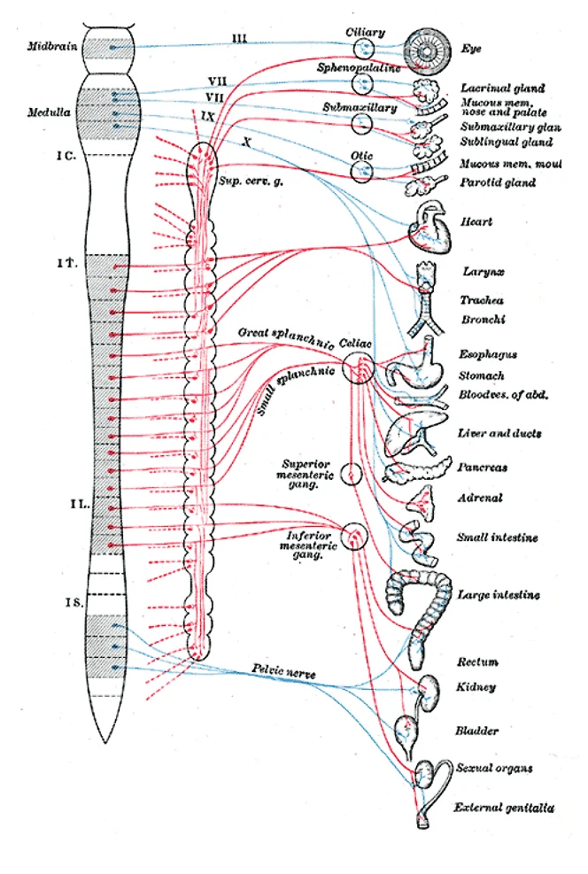
The autonomic nervous system functions as a coordinated network, allowing disparate effectors to undergo systemic change. Its architecture is often compared to an electrical circuit diagram, where simplified circuits link various receptacles and devices. In this view, the sympathetic outflow is shown originating from the thoracolumbar region. The preganglionic neurons extend to either the sympathetic trunk ganglia or the collateral ganglia. The collateral ganglia, also known as prevertebral ganglia, are situated anterior to the vertebral column. They receive inputs from splanchnic nerves and central sympathetic neurons. The three main collateral ganglia are the celiac ganglion, the superior mesenteric ganglion, and the inferior mesenteric ganglion. These structures control organs in the abdominal cavity and are considered part of the enteric nervous system. The parasympathetic component projects from the cranial nerves to the eye and intestines, while the sacral component serves the large intestine and pelvic organs. [1]
Figure 7Gray839
Long description
Diagram of efferent sympathetic nervous system. Blue, cranial and sacral outflow. Red, thoracohumeral outflow. ——, Postganglionic fibers to spinal and cranial nerves to supply vasomotors to head, trunk and limbs, motor fibers to smooth muscles of skin and fibers to sweat glands. (Modified after Meye

Figure 7: Diagram of efferent sympathetic nervous system (blue, cranial and sacral outflow).
Figure 8Gray843
Long description
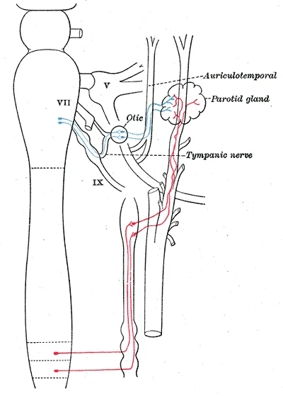
Sympathetic connections of the otic and superior cervical ganglia.

Figure 8: Sympathetic connections of the otic and superior cervical ganglia.
Figure 9Gray838
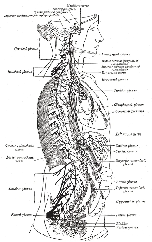
Long description
The right sympathetic chain and its connections with the thoracic, abdominal, and pelvic plexuses. (After Schwalbe.)

Figure 9: The right sympathetic chain and its connections with the thoracic, abdominal, and pelvic plexuses (After Schwalbe.).
DefinitionSynapse Classification
Synapses are classified as cholinergic if acetylcholine is released, or adrenergic if norepinephrine is released. [1]
The afferent branch of visceral reflexes differs from somatic reflexes. While somatic reflexes involve sensory receptors connecting to the central nervous system, visceral inputs often come from special senses associated with the viscera. For instance, a specific type of mechanoreceptor, called a baroreceptor, in the walls of the aorta and carotid sinuses that senses the stretch. This input is part of the afferent branch of cardiovascular reflexes, though it does not reach conscious perception. The sensory neuron is essent [2]
DefinitionReflex Types
A long reflex involves afferent branches entering the spinal cord or brain, while a short reflex is completely peripheral. [2]
The effector organs range from the iris to the urinary bladder. The thoracolumbar output reaches all these organs through sympathetic ganglia. At most targets, the response depends on the neurotransmitter and receptor present. Postganglionic sympathetic fibers release norepinephrine, with a minor exception. Postganglionic parasympathetic fibers release acetylcholine. Target cells possess adrenergic and muscarinic receptors. If norepinephrine binds to adrenergic receptors, it causes depolarization. If acetylcholine binds to muscarinic receptors, it causes hyperpolarization. [2]
| Parameter | Sympathetic Effect | Parasympathetic Effect |
|---|---|---|
| Neurotransmitter | Norepinephrine | Acetylcholine |
| Receptor | Adrenergic | Muscarinic |
| Heart Rate | 120–140 bpm | 60–80 bpm |
Without parasympathetic input, the heart works at approximately 100 beats per minute. The sympathetic system speeds this up during exercise. The parasympathetic system slows it to the resting rate. Blood pressure is primarily under sympathetic control. Smooth muscle in vessel walls has adrenergic receptors that constrict when norepinephrine is released. The parasympathetic system has no significant input to systemic blood vessels. [2]
The midbrain is positioned superiorly, containing the Edinger–Westphal nucleus. This nucleus forms part of the oculomotor complex, and its axons travel within the oculomotor nerve to innervate extraocular muscles. The preganglionic parasympathetic fibers terminate in the ciliary ganglion, located in the posterior orbit. Postganglionic fibers then project to the iris smooth muscle to control pupillary size. [1]
Inferior to the midbrain lies the upper medulla. Here, the salivatory nuclei contain neurons whose axons project through the facial and glossopharyngeal nerves. These pathways control salivary glands. Tear production is influenced by parasympathetic fibers in the facial nerve, which activate a ganglion and ultimately the lacrimal gland. [1]
The sympathetic output originates from the lateral horn of the thoracolumbar spinal cord. An axon from a central neuron projects via the ventral spinal nerve root and spinal nerve to a sympathetic ganglion. These preganglionic fibers release acetylcholine, which excites the ganglionic neuron through the nicotinic receptor. The nicotinic receptor is a ligand-gated cation channel that results in depolarization of the postsynaptic membrane. The postganglionic fiber then projects to a target effector, releasing norepinephrine to bind to an adrenergic receptor. [1] [2]
DefinitionReflex Arcs
Short reflexes do not involve the CNS, whereas somatic reflexes always involve the CNS.
Blood vessels in skeletal muscle, particularly in the lower limbs, are more likely to dilate. This allows blood flow to increase for active skeletal muscles during the fight-or-flight response. The sympathetic tone of blood vessels is caused by the lack of parasympathetic input to the systemic circulatory system. Only certain regions receive parasympathetic input that relaxes the smooth muscle wall of the blood vessels. [2]
| Component | Neurotransmitter | Receptor Type |
|---|---|---|
| Preganglionic (Sympathetic/Parasympathetic) | Acetylcholine | Nicotinic |
| Postganglionic (Parasympathetic) | Acetylcholine | Muscarinic |
| Postganglionic (Sympathetic) | Norepinephrine | Adrenergic |
Autonomic Pathway
- Preganglionic NeuronReleases ACh
- Question: Receptor?
Nicotinic
- Ganglionic Neuron
Muscarinic
- Target Organ
Sweat glands are another example, which only receive input from the sympathetic system. Acetylcholine released by postganglionic parasympathetic fibers cause the vessels to dilate, leading to the engorgement of the erectile tissue. [2]
The autonomic nervous system operates as a homeostatic reflex mechanism, maintaining physiological stability through antagonistic control. A primary example is the regulation of pupillary diameter to control retinal light exposure. In a threat scenario, such as encountering a predator, the sympathetic response triggers fight or flight, increasing pupillary diameter to allow more light into the retina for enhanced visual information processing. Conversely, the parasympathetic response promotes rest, reducing light entry to allow photoreceptor regeneration and maintain homeostasis. [2]
There are twelve cranial nerves, designated CNI through CNXII using Roman numerals. These are classified as sensory, motor, or mixed; three are solely sensory, five are strictly motor, and four are mixed. [3] [1][4]
The adrenergic system utilizes two main receptor types: alpha (α) and beta (β) adrenergic receptors. Unlike cholinergic receptors, these are not classified by drug binding but are all G protein–coupled receptors. There are two types of α-adrenergic receptors, termed α1 and α2, and three types of β-adrenergic receptors, termed β1, β2, and β3. The signaling molecule epinephrine differs from norepinephrine by the addition of a methyl group (CH3), with the prefix “nor-” indicating the absence of this group. [1]
Autonomic plexuses arise from spinal nerves, containing both sensory and motor fibers. Sensory fibers originate from dorsal root ganglia, entering the spinal cord via the dorsal nerve root to send action potentials into the central nervous system. Motor fibers originate from the anterior horn of the spinal cord, emerging via the ventral nerve root to cause skeletal muscle contraction. For instance, the radial nerve contains cutaneous sensory fibers and motor fibers for arm muscles. [3]
Higher neurological functions, such as memory and emotion, result from cerebral function. The cerebrum comprises the outer gray matter cortex and deep nuclei. The basal nuclei plan movements, the basal forebrain aids learning and memory, and the limbic cortex handles emotion and behavior. The cortex is covered by gray matter, with gyri being ridges and sulci being grooves. Brodmann’s areas, based on cytoarchitecture, localize function; areas 17 and 18 in the occipital lobe handle primary visual perception. The tegmentum of the brainstem contains nuclei for cranial nerves and regulates cardiovascular and respiratory functions. Posterior spinal regions handle sensory functions, while anterior regions handle motor functions, derived from the alar and basal plates respectively. [4]
Clinical pearlAutonomic Balance
The autonomic nervous system maintains homeostasis through opposing sympathetic and parasympathetic actions, such as regulating pupillary size to optimize retinal light exposure.
| Classification | Count | Description |
|---|---|---|
| Sensory | 3 | Solely sensory fibers |
| Motor | 5 | Strictly motor fibers |
| Mixed | 4 | Combination of sensory and motor fibers |
To understand autonomic lesions, one must first visualise the anatomical layout of the pathways. The view is a sagittal section of the head and neck, oriented with anterior to the left. This diagram labels the sympathetic and parasympathetic outflow, the ganglia, plexuses, neurotransmitters, and central pathways, specifically highlighting Horner syndrome.
DefinitionGanglia
A ganglion is a group of neuron cell bodies in the periphery. Ganglia can be categorized, for the most part, as either sensory ganglia or autonomic ganglia, referring to their primary functions. [3]
The sympathetic division relies on chain ganglia, also known as paravertebral ganglia. These are located adjacent to the vertebral column. Preganglionic sympathetic fibers are relatively short and myelinated. In contrast, postganglionic sympathetic fibers are long and unmyelinated. An axon from the central neuron that projects to a sympathetic ganglion is referred to as a preganglionic fiber. A postganglionic fiber is the axon from a ganglionic neuron that projects to the target effector. [1]
NoteAdrenal Medulla
One type of preganglionic sympathetic fiber does not terminate in a ganglion. These are the axons from central sympathetic neurons that project to the adrenal medulla. The cells in the adrenal medulla that are contacted by the preganglionic fibers are called chromaffin cells. [1]
The parasympathetic division uses terminal ganglia. The preganglionic fibers from the cranial region travel in cranial nerves. The targets of these fibers are terminal ganglia, which are located near—or even within—the target effector. These ganglia are often referred to as intramural ganglia when they are found within the walls of the target organ. The postganglionic fiber projects from the terminal ganglia a short distance to the target effector. [1]
| Division | Outflow | Ganglia Type | Location |
|---|---|---|---|
| Sympathetic | Thoracolumbar | Chain (Paravertebral) | Adjacent to vertebral column |
| Parasympathetic | Craniosacral | Terminal (Intramural) | Near or within target effector |
Neurons in the dorsal nucleus of the vagus nerve and the nucleus ambiguus project through the vagus nerve. Parasympathetic preganglionic fibers primarily influence the heart, bronchi, and esophagus in the thoracic cavity. The postganglionic fibers from the ganglia activated by the vagus nerve are often incorporated into the structure of the organ. [1]
Exam trapSweat Glands
In the sympathetic system, there are exceptions to this pattern of dual innervation. The postganglionic sympathetic fibers that contact sweat glands do not release norepinephrine, they release ACh. Sweat glands have muscarinic receptors and produce and secrete sweat in response to the presence of ACh. [1] [2]
Horner syndrome results from disruption of the sympathetic pathway. In the control of pupillary size, if light levels are low, the sympathetic system sends a signal out through the upper thoracic spinal cord to the superior cervical ganglion. The postganglionic fiber then projects to the iris. When those fibers contract, the pupil dilates. A lesion anywhere along this pathway from the hypothalamus to the eye causes ptosis, miosis, and anhidrosis. [2]
Horner Syndrome Pathway
- StartSympathetic outflow from upper thoracic cord
- Question: Lesion site?
Central
- Brainstem or spinal cord
Peripheral
- Superior cervical ganglion
| Feature | Sympathetic | Parasympathetic |
|---|---|---|
| Preganglionic Length | Short | Long |
| Postganglionic Length | Long | Short |
| Neurotransmitter (Post) | Norepinephrine (mostly) | Acetylcholine |
| Ganglia Location | Chain/Prevertebral | Terminal/Intramural |
- The autonomic nervous system has two main divisions: sympathetic (fight-or-flight) and parasympathetic (rest-and-digest). The sympathetic system prepares the body for emergency situations, increasing heart rate, dilating pupils, and redirecting blood flow to muscles, while the parasympathetic system conserves energy, slowing heart rate, and stimulating digestion.
- Cholinergic receptors include nicotinic (ligand-gated cation channel) and muscarinic (G protein-coupled receptor) types. Nicotinic receptors are found at autonomic ganglia, the neuromuscular junction, and many CNS sites, while muscarinic receptors are located on target organs in the periphery. [1]
- Nicotine binds only to nicotinic receptors, while muscarine binds only to muscarinic receptors, with no cross-reactivity. This specificity is similar to locks and keys—each receptor type has its own unique binding site that only specific molecules can activate.
- Adrenaline and epinephrine are two names for the same molecule, with epinephrine preferred in the US and adrenaline in Great Britain. The term "adrenaline" comes from the adrenal gland (ad- = "on top of"; renal = "kidney"), which secretes this hormone. [1]
- Norepinephrine and noradrenaline are also two names for the same molecule. Both terms refer to the neurotransmitter released by most postganglionic sympathetic fibers. [1]
- Acetylcholine is primarily a neurotransmitter released at synapses, while epinephrine and norepinephrine can act as both neurotransmitters and hormones. Postganglionic sympathetic fibers release norepinephrine as a neurotransmitter, but the adrenal medulla releases epinephrine and norepinephrine into circulation as hormones. [1]
- Visceral reflexes have a two-step efferent pathway (preganglionic to postganglionic fibers), unlike somatic reflexes which have direct motor neuron output. This two-neuron chain allows for greater control and modulation of autonomic responses.
- There are 31 pairs of spinal nerves: 8 cervical (C1-C8), 12 thoracic (T1-T12), 5 lumbar (L1-L5), 5 sacral (S1-S5), and 1 coccygeal. These nerves emerge from the vertebral column through intervertebral foramina, with C1 emerging between the first cervical vertebra and the occipital bone.
- The cervical plexus (C1-C5) gives rise to the phrenic nerve, which innervates the diaphragm. Damage to the cervical plexus or phrenic nerve can lead to diaphragmatic paralysis and respiratory compromise.
- The brachial plexus (C4-T1) reorganizes to form the nerves of the arm, including the radial, ulnar, and median nerves. Injuries to the brachial plexus can result in loss of function in the upper limb.
- Dorsal and ventral are often used interchangeably with posterior and anterior, particularly in reference to nerves and spinal cord structures. In spinal cord terminology, dorsal root contains sensory fibers while ventral root contains motor fibers. [4]
The autonomic nervous system (ANS) is divided into sympathetic and parasympathetic divisions, which generally have opposing effects on target organs. The sympathetic system prepares the body for "fight-or-flight" responses, increasing heart rate, blood pressure, and respiratory rate, while the parasympathetic system promotes "rest-and-digest" functions, slowing heart rate and stimulating digestion. [3]
The ANS uses two main neurotransmitters: acetylcholine (ACh) and norepinephrine (also called noradrenaline). ACh is released by both preganglionic sympathetic and all parasympathetic fibers, as well as by postganglionic parasympathetic fibers. Norepinephrine is released by most postganglionic sympathetic fibers. The adrenal medulla releases epinephrine (adrenaline) and norepinephrine (noradrenaline) into the bloodstream, allowing these substances to act as hormones with widespread effects throughout the body. [1]
The cholinergic receptors include nicotinic and muscarinic types. Nicotinic receptors are ligand-gated cation channels found at ganglia and neuromuscular junctions, while muscarinic receptors are G protein-coupled receptors located on target organs.
The sympathetic nervous system originates in the lateral horn of the spinal cord from T1 to L2 levels. Preganglionic fibers are relatively short and synapse in ganglia close to the spinal cord. The parasympathetic nervous system originates in the brainstem (cranial nerves III, VII, IX, X) and sacral spinal cord (S2-S4). Preganglionic fibers are long and synapse in ganglia near or within target organs. Visceral reflexes have a two-step efferent pathway (preganglionic to postganglionic fibers), un [2]
The spinal cord contains 31 pairs of spinal nerves, each with dorsal (sensory) and ventral (motor) roots. The dorsal root ganglion contains the cell bodies of sensory neurons, while the ventral root contains both somatic and autonomic motor fibers. There are 8 cervical (C1-C8), 12 thoracic (T1-T12), 5 lumbar (L1-L5), 5 sacral (S1-S5), and 1 coccygeal pair of spinal nerves. The brachial plexus (C4-T1) forms the nerves of the upper limb, including the radial, ulnar, and median nerves. [4]
The cerebrum is the largest portion and contains the cerebral cortex, which is responsible for higher cognitive functions.
Students frequently confuse the anatomical location of autonomic ganglia with the location of the neurons themselves. A critical distinction is that the cell body resides within the ganglion, whereas the term 'postganglionic' applies strictly to the axon extending from that cell body to the effector. This semantic precision is vital when describing neural pathways, as the neuron comprises the entire cell structure, not just the fibre.
Another frequent error involves the myelination status of sympathetic rami communicantes. Students often assume all autonomic fibres are unmyelinated. However, the preganglionic sympathetic fibres travelling from the spinal nerve to the chain ganglion are myelinated and are therefore referred to as white rami communicantes. Conversely, the postganglionic fibres returning from the ganglion to the spinal nerve via gray rami communicantes are unmyelinated axons. This difference in myelination explains the colour distinction and affects conduction velocity. [1]
Misidentifying the level of sympathetic innervation is also common. For instance, when considering the trachea, students might guess a cervical origin. In reality, spinal nerve T1 synapses with the T1 chain ganglion to innervate the trachea. This illustrates the direct junction type where the sympathetic nerve projects to the chain ganglion at the same level as the target effector.
Confusion between cranial and spinal sensory ganglia is prevalent. While dorsal root ganglia are associated with spinal nerves, cranial nerve ganglia are analogous structures associated with cranial nerves. A key anatomical difference is that the roots of cranial nerves are within the cranium, whereas the ganglia are located outside the skull. For example, the trigeminal ganglion is superficial to the temporal bone, while its associated nerve attaches to the mid-pons region of the brain stem. [3]
Students often overlook the specific composition of the lumbar and sacral plexuses. The lumbar plexus arises from all the lumbar spinal nerves and gives rise to nerves innervating the pelvic region and the anterior leg. The femoral nerve is a major branch, giving rise to the saphenous nerve as a branch that extends through the anterior lower leg. In contrast, the sacral plexus comes from the lower lumbar nerves L4 and L5 and the sacral nerves S1 to S4. [3]
The sciatic nerve is frequently misattributed to the lumbar plexus alone. It is the most significant systemic nerve to come from the sacral plexus. It is a combination of the tibial nerve and the fibular nerve. Understanding this distinction is crucial for localising lesions, as sciatica is the result of compression or irritation of the nerve or any of the spinal nerves giving rise to it.
Finally, students may confuse the functional roles of frontal lobe subregions. The precentral gyrus is the primary motor cortex, where cells are the upper motor neurons that instruct cells in the spinal cord to move skeletal muscles. The premotor area is responsible for thinking of a movement to be made, distinct from the execution of that movement. Broca’s area is responsible for the production of language, or controlling movements responsible for speech, and in the vast majority of people, it is located only on the left side.
Exam trapGanglion vs Neuron
Remember that the cell body is in the ganglion, and only the fiber is postganglionic. Typically, the term neuron applies to the entire cell. [1]
In the operating theatre, understanding the sympathetic chain is essential for procedures involving thoracic or lumbar regions. The distinction between white and gray rami communicantes guides surgeons during sympathectomy or ganglion blocks. Recognising that white rami are myelinated preganglionic fibres helps in predicting the speed of block onset and the specific levels affected. For example, targeting T1 requires understanding its direct projection to the T1 chain ganglion to affect the trachea.
On imaging, particularly MRI of the brainstem, identifying the pons is fundamental. The word pons comes from the Latin word for bridge, visible on the anterior surface as the thick bundle of white matter attached to the cerebellum. This structure is the main connection between the cerebellum and the brain stem. Radiologists look for the bridge-like white matter on the anterior surface, noting that the gray matter beneath is a continuation of the tegmentum from the midbrain. [4]
Bedside neurological examinations rely on distinguishing the functions of the midbrain and pons. The midbrain is a small region between the thalamus and pons, separated into the tectum and tegmentum. The cerebral aqueduct passes through the center of the midbrain, such that these regions are the roof and floor of that canal. Lesions here can affect eye movements via the frontal eye fields or motor control via the precentral gyrus.
In examinations, questions often test the embryological and anatomical continuity of the spinal cord. The length of the spinal cord is divided into regions that correspond to the regions of the vertebral column. The name of a spinal cord region corresponds to the level at which spinal nerves pass through the intervertebral foramina. Students must know that the spinal cord does not grow significantly longer after the first or second year, but the skeleton continues to grow.
The cerebellum’s role in motor coordination is frequently tested. The cerebellum is largely responsible for comparing information from the cerebrum with sensory feedback from the periphery through the spinal cord. It accounts for approximately 10 percent of the mass of the brain. It is connected to the brain stem, primarily at the pons, where it receives a copy of the descending input from the cerebrum to the spinal cord. It can compare this with sensory feedback input through the medulla and send output through the midbrain that can correct motor commands for coordination. [2]
Understanding the basal ganglia pathways is also relevant for movement disorders. The switch between the two pathways is the substantia nigra pars compacta, which projects to the striatum and releases the neurotransmitter dopamine. Dopamine receptors are either excitatory (D1-type receptors) or inhibitory (D2-type receptors). The direct pathway is activated by dopamine, and the indirect pathway is inhibited by dopamine. When the substantia nigra pars compacta is firing, it signals to the basal nuclei that the body is in an active state, and movement will be more likely.
DefinitionPons Anatomy
The pons is visible on the anterior surface of the brain stem as the thick bundle of white matter attached to the cerebellum. It is the main connection between the cerebellum and the brain stem.
The cell body is in the ganglion, and only the fiber is postganglionic. White rami communicantes are myelinated preganglionic sympathetic fibres. Gray rami communicantes are unmyelinated postganglionic sympathetic fibres. Cranial nerve ganglia are outside the skull, while their roots are within the cranium. [3] The lumbar plexus arises from all lumbar spinal nerves. The sacral plexus comes from L4, L5, and S1 to S4. The sciatic nerve combines the tibial and fibular nerves. [3] The pons is the main connection between the cerebellum and the brain stem.
Answer each one in your head first, then open the answer.
What is the myelination status of white rami communicantes?
Show answerHide answer
MyelinatedWhich spinal nerve level innervates the trachea via the T1 chain ganglion?
Show answerHide answer
T1Where is the trigeminal ganglion located relative to the temporal bone?
Show answerHide answer
Superficial to the temporal boneWhat two nerves combine to form the sciatic nerve?
Show answerHide answer
Tibial nerve and fibular nerveWhat percentage of the brain mass does the cerebellum account for?
Show answerHide answer
10 percent
- Betts JG, Young KA, Wise JA, et al. Divisions of the Autonomic Nervous System. OpenStax, Anatomy and Physiology 2e (CC BY-NC-SA 4.0). Accessed .
- Betts JG, Young KA, Wise JA, et al. Autonomic Reflexes and Homeostasis. OpenStax, Anatomy and Physiology 2e (CC BY-NC-SA 4.0). Accessed .
- Betts JG, Young KA, Wise JA, et al. The Peripheral Nervous System. OpenStax, Anatomy and Physiology 2e (CC BY-NC-SA 4.0). Accessed .
- Betts JG, Young KA, Wise JA, et al. The Central Nervous System. OpenStax, Anatomy and Physiology 2e (CC BY-NC-SA 4.0). Accessed .
Figure credits
- Figure 1: Henry Vandyke Carter, via Wikimedia Commons, Public domain, Public domain.
- Figure 2: Henry Vandyke Carter, via Wikimedia Commons, Public domain, Public domain.
- Figure 3: Henry Vandyke Carter, via Wikimedia Commons, Public domain, Public domain.
- Figure 4: Henry Vandyke Carter, via Wikimedia Commons, Public domain, Public domain.
- Figure 5: Henry Vandyke Carter, via Wikimedia Commons, Public domain, Public domain.
- Figure 6: Henry Vandyke Carter, via Wikimedia Commons, Public domain, Public domain.
- Figure 7: Henry Vandyke Carter, via Wikimedia Commons, Public domain, Public domain.
- Figure 8: Henry Vandyke Carter, via Wikimedia Commons, Public domain, Public domain.
- Figure 9: Henry Vandyke Carter, via Wikimedia Commons, Public domain, Public domain.
Educational content for medical students and doctors. It is not a substitute for clinical judgement or local guidelines.