Sensory pathways
Sensory pathways transmit electrochemical signals from receptors to the CNS for integration and perception.
Sensory receptors serve a major role in helping us learn about the environment around us, or about the state of our internal environment. Stimuli from varying sources, and of different types, are received and changed into the electrochemical signals of the nervous system. This occurs when a stimulus changes the cell membrane potential of a sensory neuron. The stimulus causes the sensory cell to produce an action potential that is relayed into the central nervous system (CNS), where it is integra [1]
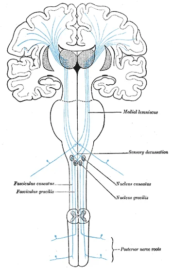
Figure 1Gray759
Long description
The sensory tract. (Modified from Poirier.)

Figure 1: The sensory tract (Modified from Poirier.).
Once any sensory cell transduces a stimulus into a nerve impulse, that impulse has to travel along axons to reach the CNS. In many of the special senses, the axons leaving the sensory receptors have a topographical arrangement, meaning that the location of the sensory receptor relates to the location of the axon in the nerve. For example, in the retina, axons from RGCs in the fovea are located at the center of the optic nerve, where they are surrounded by axons from the more peripheral RGCs. [1]
The nerves that convey sensory information from the periphery to the CNS are either spinal nerves, connected to the spinal cord, or cranial nerves, connected to the brain. [1]
Cranial nerves convey specific sensory information from the head and neck directly to the brain. That said, sensory pathways are complex and cranial nerves may project ipsilaterally, contralaterally, bilaterally, or a combination of these. Some cranial nerves contain only sensory axons, such as the olfactory, optic, and vestibulocochlear nerves. Other cranial nerves contain both sensory and motor axons, including the trigeminal, facial, glossopharyngeal, and vagus nerves (however, the vagus nerve is not associated with the somatic nervous system). The general senses of somatosensation for the face travel through the trigeminal system. [1]
Figure 2Gray760
Long description
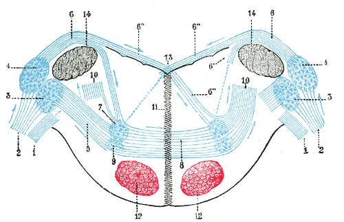
Terminal nuclei of the cochlear nerve, with their upper connections. (Schematic.) The vestibular nerve with its terminal nuclei and their efferent fibers have been suppressed. On the other hand, in order not to obscure the trapezoid body, the efferent fibers of the terminal nuclei on the right side

Figure 2: Terminal nuclei of the cochlear nerve, with their upper connections.
DefinitionTransduction
The process by which a stimulus changes the cell membrane potential of a sensory neuron to produce an action potential.
Clinical pearlTopography
Axons from sensory receptors often maintain a topographical arrangement, linking receptor location to axon position in the nerve.
| Nerve Type | Examples | Projection Pattern |
|---|---|---|
| Sensory Only | Olfactory, Optic, Vestibulocochlear | Mostly Ipsilateral |
| Mixed | Trigeminal, Facial, Glossopharyngeal, Vagus | Mostly Ipsilateral |
Sensory pathways transmit information from receptors to the cerebral cortex via chains of neurons. Most pathways involve three orders of neurons, though olfaction is a notable exception. The first-order neuron carries the signal from the periphery to the central nervous system. The third-order neuron projects from the thalamus to the primary sensory cortex.
DefinitionDecussation
The crossing of nerve fibres from one side of the central nervous system to the other, ensuring that the left hemisphere processes sensory input from the right side of the body.
The dorsal column-medial lemniscus pathway conveys fine touch and proprioception. First-order neurons have cell bodies in the dorsal root ganglia and ascend ipsilaterally in the dorsal columns. Second-order neurons are located in the nucleus gracilis and nucleus cuneatus in the medulla. These neurons decussate as internal arcuate fibres and ascend as the medial lemniscus. Third-order neurons reside in the ventral posterolateral (VPL) nucleus of the thalamus and project to the primary somatosensory cortex. Lesions above the decussation cause contralateral loss of sensation.
Retinal ganglion cells form the first-order neurons. Axons form the optic nerve. At the optic chiasm, fibres from the nasal retina decussate. The optic tract contains second-order neurons projecting to the lateral geniculate nucleus (LGN). Third-order neurons in the LGN project via the optic radiations to the primary visual cortex.
The auditory pathway is largely bilateral. First-order neurons are bipolar cells in the spiral ganglion. Second-order neurons in the cochlear nuclei project to the superior olivary complex, with some decussation. Third-order neurons in the medial geniculate body (MGB) project to the auditory cortex. The MGB is part of the auditory thalamus. [2]
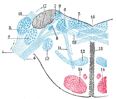
First-order neurons are in the vestibular ganglion. Second-order neurons in the vestibular nuclei project to the cerebellum, thalamus, and spinal cord. Thalamic projections (third-order) reach the parietal cortex.
First-order neurons are in the geniculate, petrosal, and nodose ganglia. Second-order neurons in the nucleus of the solitary tract project to the ventral posteromedial (VPM) nucleus of the thalamus. Third-order neurons project to the insular cortex.
Olfaction differs significantly. Olfactory receptor neurons are located in a small region within the superior nasal cavity [1]. These are the first-order neurons. Their axons pass through the cribriform plate to synapse in the olfactory bulb. There is no thalamic relay for the primary pathway. Second-order neurons (mitral and tufted cells) project directly to the piriform cortex and amygdala. The olfactory epithelium contains bipolar sensory neurons. [1]
| Sense | 1st Order Site | 2nd Order Site | Decussation | 3rd Order Site |
|---|---|---|---|---|
| Somatosensory | Dorsal Root Ganglion | Medulla (Nucleus Gracilis/Cuneatus) | Medulla | VPL Thalamus |
| Visual | Retina | Lateral Geniculate Nucleus | Optic Chiasm | V1 Cortex |
| Auditory | Spiral Ganglion | Cochlear Nuclei/Superior Olive | Brainstem | MGB Thalamus |
| Olfactory | Olfactory Epithelium | Olfactory Bulb | None | Piriform Cortex |
The transmission of sensory information from the periphery to the cerebral cortex involves a complex, multi-synaptic journey through the central nervous system. Understanding this course requires distinguishing between the initial detection of a stimulus and its subsequent interpretation. Sensation is defined as the activation of sensory receptor cells at the level of the stimulus, whereas perception is the central processing of these stimuli into a meaningful pattern[1]. While perception is dependent on sensation, not all sensations reach the level of conscious perception[1].
This diagram presents a coronal section of the brainstem and diencephalon, oriented with the anterior aspect at the top and the posterior at the bottom. The image highlights the decussation of sensory fibres in the medulla, showing the internal arcuate fibres crossing the midline to form the medial lemniscus. Superiorly, the diagram illustrates the synapse within the ventral posterolateral nucleus of the thalamus, where second-order neurons relay information to third-order neurons projecting to the primary somatosensory cortex. The labelled structures include the gracile and cuneate nuclei, the medial lemniscus, the thalamus, and the internal capsule, demonstrating the precise anatomical relationships required for somatic sensation. [1]
Figure 3Gray762
Long description
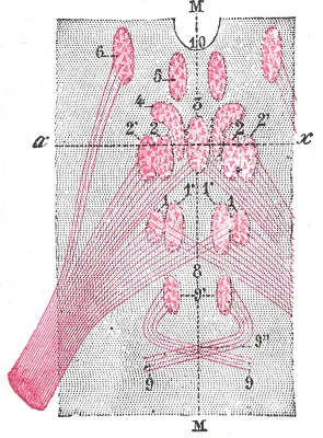
Figure showing the different groups of cells, which constitute, according to Perlia, the nucleus of origin of the oculomotor nerve. 1. Posterior dorsal nucleus. 1’. Posterior ventral nucleus. 2. Anterior dorsal nucleus. 2’. Anterior ventral nucleus. 3. Central nucleus. 4. Nucleus of Edinger and West

Figure 3: Figure showing the different groups of cells, which constitute, according to Perlia, the nucleus of origin of the oculomotor nerve (1).
Figure 4Gray761
Long description
Terminal nuclei of the vestibular nerve, with their upper connections. (Schematic.) 1. Cochlear nerve, with its two nuclei. 2. Accessory nucleus. 3. Tuberculum acusticum. 4. Vestibular nerve. 5. Internal nucleus. 6. Nucleus of Deiters. 7. Nucleus of Bechterew. 8. Inferior or descending root of acous

Figure 4: Terminal nuclei of the vestibular nerve, with their upper connections (Schematic.) 1.
Sensory receptors are the cells or structures that detect sensations[1]. These receptors can be categorized by the type of stimuli they transduce. For instance, mechanoreceptors interpret physical stimuli such as pressure, vibration, sound, and body position[1]. Thermoreceptors sense temperature deviations above or below normal body temperature[1].
Once activated, neural signals travel via specific pathways. In the case of hearing and balance, signals from the inner ear are relayed to the brain stem through separate fiber bundles[1]. The inner ear, composed of the cochlea and vestibule, contains sensory neurons in the spiral ganglia[1]. These distinct bundles travel together from the inner ear to the brain stem as the vestibulocochlear nerve[1].
For somatic sensation, the pathway typically involves a three-neuron chain. First-order neurons carry signals from the periphery to the spinal cord or brainstem. Second-order neurons decussate and ascend to the thalamus. Third-order neurons project from the thalamus to the cortex.
DefinitionSensation vs Perception
Sensation is the activation of sensory receptor cells at the level of the stimulus, while perception is the central processing of sensory stimuli into a meaningful pattern[1].
| Receptor Type | Stimulus Detected | Example |
|---|---|---|
| Mechanoreceptor | Pressure, vibration, sound, balance | Touch, hearing |
| Nociceptor | Chemicals from tissue damage | Pain |
| Thermoreceptor | Temperature changes | Heat, cold |
| Chemoreceptor | Chemical stimuli | Taste, smell |
The thalamus modulates attention, language processing, and working memory via these pathways. This regulatory power ensures that sensory input is integrated appropriately with higher cortical functions, allowing for coherent perception rather than isolated sensation.
Figure 5Sensory Pathways III

Long description
Flow diagram from sensory receptors to the cerebral cortex

Figure 5: Flow diagram from sensory receptors to the cerebral cortex.
Sensory modalities describe the specific ways in which information is encoded by the nervous system, a process fundamentally similar to transduction. Each distinct sense represents a unique modality, and while there are five major senses, these can be subdivided into as many as seventeen specific sensory modalities. An individual sensory modality represents the sensation of a specific type of stimulus. For instance, the general sense of touch, technically known as somatosensation, is not a s [1]
The classification of modalities is largely determined by the mechanism of transduction. The chemical senses, specifically taste and smell, rely on chemical detection. In contrast, the general sense of touch often includes chemical sensation in the form of nociception, or pain. Pressure, vibration, muscle stretch, and the movement of hair by an external stimulus are all detected by mechanoreceptors. Hearing and balance are also mediated by mechanoreceptors, while vision involves the activation of photoreceptors. [1]
DefinitionSensory Modality
A specific way that information is encoded, defined by the type of stimulus and the transduction mechanism involved.
Beyond somatic senses, the general senses include visceral senses. These are distinct from somatic nervous system function because they do not normally rise to the level of conscious perception. This distinction is crucial for understanding why certain internal stimuli remain subconscious while external stimuli become conscious. [1]
The olfactory system provides a detailed example of specialized sensory anatomy. These neurons basally project an axon to the olfactory bulb, while apically, they contact the lumen of the nasal cavity with the terminal knob of their apical dendrite. Between the cell bodies and the basal lamina lies a thin layer of globose and horizontal basal cells, which contain adult neuronal stem cells. These stem cells are essential for maintaining the function and histology of the olfactory epithelium, as olfactory neurons are prone to damage and infection due to their exposure to ambient air. [3]
| Modality | Primary Stimulus | Transduction Mechanism |
|---|---|---|
| Somatosensation | Pressure, Vibration, Pain | Mechanoreceptors, Nociceptors |
| Vision | Light | Photoreceptors |
| Hearing/Balance | Mechanical Movement | Mechanoreceptors |
| Taste/Smell | Chemicals | Chemoreceptors |
Clinical pearlClinical Correlation
Understanding the distinction between somatic and visceral senses helps explain why visceral pain is often poorly localized, as these signals do not typically reach conscious perception under normal conditions.
The olfactory epithelium's unique structure, with its exposed stem cells and direct connection to the bulb, highlights the vulnerability and regenerative capacity of this sensory system. The presence of microvillar cells, despite their unknown function, adds another layer of complexity to our understanding of olfactory transduction.
The sensory pathways consist of a series of neurons that transmit sensory information from peripheral receptors to the cerebral cortex. Lesions at different levels of these pathways result in distinct clinical deficits.
Peripheral nerves contain a mixture of afferent sensory axons and efferent motor axons. When a peripheral nerve is injured, both sensory and motor functions may be impaired depending on the specific nerve affected. The deficit follows the distribution of the nerve's sensory territory.
The dorsal root contains only the axons of sensory neurons. A lesion to a dorsal root results in loss of all sensory modalities in the dermatome supplied by that root, while motor function remains intact as the ventral root is unaffected. [1]
| Lesion Level | Resulting Sensory Deficit |
|---|---|
| Posterior (dorsal) column | Loss of fine touch, vibration, and proprioception (dorsal column-medial lemniscus pathway) |
| Lateral spinothalamic tract | Loss of pain and temperature (contralateral, 1-2 segments below lesion) |
| Anterior (ventral) spinothalamic tract | Loss of crude touch and pressure (contralateral, 1-2 segments below lesion) |
| Central cord syndrome | Loss of pain and temperature (bilaterally in upper extremities if lesion is cervical) |
| Brown-Séquard syndrome | Ipsilateral loss of proprioception and vibration, contralateral loss of pain and temperature |
Lesions in the brainstem can affect specific sensory tracts as they ascend or descend. For example, a lesion in the medial lemniscus would cause loss of fine touch and proprioception contralaterally, while a lesion in the spinothalamic tract would cause loss of pain and temperature contralaterally.
The thalamus serves as a relay station for sensory information to the cortex. A thalamic lesion typically causes contralateral sensory loss affecting all modalities, often with a characteristic "thalamic pain" syndrome.
Lesions in the primary sensory cortex (e.g., postcentral gyrus for somatosensation) cause contralateral sensory deficits specific to the modality processed in that area. For example, a lesion in the primary somatosensory cortex would cause contralateral loss of fine touch, vibration, and proprioception.
Sensory testing relies on the principle that physical stimuli are converted into electrochemical signals. These physical alterations increase ion flow across the cell membrane, potentially generating an action potential or a graded potential within the sensory neuron. This initial transduction is the fundamental first step in any sensory pathway. [1]
In the context of clinical neurology, we assess these pathways by evaluating specific modalities. For instance, the olfactory system demonstrates how physiochemical properties of volatile compounds are transduced into electrochemical membrane potentials at the apical surface membrane of olfactory sensory neurons. This interface allows the brain to construct perceptions such as the aroma of black raspberries or pain grillé. The apical surface is significantly enlarged by cilia or microvilli emerging from the terminal knob of the apical dendrite, protruding into the mucus. This enlargement increases the surface area for detection. Signal termination is complex, though the dwell-time of the receptor-odorant complex may be too short for some inactivation processes under physiological conditions. [3]
Visual sensory testing also involves distinct transduction mechanisms. In contrast, ciliary photoreceptors prefer phosphodiesterase-mediated signal transduction with cGMP as a second messenger. Understanding this distinction is vital for interpreting visual pathway lesions. [3]
DefinitionTransduction
The conversion of a physical stimulus into an electrochemical signal by sensory receptors, initiating the neural impulse.
Clinical pearlOlfactory Mechanism
Olfactory depolarization relies on high intracellular chloride, unlike most neurons where chloride is inhibitory.
The thalamus plays a critical role in gating and modulating these sensory inputs. Corticothalamic pathways allow the thalamus to provide contextual modulation to cortical computations[1].
Exam trapSensory Attenuation
Self-generated sounds are perceived as quieter due to corollary discharge from motor areas via CT pathways, not due to reduced stimulus intensity.
Synaptic plasticity also influences sensory processing. Such plasticity allows the system to adapt its sensitivity based on recent activity.
| System | Primary Transduction Feature | Key Ion/Second Messenger |
|---|---|---|
| Olfactory | High intracellular chloride | Chloride efflux |
| Ciliary Photoreceptors | Phosphodiesterase activation | cGMP |
| Rhabdomeric/Retinal | Phosphoinositide signalling | Phosphoinositides |
DefinitionGustatory Pathways
Taste sensation is mediated by three cranial nerves: the facial, glossopharyngeal, and vagus nerves, each serving distinct regions of the tongue.
The facial nerve (CN VII) provides taste sensation to the anterior two thirds of the tongue. The glossopharyngeal nerve (CN IX) innervates the posterior one third of the tongue. These neurons are activated when taste molecules bind to gustatory cells, triggering neurotransmitter release onto sensory neuron dendrites.
DefinitionOlfactory Anatomy
Olfactory neurons extend axons through the cribriform plate to the olfactory bulb, bypassing the thalamus before reaching the cortex.
Olfactory neuron axons pass through olfactory foramina in the cribriform plate of the ethmoid bone. These axons form the olfactory tract, which connects to the olfactory bulb on the ventral frontal lobe. From the bulb, signals travel to the primary olfactory cortex in the inferior medial temporal lobe. Other projections reach the limbic system and hypothalamus, linking smell to memory and emotion. Crucially, smell is the only sensory modality that does not synapse in the thalamus before connecting to the cerebral cortex. [1]
DefinitionSemicircular Canals
The three semicircular canals detect rotational head movements via hair cells in the ampullae.
One canal lies in the horizontal plane, while the anterior and posterior canals are vertical. Each canal connects to the vestibule at an enlarged region called the ampulla. Hair cells within the ampulla respond to rotational movement, such as turning the head. Their stereocilia extend into the cupula, which deflects opposite [1]
DefinitionSomatosensory Transduction
Free nerve endings transduce pain and temperature using nociceptors and thermoreceptors.
Pain and temperature signals are transduced by free nerve endings. Thermoreceptors respond to local temperatures differing from body temperature. Some thermoreceptors detect cold, while others detect heat. Nociceptors respond to potentially damaging mechanical, chemical, or thermal stimuli. This binding activates nociceptors, creating the sensation of heat from spicy foods. [1]
Students frequently confuse the structural classification of sensory receptors with their functional location. A common error is assuming that all sensory neurons have free nerve endings. In reality, receptors are classified by structure into three distinct types: free nerve endings, encapsulated endings, and specialized receptor cells. Free nerve endings, such as those for pain and temperature in the dermis, lack connective tissue wrapping. In contrast, lamellated corpuscles are neurons with encapsulated nerve endings that respond to pressure and touch. Specialized receptor cells, like photoreceptors in the retina, have distinct structural components for specific stimuli. Another frequent mistake is misclassifying receptors by location. Exteroceptors are located near external stimuli, such as somatosensory receptors in the skin. Interoceptors interpret stimuli from internal organs, such as those sensing blood pressure in the aorta or carotid sinus. Proprioceptors are located near moving body parts, such as muscles, to interpret tissue positions. [1]
A second major pitfall involves the anatomy of the eye. Students often overlook the layered structure of the eyeball. The eye is a hollow sphere composed of three tissue layers: the fibrous tunic, vascular tunic, and neural retina. The fibrous tunic includes the sclera and cornea. The sclera accounts for five sixths of the surface of the eye. The transparent cornea covers the anterior part and allows light to enter. The vascular tunic contains the choroid, ciliary body, and iris. The choroid provides blood supply to the eyeball and is posterior to the ciliary body. The ciliary body is a muscular structure attached to the lens by suspensory ligaments. [1]
A third pitfall concerns visual acuity and the fovea. Students may assume uniform sensitivity across the retina. However, photoreceptors are located behind axons, retinal ganglion cells, bipolar cells, and blood vessels. Significant light is absorbed by these structures before reaching photoreceptors. The fovea is a small area at the exact center of the retina where supporting cells and blood vessels are absent. Only photoreceptors remain here, allowing the least amount of light to be absorbed b. In addition, each photoreceptor cell of the fovea is connected to a single RGC. [1]
Exam trapReceptor Classification
Do not mix structural and location-based classifications. Free nerve endings are a structural type, while exteroceptors are a location-based type.
At the bedside, understanding receptor types aids in neurological examination. Differentiating between free nerve endings and encapsulated corpuscles helps localise sensory deficits. For instance, testing for pain and temperature assesses free nerve endings in the dermis. Testing for pressure and touch involves lamellated corpuscles. Recognising proprioceptors allows assessment of joint position sense, crucial for detecting sensory ataxia. Interoceptor assessment, such as monitoring blood pressure via carotid sinus receptors, is vital in cardiovascular evaluation.
In the operating theatre, knowledge of the ear’s anatomy is essential. The auricle, or pinna, directs sound waves toward the auditory canal. The canal enters the skull through the external auditory meatus of the temporal bone. The tympanic membrane vibrates when struck by sound waves. These structures constitute the external ear. The middle ear contains ossicles spanning a space. Surgeons must respect these relations to avoid iatrogenic hearing loss. [1]
On imaging, identifying the eye’s layers prevents misinterpretation. The sclera’s large surface area is visible as the white of the eye. The cornea’s transparency is key for anterior segment imaging. The choroid’s vascularity is apparent on angiography. The ciliary body’s muscular nature is relevant in glaucoma surgery. Understanding the fovea’s avascular zone is critical in interpreting optical coherence tomography scans for macular diseases.
In examinations, students must recall specific anatomical details. The sclera accounts for five sixths of the eye’s surface. The fovea lacks supporting cells and blood vessels. Photoreceptors in the fovea connect to single retinal ganglion cells. These facts are frequently tested.
DefinitionFovea
A small central area of the retina lacking supporting cells and blood vessels, containing only photoreceptors for maximum visual acuity.
| Type | Structure/Location | Example |
|---|---|---|
| Free Nerve Ending | Neuron with dendrites embedded in tissue | Pain and temperature receptors in dermis |
| Encapsulated Ending | Sensory endings in connective tissue | Lamellated corpuscles for pressure and touch |
| Specialized Cell | Distinct structural components | Photoreceptors in retina |
| Exteroceptor | Near external stimulus | Somatosensory receptors in skin |
| Interoceptor | Internal organs/tissues | Blood pressure receptors in aorta/carotid sinus |
| Proprioceptor | Near moving body parts | Muscle receptors for tissue position |
Sensory Pathway Assessment
- Patient PresentationSensory deficit identified
- Question: Test Modality
Pain/Temperature
- Assess Free Nerve EndingsCheck dermal response
Pressure/Touch
- Assess Encapsulated EndingsCheck lamellated corpuscles
Position/Movement
- Assess ProprioceptorsCheck muscle/joint sense
Sensory receptors are classified structurally as free nerve endings, encapsulated endings, or specialized cells. Free nerve endings mediate pain and temperature sensations in the skin. Encapsulated endings, like lamellated corpuscles, respond to pressure and touch. Specialized receptor cells, such as photoreceptors, interpret specific stimuli like light. Exteroceptors detect external stimuli, such as somatosensory receptors in the skin. Interoceptors sense internal conditions, like blood pressure in the aorta. Proprioceptors monitor body position and movement, located near muscles. The fovea has the highest visual acuity due to the absence of supporting cells and blood vessels.
Answer each one in your head first, then open the answer.
Which receptor type is responsible for pain and temperature in the dermis?
Show answerHide answer
Free nerve endingsWhat structure accounts for five sixths of the eye's surface?
Show answerHide answer
The scleraWhere are proprioceptors located?
Show answerHide answer
Near moving parts of the body, such as musclesWhy is visual acuity greatest at the fovea?
Show answerHide answer
It lacks supporting cells and blood vessels, minimizing light absorptionWhat is the function of the ciliary body?
Show answerHide answer
It is a muscular structure attached to the lens by suspensory ligaments
- Betts JG, Young KA, Wise JA, et al. Sensory Perception. OpenStax, Anatomy and Physiology 2e (CC BY-NC-SA 4.0). Accessed .
- Antunes FM, Malmierca MS. Corticothalamic Pathways in Auditory Processing: Recent Advances and Insights From Other Sensory Systems. Frontiers in neural circuits 2021;15:721186. doi:10.3389/fncir.2021.721186. Accessed .
- Elsaesser R, Paysan J. The sense of smell, its signalling pathways, and the dichotomy of cilia and microvilli in olfactory sensory cells. BMC neuroscience 2007;8 Suppl 3:S1. doi:10.1186/1471-2202-8-s3-s1. Accessed .
Figure credits
- Figure 1: Henry Vandyke Carter, via Wikimedia Commons, Public domain, Public domain.
- Figure 2: Henry Vandyke Carter, via Wikimedia Commons, Public domain, Public domain.
- Figure 3: Henry Vandyke Carter, via Wikimedia Commons, Public domain, Public domain.
- Figure 4: Henry Vandyke Carter, via Wikimedia Commons, Public domain, Public domain.
- Figure 5: Btarski, via Wikimedia Commons, CC BY-SA 3.0, CC BY-SA 3.0.
Educational content for medical students and doctors. It is not a substitute for clinical judgement or local guidelines.