Motor pathways
Motor pathways control voluntary skeletal muscle movement through cortical and spinal connections.
The motor system begins with the prefrontal cortex in the frontal lobe, which is responsible for higher functions such as working memory. The prefrontal areas, defined as regions of the frontal lobe that do not elicit movement when electrically stimulated, are primarily located in the anterior part of the frontal lobe. These areas project into secondary motor cortices, including the premotor cortex and supplemental motor area, which help plan movements. [1]
The primary motor cortex contains large Betz cells that initiate movements and project through two main descending pathways: the corticobulbar tract and the corticospinal tract. The corticobulbar tract connects to motor nuclei in the brain stem, while the corticospinal tract connects to motor neurons in the ventral horn of the spinal cord. These pathways are responsible for conscious or voluntary movements of skeletal muscles. [1]
The corticobulbar tract consists of ipsilateral axons, meaning they project from the cortex to the motor nucleus on the same side of the nervous system. In contrast, the corticospinal tract is largely contralateral, with fibers crossing the midline to synapse on the opposite side of the body. Therefore, the right motor cortex controls muscles on the left side of the body, and vice versa. [1]
The extrapyramidal system, influenced by subcortical structures like the basal nuclei, modulates movements, particularly those involving the spine and cranium. The skeletomotor system is largely based on a two-cell projection from the precentral gyrus to skeletal muscles, consisting of upper motor neurons (UMN) and lower motor neurons (LMN). Voluntary movements require both UMNs and LMNs to be active. [1] [2]
Motor units consist of a motor neuron and all the muscle fibers it innervates, with sizes varying from 10 fibers in extraocular muscles to 1000 fibers in the quadriceps. The number of fibers in a motor unit corresponds to the precision of control needed for that muscle. Muscles requiring finer motor control have more motor units and require a larger topographical field in the primary motor cortex. [1]
Figure 1Gray764
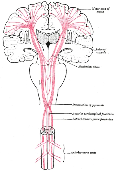
Long description
The motor tract. (Modified from Poirier.)

Figure 1: The motor tract (Modified from Poirier.).
Figure 21426 Corticospinal Pathway
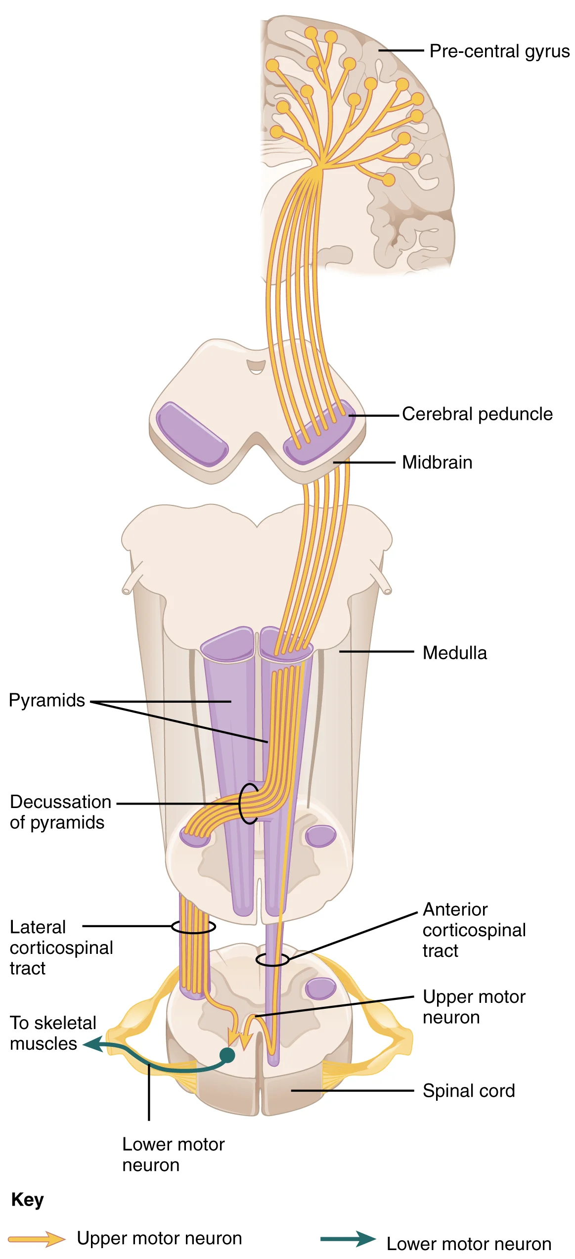
Long description
The major descending tract that controls skeletal muscle movements is the corticospinal tract. It is composed of two neurons, the upper motor neuron and the lower motor neuron. The upper motor neuron has its cell body in the primary motor cortex of the frontal lobe and synapses on the lower motor ne

Figure 2: The major descending tract that controls skeletal muscle movements is the corticospinal tract.
The motor pathway involves a sequence of neurons that transmit signals from the brain to skeletal muscles. First-order neurons originate in the primary motor cortex, located in the precentral gyrus of the frontal lobe. These neurons send axons that form the corticospinal tract, which descends through the brainstem and spinal cord. The corticospinal tract contains two main components: the lateral corticospinal tract and the anterior corticospinal tract. The lateral corticospinal tract, which decussates (crosses over) in the lower medulla, is where most of the fibers in the corticospinal tract cross over to the opposite side of the brain. [1]
Second-order neurons are located in the ventral horn of the spinal cord and receive input from the first-order neurons. These lower motor neurons are large, multipolar neurons with dendrites surrounding the cell body and long axons that extend through the ventral nerve root to join spinal nerves. The axons of second-order neurons can be extremely long, up to a meter in length, such as those innervating muscles in the first digits of the feet. Third-order neurons are the muscle fibers themselves, which receive signa [1]
The corticospinal tract descends through the internal capsule and brainstem, with the decussation point in the medulla indicated.
The decussation of the lateral corticospinal tract in the medulla is indicated.
DefinitionMotor pathway
A sequence of neurons transmitting signals from the brain to skeletal muscles, involving first-order neurons in the motor cortex, second-order neurons in the spinal cord, and third-order neurons in muscle fibers.
The premotor cortex and supplemental motor area are adjacent to the primary motor cortex and assist in planning and coordinating movements. The premotor cortex controls core muscles for posture during movement, while the supplemental motor area plans sequential movements based on prior experience. These areas become active before movement initiation, preparing the body for action. [1]
The cerebellum plays a crucial role in motor control by comparing cerebral motor commands with proprioceptive feedback. Corticospinal fibers have branches that synapse in the pons and project to the cerebellum, while proprioceptive sensations from the dorsal column system also project to the cerebellum. This comparison allows for constant refinement of motor commands during movement. [1]
The primary motor cortex is located in the precentral gyrus. Neurons here initiate voluntary movement. The corticospinal tract descends through the deep white matter of the cerebrum. It passes between the caudate nucleus and putamen within the internal capsule. This bundle is critical for motor signal transmission. Next, the tract passes through the midbrain as part of the cerebral peduncles. It then traverses the pons. Upon entering the medulla, the fibres form the pyramids. The defining landmark is the pyramidal decussation at the medullary-spinal border. Here, most fibres cross to the opposite side. This crossing ensures that the left brain controls the right body. After decussation, the tract continues into the spinal cord. [1]
The primary motor cortex is arranged in a topographical map known as the motor homunculus[1]. This map reflects the relative space allotted to different body parts. The greatest amount of cortical space is given to muscles that perform fine, agile movements[1]. These include the muscles of the fingers and the lower face. In contrast, power muscles like the buttock and back occupy much less space[1]. The neurons for the feet and lower legs are in the medial wall of the precentral gyrus. The thighs, trunk, and shoulder are at the crest of the longitudinal fissure. The hand and face are located on the lateral face of the gyrus[1]. This organisation means that small cortical areas control large muscle groups, while large areas control precise movements.
DefinitionMotor Homunculus
A distorted map of the body in the primary motor cortex, where the size of each region corresponds to the precision of movement rather than the physical size of the muscle.
The corticospinal tract is the main pathway for voluntary motor control. It descends from the cortex through the deep white matter of the cerebrum[1]. It passes between the caudate nucleus and putamen of the basal nuclei as the internal capsule[1]. The tract then passes through the midbrain as the cerebral peduncles[1]. After this, it burrows through the pons[1]. Upon entering the medulla, the tracts make up the pyramids[1]. The pyramidal decussation is where most fibres cross over[1]. At this point, the tract separates into parts controlling different musculature domains[1]. This pathway allows the cerebral cortex to send commands directly to the spinal cord.
| Structure | Location | Function |
|---|---|---|
| Internal Capsule | Between caudate and putamen | Transmits motor fibres from cortex |
| Cerebral Peduncles | Midbrain | Part of descending motor tract |
| Pyramids | Medulla | Large white matter tracts |
| Pyramidal Decussation | Medullary-spinal border | Crossing of most fibres to opposite side |
The cerebellum plays a crucial role in correcting motor commands. For example, walking in water requires larger steps to overcome resistance[1]. The cerebellum alters the motor command via the rubrospinal tract[1]. It calculates the appropriate response to environmental changes[1]. When the cerebellum fails, coordination and balance are severely affected[1]. Alcohol overconsumption is a dramatic example of this dysfunction[1]. The cerebellum ensures that movements are smooth and coordinated. It works alongside spinal reflexes to modulate basic commands[1].
Clinical pearlClinical Pearl
Alcohol intoxication demonstrates cerebellar dysfunction, leading to loss of coordination and balance due to impaired calculation of motor responses.
The spinal cord receives motor commands from the brain. The dorsal horns are mainly devoted to sensory functions. The ventral and lateral horns are associated with motor functions. The spinal cord retains a tube-like structure with gray matter surrounding the central canal. White matter is on the surface in three columns. This simple configuration allows for efficient transmission of signals. The brain develops into complex nuclei, but the spinal cord remains relatively simple. [2]
NoteNote
The spinal cord's gray matter is arranged in horns, with ventral horns handling motor output and dorsal horns handling sensory input.
Reflexes combine spinal sensory and motor components. Deep tendon reflexes are stretch reflexes elicited by a strong tap to a tendon. Superficial reflexes are elicited through gentle skin stimulation. The corneal reflex is a specialized protective reflex[1]. Sensory input travels through the trigeminal nerve[1]. The motor response travels through the facial nerve[1]. This innervates the orbicularis oculi bilaterally[1]. The reflex is tested using an air pu [2]
DefinitionCorneal Reflex
A protective blink reflex triggered by tactile stimulation of the cornea, involving the trigeminal nerve for sensation and the facial nerve for motor response.
The withdrawal reflex from a hot stove involves rapid muscle contraction. The biceps brachii contracts while the triceps brachii relaxes[1]. Skeletal muscles do not actively relax; the motor neuron must be inhibited[1]. An interneuron in the dorsal horn inhibits the triceps motor neuron[1]. This interneuron receives input from the sensory neuron detecting heat[1]. The neurotransmitter hyperpolarizes the motor neuron[1]. This makes it less likely to initiate an action potential[1]. The triceps brachii then relaxes[1]. This antagonistic relaxation allows faster withdrawal and prevents tissue damage[1].
Exam trapCommon Trap
Students often think muscles actively relax. In reality, relaxation occurs when the motor neuron is inhibited by an interneuron, stopping the excitatory signal.
The motor system maintains posture through continuous feedback. If a patient allows an arm to pronate when eyes are closed, it indicates motor system failure. This test checks the ability to maintain supination. It reveals subtle deficits in motor control. Such failures suggest underlying pathology in the motor pathways. Understanding these pathways is essential for neurological examination. [2]
NoteNote
The drift test assesses motor system integrity by observing if the arm pronates when the patient maintains supination with eyes closed.
The motor system is broadly divided into two functional components: the pyramidal system, which controls voluntary movement, and the extrapyramidal system, which regulates posture, balance, and muscle tone. The pyramidal system is defined by the presence of the corticospinal tracts, which pass through the pyramids of the medulla. In contrast, the extrapyramidal system comprises descending connections outside this pathway, originating primarily from the brain stem to influence movement. [1]
The large, prominent bundle in the anterior medulla is the pyramid, containing the corticospinal fibres responsible for voluntary motor control. Lateral to the pyramid lies the lateral corticospinal tract, which is the primary pathway for moving the muscles of the arms and legs. This tract is part of the voluntary motor system. Surrounding these central structures are the extrapyramidal pathways. These include the rubrospinal tract, which originates from the red nucleus in the midbrain and sends corrective commands to the spinal cord. The vestibular nuclei in the medulla and the reticular formation throughout the brain stem also contribute tracts to this system. These extrapyramidal tracts are responsible for maintaining balance, posture, and muscle tone. The segregation of these systems allows for distinct clinical testing; voluntary movement is tested separately from reflexes and tone. [1]
The lateral corticospinal tract is specifically responsible for the fine motor control of the appendicular muscles. This is reflected in the anatomy of the spinal cord, where the ventral horns are wider in the cervical and lumbar regions. The cervical enlargement is particularly large because it controls the fine musculature of the upper limbs, especially the fingers. The lumbar enlargement is less significant in appearance due to less fine motor control in the lower limbs. [1]
Reflexes represent the simplest circuits within the somatic nervous system, involving only the sensory fiber entering the spinal cord and the motor neuron projecting to the muscle. A common example is the stretch reflex, where a skeletal muscle is stretched, activating a muscle spindle receptor. This causes direct contraction of the muscle to maintain constant length. The knee jerk reflex, elicited by striking the patellar ligament, is a standard clinical test for this pathway. Another superfici [1]
The motor exam assesses these neurons and the muscles they control. Muscle tone is assessed by moving the limb through a passive range of motion. Skeletal muscle should have a resting tension representing a slight contraction. A lack of muscle tone, known as hypotonicity or flaccidity, may indicate that the lower motor neuron is not conducting action potentials. This assessment distinguishes between voluntary control deficits and reflex or tone abnormalities, allowing for precise localisation of lesions within the motor pathways. [2]
Lesions at different levels of the motor pathway result in distinct clinical presentations. Understanding these differences is crucial for localizing neurological deficits.
| Lesion Level | Clinical Features | Examples |
|---|---|---|
| Upper Motor Neuron (UMN) | Spasticity, preserved muscle tone | Stroke, cerebral palsy, multiple sclerosis [2] |
| Lower Motor Neuron (LMN) | Flaccid paralysis, hyporeflexia or areflexia, fasciculations, fibrillations | Peripheral nerve injuries, poliomyelitis [2] |
| Cerebellar | Ataxia, dysmetria, intention tremor, dysdiadochokinesia | Cerebellar stroke, degenerative cerebellar diseases |
| Basal Ganglia | Rigidity, bradykinesia, resting tremor, postural instability | Parkinson's disease, Huntington's disease |
The pyramidal pathway consists of upper motor neurons whose axons form the lateral corticospinal tract, which crosses at the pyramidal decussation to control appendicular muscles. A UMN lesion results in spastic paralysis with increased muscle tone and hyperreflexia. The pronator drift test is used to assess for UMN lesions - when patients extend both arms with palms facing up, an affected arm will drift downward and pronate. [2]
The extrapyramidal system includes basal ganglia and related structures that regulate movement. Lesions here typically cause disorders of muscle tone and coordination rather than weakness. Parkinson's disease demonstrates rigidity, bradykinesia, and resting tremor due to dopaminergic neuron loss in the substantia nigra.
Cerebellar lesions produce incoordination rather than weakness. Patients with cerebellar damage show intention tremor (worsening as they approach a target), dysmetria (overshooting or undershooting targets), and dysdiadochokinesia (impaired rapid alternating movements).
LMN lesions cause flaccid paralysis with decreased or absent muscle tone and reflexes. Other signs include fibrillation and fasciculation due to denervation of muscle fibers. Unlike UMN lesions, LMN lesions result in muscle atrophy over time. [2]
Clinical testing of motor pathways involves a systematic assessment of both upper and lower motor neuron function. The examination begins with observation of the patient at rest and during movement, looking for any abnormalities in muscle tone, posture, or involuntary movements. For upper motor neuron lesions, one would expect to find spasticity, increased reflexes, and a positive Babinski sign. The Babinski sign is tested by stroking the lateral aspect of the foot from the heel to the toe, with [2]
Testing of motor strength follows a standardized pattern, typically from proximal to distal muscles. The Medical Research Council (MRC) scale is commonly used, grading muscle power from 0 (no contraction) to 5 (normal strength). For example, shoulder abduction (deltoid) is tested against resistance, with grade 5 being full range of movement against strong resistance, while grade 3 is movement against gravity only. Lower limb strength is assessed by hip flexion (iliopsoas), knee extension (quadri [2]
Coordination is tested using finger-nose-finger and heel-shin tests, which assess cerebellar function. The patient is asked to touch their nose with their index finger and then touch the examiner's finger, repeating this sequence. For the heel-shin test, the patient runs their heel down the opposite shin. These tests evaluate for intention tremor, dysmetria, and past-pointing, which indicate cerebellar dysfunction[1].
Gait assessment is crucial for detecting motor pathway abnormalities. The patient is asked to walk normally, then on their heels, then on their toes, and in a tandem fashion (heel-to-toe). A normal gait shows smooth, coordinated movement, while abnormalities such as a wide-based gait, foot drop, or steppage gait may suggest specific neurological deficits[1]. [2]
DefinitionUpper motor neuron lesion
A lesion affecting the motor pathways from the cerebral cortex to the spinal cord, characterized by spasticity, hyperreflexia, and a positive Babinski sign.
The somatic nervous system is defined by its control over skeletal muscles, distinguishing it from autonomic pathways. While 'voluntary' implies conscious intent, the system also manages unconscious movements, such as the automatic regulation of breathing during focused tasks. This dual nature is exemplified by respiratory muscles, which sustain unconscious breathing but are co-opted for voluntary speech. [1]
Sensory discrimination relies on two distinct mechanisms: identifying submodalities and locating stimuli. The two-point discrimination test specifically assesses the density of sensory endings and the size of receptive fields in the skin. Fine touch sensitivity, crucial for perceiving texture and detailed shape, is highest in the fingertips. [2]
To perform two-point discrimination, the examiner uses either standard forceps or specialized calipers to apply stimuli simultaneously. The patient must keep their eyes closed to prevent visual compensation. The examiner alternates between applying two distinct points and a single point to the skin. The patient indicates whether they perceive one or two stimuli. This method determines the threshold distance at which two points are distinguished as separate. [2]
Clinical pearlClinical Relevance
High fingertip sensitivity allows for precise manipulation and object recognition, a key skill for surgeons and musicians.
Exam trapCommon Misconception
Do not assume all somatic movements are voluntary; breathing is a primary example of unconscious somatic control.
DefinitionReceptive Field
The area of skin where stimulation affects the firing rate of a sensory neuron, inversely related to sensory acuity.
NoteAssessment Technique
Always verify patient compliance by checking for eye-opening or peeking during the two-point discrimination test.
Clinical pearlAnatomical Correlation
Smaller receptive fields in the fingertips correlate with higher cortical representation in the sensory homunculus.
Exam trapEquipment Error
Using blunt forceps instead of sharp calipers may artificially lower the discrimination threshold due to pressure sensation.
DefinitionTwo-Point Discrimination
A psychophysical test measuring the minimum distance between two stimuli required for them to be perceived as distinct.
NotePhysiological Basis
The test reflects lateral inhibition in the dorsal column-medial lemniscus pathway, enhancing spatial resolution.
Clinical pearlRegional Variation
Discrimination thresholds are significantly higher on the back and limbs compared to the hands and face.
Exam trapInterpretation Pitfall
A failure to distinguish points may indicate peripheral nerve damage, not just cortical lesions.
DefinitionSomatic Sensation
Sensory input from the body surface and musculoskeletal system, processed for conscious perception and voluntary response.
NotePractical Application
Regular monitoring of two-point discrimination is essential in carpal tunnel syndrome follow-up to assess median nerve recovery.
Students frequently confuse the lateral and anterior corticospinal tracts regarding their laterality and function. The lateral corticospinal tract is the primary pathway for voluntary movement of the distal limbs and is entirely contralateral, meaning it crosses in the medullary pyramids. In contrast, the anterior corticospinal tract is not entirely contralateral. Some collateral branches of the tract will project into the ipsilateral ventral horn to control synergistic muscles on that side of the body, or to inhibit antagonistic muscles through interneurons within the ventral horn. These coordinating axons in the anterior corticospinal tract are often considered bilateral, as they are both ipsilateral and contralateral. A common error is assuming all corticospinal fibres cross immediately; the anterior tract specifically coordinates postural muscles in broad movements of the body. [1]
Another significant pitfall involves the distinction between somatic and visceral sensory pathways. Somatic senses are those that usually make up the conscious perception of the how the body interacts with the environment. These are incorporated mostly into the skin, muscles, or tendons. Conversely, visceral senses come from nervous tissue incorporated into the majority of organs such as the heart or stomach. Students often mistakenly apply somatic reflex arcs to visceral regulation. Visceral senses are most often below the limit of conscious perception because they are involved in homeostatic regulation through the autonomic nervous system. Understanding this distinction is crucial for interpreting sensory deficits correctly. [2]
Confusion also arises regarding the specific roles of the extrapyramidal tracts, particularly the tectospinal and reticulospinal tracts. The tectospinal tract projects from the midbrain to the spinal cord and is important for postural movements that are driven by the superior colliculus. The name of the tract comes from an alternate name for the superior colliculus, which is the tectum. It is incorrect to attribute fine motor control of the hands to this tract. Similarly, the reticulospinal tract connects the reticular system, a diffuse region of gray matter in the brain stem, with the spinal cord. This tract influences trunk and proximal limb muscles related to posture and locomotion. It also contributes to muscle tone and influences autonomic functions. Mistaking these for voluntary motor pathways leads to errors in localising lesions. [1]
The vestibulospinal tract is another area of frequent misunderstanding. This tract connects the brain stem nuclei of the vestibular system with the spinal cord. This allows posture, movement, and balance to be modulated on the basis of equilibrium information provided by the vestibular system. It does not carry voluntary motor commands but rather reflexive adjustments to gravity and head position. [1]
Finally, students often misinterpret the clinical significance of the Romberg test. A final subtest of sensory perception that concentrates on the sense of proprioception is known as the Romberg test. The patient is asked to stand straight with feet together. Once the patient has achieved their balance in that position, they are asked to close their eyes. Without visual feedback that the body is in a vertical orientation relative to the surrounding environment, the patient must rely on the proprioceptive stimuli of joint and muscle position, as well as information from the inner ear, to maintain balance. This test can indicate deficits in dorsal column pathway proprioception, as well as problems with proprioceptive projections to the cerebellum through the spinocerebellar tract. Failing to distinguish between cerebellar ataxia and sensory ataxia is a critical diagnostic error. [2]
At the bedside, knowledge of motor pathways is essential for localising neurological lesions. When examining a patient with weakness, the clinician must determine if the lesion is upper or lower motor neuron in origin. The presence of spasticity and hyperreflexia suggests damage to the corticospinal tracts, while flaccidity and atrophy suggest lower motor neuron involvement. The Romberg test is a standard bedside tool to assess proprioceptive integrity. If a patient sways significantly upon closing their eyes, it indicates a deficit in the dorsal columns or spinocerebellar tracts, as described in the test protocol.
In the operating theatre, understanding the anterior corticospinal tract is vital for surgeries involving the cervical spine. Because the anterior corticospinal tract coordinates postural muscles in broad movements of the body, damage to this tract can result in significant postural instability. Surgeons must be aware that some collateral branches of the tract will project into the ipsilateral ventral horn to control synergistic muscles on that side of the body. Preservation of these bilateral connections is crucial for maintaining trunk stability during and after surgery. [1]
On imaging, radiologists use the anatomy of these tracts to interpret MRI scans. Lesions in the medulla affecting the pyramids will impact the lateral corticospinal tract, leading to contralateral weakness. Lesions in the midbrain may affect the tectospinal tract, which projects from the midbrain to the spinal cord and is important for postural movements that are driven by the superior colliculus. Identifying the level of the lesion helps predict the specific motor deficits, such as head and neck coordination issues versus limb weakness.
In examinations, students are often tested on the functional differences between these pathways. For instance, questions may ask about the role of the reticulospinal tract. This tract connects the reticular system, a diffuse region of gray matter in the brain stem, with the spinal cord. This tract influences trunk and proximal limb muscles related to posture and locomotion. Recognising that this tract is involved in posture rather than fine voluntary movement is a key examination point. Similarly, understanding that the vestibulospinal tract allows posture, movement, and balance to be modulated on the basis of equilibrium information provided by the vestibular system is frequently tested.
Furthermore, clinical scenarios may involve the effects of substances on motor control. Alcohol inhibits the ability of the cerebellum to interpret proprioceptive feedback, making it more difficult to coordinate body movements, such as walking a straight line, or guide the movement of the hand to touch the tip of the nose. This illustrates the integration of sensory feedback with motor output, a concept frequently assessed in clinical vignettes. [1]
Clinical pearlClinical Pearl
The Romberg test distinguishes sensory ataxia from cerebellar ataxia; worsening balance with eyes closed indicates proprioceptive loss, not cerebellar dysfunction.
| Tract | Origin | Primary Function |
|---|---|---|
| Lateral Corticospinal | Cerebral Cortex | Voluntary movement of distal limbs (contralateral) |
| Anterior Corticospinal | Cerebral Cortex | Postural muscles in broad movements (bilateral) |
| Tectospinal | Superior Colliculus | Postural movements driven by visual stimuli |
| Reticulospinal | Reticular System | Trunk and proximal limb posture and locomotion |
| Vestibulospinal | Vestibular Nuclei | Balance and equilibrium modulation |
The lateral corticospinal tract is entirely contralateral and controls voluntary distal limb movement. The anterior corticospinal tract is bilateral and coordinates postural muscles in broad movements of the body. Somatic senses are those that usually make up the conscious perception of the how the body interacts with the environment. Visceral senses are most often below the limit of conscious perception because they are involved in homeostatic regulation through the autonomic nervous system. The tectospinal tract projects from the midbrain to the spinal cord and is important for postural movements that are driven by the superior colliculus. The reticulospinal tract influences trunk and proximal limb muscles related to posture and locomotion. The vestibulospinal tract allows posture, movement, and balance to be modulated on the basis of equilibrium information provided by the vestibular system. Alcohol inhibits the ability of the cerebellum to interpret proprioceptive feedback, making it more difficult to coordinate body movements.
Answer each one in your head first, then open the answer.
Which tract is considered bilateral because it controls synergistic muscles on both sides?
Show answerHide answer
Anterior corticospinal tractWhat does the Romberg test primarily assess?
Show answerHide answer
Proprioception and dorsal column pathway integrityWhich tract connects the reticular system with the spinal cord?
Show answerHide answer
Reticulospinal tractWhat is the origin of the tectospinal tract?
Show answerHide answer
Superior colliculus (tectum)How does alcohol affect motor coordination?
Show answerHide answer
It inhibits the cerebellum's ability to interpret proprioceptive feedback
- Betts JG, Young KA, Wise JA, et al. Motor Responses. OpenStax, Anatomy and Physiology 2e (CC BY-NC-SA 4.0). Accessed .
- Betts JG, Young KA, Wise JA, et al. The Sensory and Motor Exams. OpenStax, Anatomy and Physiology 2e (CC BY-NC-SA 4.0). Accessed .
Figure credits
Educational content for medical students and doctors. It is not a substitute for clinical judgement or local guidelines.