Neurons and neuroglia
Neurons and neuroglia: structure, function, and clinical relevance for medical students.
A typical neuron has a large cell body (soma) with short branching extensions called dendrites, which receive chemical signals from other neurons, and a single long axon that relays signals away to other neurons, muscles, or glands. The axon terminal branches into several endings, each ending in a synaptic end bulb that forms the connection with the target cell at the synapse. Most neurons are multipolar, having one axon and two or more dendrites; exceptions include pseudo-unipolar sensory neurons (whose cell bodies lie in ganglia) and a few bipolar cells. Anaxonic neurons are very small and their processes cannot be distinguished as axon or dendrite under standard histology. [1]
Dendrites receive input; the axon conducts the action potential away from the soma. At the synapse, the synaptic end bulb of the presynaptic neuron releases chemical signals that act on the postsynaptic target cell. The synapse is the functional junction where information passes from one neuron to the next. [1]
Nervous tissue is composed of two principal cell types: neurons and glial cells (neuroglia). Neurons are the electrically active cells responsible for computation and communication; they receive stimuli and generate action potentials that travel rapidly over long distances. Glial cells provide essential support, maintaining the environment around neurons; without them, neurons cannot function. Neurons are located throughout the central nervous system (CNS: brain and spinal cord) and peripheral n
Axonal transport moves materials between the cell body and the axon terminal. This is essential because the axon terminal is far from the soma and cannot synthesise proteins locally. Transport occurs in both directions: anterograde (from soma to terminal) and retrograde (from terminal to soma). This mechanism underlies the spread of certain pathogens (e.g., rabies virus) and neurotoxins.
When an axon is cut, the distal segment degenerates (Wallerian degeneration). In the PNS, Schwann cells support regeneration by forming a guiding tube; in the CNS, oligodendrocytes do not support regeneration, and inhibitory factors prevent axon regrowth. Clinically, peripheral nerve injuries may recover, whereas CNS injuries typically do not. [1]
The glial cells are the supporting cells of nervous tissue. They outnumber neurons and maintain homeostasis, form myelin, and provide defence. The five main types are listed below, with their locations and functions.
Astrocytes are the most abundant glial cells in the CNS. They maintain the chemical environment around neurons, regulate the blood-brain barrier (BBB), and take up excess neurotransmitters and ions. They have star-shaped processes that contact both neurons and blood vessels. Astrocytes are crucial for BBB integrity; they also respond to CNS injury by forming glial scars. [1]
Oligodendrocytes are the myelinating glia of the CNS. Each oligodendrocyte extends multiple processes that wrap around several axon segments, myelinating many axons simultaneously. They are found in both white and grey matter of the brain and spinal cord. Their myelin sheath speeds action potential conduction; damage to oligodendrocytes (e.g., in multiple sclerosis) slows or blocks conduction. [1]
Neurons are located throughout the central nervous system (CNS: brain and spinal cord) and peripheral n Ependymal cells line the ventricles of the brain and the central canal of the spinal cord. They are ciliated, helping to move CSF. [1]
Nervous tissue is composed of two principal cell types: neurons and glial cells (neuroglia). Neurons are the electrically active cells responsible for computation and communication; they receive stimuli and generate action potentials that travel rapidly over long distances. Glial cells provide essential support, maintaining the environment around neurons; without them, neurons cannot function. Neurons are located throughout the central nervous system (CNS: brain and spinal cord) and peripheral n
Satellite cells are supporting glial cells of the PNS, found in ganglia. They surround the cell bodies of peripheral neurons, regulating the chemical environment around them, similar to the role of astrocytes in the CNS. They provide structural support and may influence neuronal metabolism. [1]
Schwann cells insulate peripheral axons with myelin. Unlike oligodendrocytes, each Schwann cell wraps around only one axon segment; the entire cell surrounds that single segment, with its nucleus and cytoplasm at the edge of the myelin sheath. Schwann cells also guide axon regeneration after peripheral nerve injury)Skip. [1]
| Cell type | Location | Main function |
|---|---|---|
| Astrocyte | CNS | Maintains chemical environment; regulates blood-brain barrier |
| Oligodendrocyte | CNS | Myelinates multiple axon segments |
| Ependymal cell | CNS | Produces cerebrospinal fluid |
| Microglia | CNS | Phagocytosis; immune surveillance [1] |
| Satellite cell | PNS | Supports neuron cell bodies in ganglia |
| Schwann cell | PNS | Myelinates one axon segment; aids regeneration |
Neurons are the fundamental cells of nervous tissue, responsible for transmitting electrical signals that communicate information about sensations, produce movements, and induce thought processes[1]. These cells have a distinctive three-dimensional shape that enables the formation of numerous connections within the nervous system[1]. The main part of a neuron is the cell body, also known as the soma, which contains the nucleus and most major organelles[1].
Neurons possess specialized extensions of their cell membranes called processes[1]. Typically, neurons have one axon—a fiber that emerges from the cell body and projects to target cells—which can branch repeatedly to communicate with multiple target cells[1]. The axon propagates nerve impulses to other cells, while dendrites receive information from other neurons at specialized contact points called synapses[1].
Neurons can be classified based on the number of processes attached to the cell body[1]. Multipolar neurons have several dendrites and a single prominent axon[1]. Some neurons, like Purkinje cells found in the cerebellum, are named after their discoverer or their functional role[1].
Neuroglia, or glial cells, are the supporting cells of nervous tissue[1]. The term "glia" comes from the Greek word for "glue," reflecting their initial perceived role as connective elements[1]. Astrocytes, named for their star-like appearance under the microscope, are abundant in the central nervous system[1]. They have numerous processes that interact with neurons, blood vessels, and the pia mater[1]. Astrocytes support neurons by maintaining extracellular chemical concentrations, removing excess signaling molecules, responding to tissue damage, and contributing to the blood-brain barrier[1].
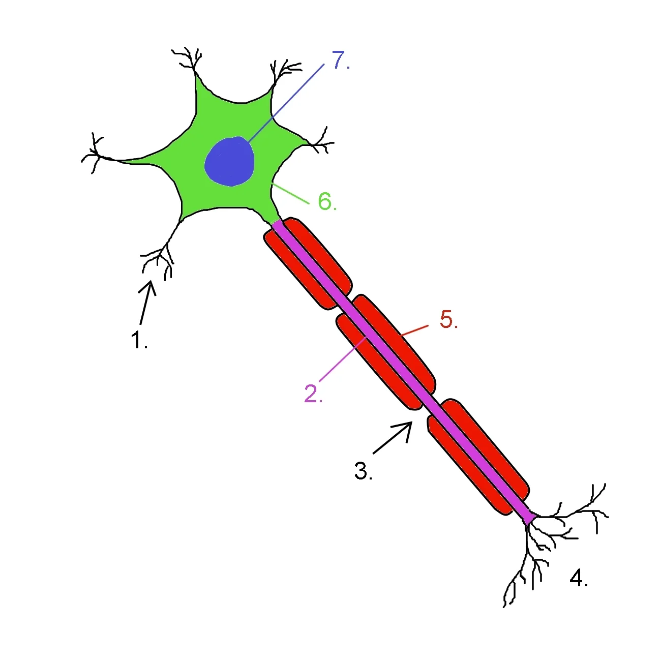
Extending from the soma are multiple dendritic branches that receive incoming signals. The axon may be partially covered by myelin sheaths formed by glial cells.
Figure 1Neuron0
Long description
Diagram of a neuron1 Dendrite2 Axon3 Nodes of Ranvier4 Axon Terminals5 Schwann cell (Myelin Sheath)6 Cell body7 Nucleus

Figure 1: Diagram of a neuron1 Dendrite2 Axon3 Nodes of Ranvier4 Axon Terminals5 Schwann cell (Myelin Sheath)6 Cell body7 Nucleus.
In cross-section, the central nervous system (CNS) shows a clear separation of grey matter (neuronal cell bodies) and white matter (myelinated axons). In the spinal cord, grey matter forms a central H-shaped column with dorsal and ventral horns; the surrounding white matter is organised into three pairs of funiculi (dorsal, lateral, ventral) containing ascending and descending tracts. In the brain, grey matter lies on the surface as the cortex, with deeper grey matter nuclei (e.g., basal ganglia, thalamus) embedded in white matter. The white matter itself is arranged into three fibre types: commissural fibres (crossing the midline), association fibres (connecting cortical areas within one hemisphere), and projection fibres (running vertically between cortex and lower centres). Laminae refer to the layered arrangement of cell bodies, most famously the six layers of the neocortex, each with distinct cell types and connections.
The central cell body (soma) contains the nucleus and is surrounded by branching dendrites that receive signals. A single axon emerges from the axon hillock and is wrapped in myelin sheaths, with gaps (nodes of Ranvier) between adjacent glial cells.
Neurons are structurally classified by the number of processes extending from the cell body. Multipolar neurons have one axon and many dendrites and are the most common type in the CNS. Bipolar neurons have one axon and one dendrite, found in special senses like the retina. Unipolar (pseudounipolar) neurons have a single process that splits into two branches, one going to the periphery and one to the CNS, typical of sensory ganglia. Functionally, neurons are sensory (afferent), motor (efferent), [1]
Neuroglia are the supporting cells of the nervous system. There are six types of glial cells: four in the CNS and two in the PNS. In the CNS, astrocytes maintain the blood-brain barrier (BBB) and support neuronal metabolism; oligodendrocytes insulate CNS axons with myelin; microglia are the resident immune cells; and ependymal cells line the ventricles and produce cerebrospinal fluid (CSF). Ependymal cells appear similar to epithelial cells, making a single layer of cells with little intracellular space and tight connections between adjacent cells, and have cilia on their apical surface to help move CSF through the ventricular space. In the PNS, Schwann cells myelinate peripheral axons, and satellite cells surround the cell bodies of neurons in sensory and autonomic ganglia, providing support similar to that of astrocytes in the CNS—except, of course, for establishing the BBB. [1]
Myelin is a lipid-rich substance that insulates axons. The myelin sheath can be thought of as similar to the pastry wrapped around a hot dog for “pigs in a blanket”: the glial cell is wrapped around the axon several times with little to no cytoplasm between the glial cell layers. For oligodendrocytes, the rest of the cell is separate from the myelin sheath as a cell process extends back toward the cell body; a few other processes provide the same insulation for other axon segments in the area. For Schwann cells, the outermost layer of the cell membrane contains cytoplasm and the nucleus as a bulge on one side of the myelin sheath. Myelin sheaths can extend for one or two millimetres, depending on the diameter of the axon; axon diameters can be as small as 1 to 20 micrometres, meaning the length of a myelin sheath can be 100–1000 times the diameter of the axon. [1]
Axonal transport moves materials between the cell body and the axon terminal. Fast anterograde transport carries vesicles and organelles along microtubules by kinesin; fast retrograde transport returns recycled materials and signalling molecules by dynein. Slow transport moves cytoskeletal proteins and soluble enzymes. When an axon is cut, the distal segment undergoes Wallerian degeneration, breaking down and being phagocytosed; the proximal stump forms a growth cone. In the PNS, Schwann cells support regeneration by forming a tube that guides the growing axon; in the CNS, regeneration is poor due to inhibitory factors and glial scar formation.
Glucose—the primary energy source—is allowed, as are amino acids; water and some other small particles, like gases and ions, can enter, but most everything else cannot, including white blood cells. The BBB protects the CNS from toxic or pathogenic substances but also keeps out [1]
Neurons form the fundamental units of the nervous system, connecting to create complex networks that transmit information throughout the body. These connections can be classified based on the direction of information flow. Afferent neurons, also known as sensory neurons, carry signals from sensory receptors towards the central nervous system (CNS). They detect stimuli from the external environment or internal body states and transmit this information as nerve impulses. In contrast, efferent neurons, or motor neurons, convey signals away from the CNS to effector organs such as muscles and glands. These neurons enable the body to respond to stimuli by initiating movement or secretion.
The connections between neurons occur at specialized junctions called synapses. At a synapse, the axon terminal of one neuron (the presynaptic neuron) communicates with the dendrites or cell body of another neuron (the postsynaptic neuron). This communication typically occurs through the release of chemical neurotransmitters, although electrical synapses also exist. The efficiency of synaptic transmission can be modulated by various factors, including the availability of neurotransmitters and the sensitivity of the postsynaptic receptors.
Neuroglia, or glial cells, play a crucial supporting role in neural connections. Astrocytes, the most abundant glial cells, help form the blood-brain barrier and provide metabolic support to neurons. Oligodendrocytes in the CNS and Schwann cells in the peripheral nervous system (PNS) produce myelin sheaths that insulate axons, allowing for rapid saltatory conduction of nerve impulses. Microglia act as the resident immune cells of the CNS, while ependymal cells line the ventricles and central canal of the spinal cord, producing and circulating cerebrospinal fluid. [1]
Surrounding the neuron are various glial cells: astrocytes with their star-shaped processes providing support, oligodendrocytes extending processes to myelinate axons, and microglia with small cell bodies and branching processes.
DefinitionSynapse
The specialized junction between two neurons where information is transmitted from a presynaptic to a postsynaptic cell, typically via chemical neurotransmitters.
The blood supply to the central nervous system is essential for providing oxygen and nutrients to neurons and neuroglia while maintaining the unique environment of the brain. The blood-brain barrier (BBB) is a physiological barrier that keeps many substances that circulate in the rest of the body from getting into the central nervous system, restricting what can cross from circulating blood into the CNS. This selective permeability is crucial for protecting neural tissue from potentially harmful substances while allowing necessary nutrients to pass. [1]
Nutrient molecules, such as glucose or amino acids, can pass through the BBB, but other molecules cannot. This creates a significant challenge for drug delivery to the CNS, as pharmaceutical companies must design drugs that can cross the BBB while still having an intended effect on the nervous system. The blood supply to neurons and neuroglia is organized into a network of arteries, capillaries, and veins that ensures adequate perfusion to all regions of the brain and spinal cord. [1]
From this circle, major branches such as the middle cerebral artery and anterior cerebral artery extend to supply different cortical regions.
The microglia, a type of neuroglia, have a unique relationship with the blood supply. These smaller glial cells may originate as white blood cells called macrophages that become part of the CNS during early development. While their origin is not conclusively determined, their function is related to what macrophages do in the rest of the body. Microglia are the cells in the CNS that can ingest and digest diseased or damaged cells or pathogens in normal, healthy tissue, and they are therefore also referred to as CNS-resident macrophages. Their activity is dependent on the nutrients delivered through the cerebral blood supply. [1]
Neurons and neuroglia work together to maintain the nervous system's structure and function. Neurons are the primary functional units responsible for transmitting electrical and chemical signals throughout the body. They possess specialized structures that enable this communication, including dendrites that receive signals, a cell body that integrates information, and an axon that transmits signals to other cells.
Neuroglia, or glial cells, provide essential support functions for neurons. They outnumber neurons by approximately 10 to 1 in the central nervous system. Astrocytes maintain the blood-brain barrier, regulate the extracellular environment, and provide metabolic support to neurons. Oligodendrocytes in the central nervous system and Schwann cells in the peripheral nervous system produce myelin sheaths that insulate axons, allowing for rapid saltatory conduction of nerve impulses. Microglia act as the resident immune cells of the central nervous system, responding to injury and infection. [1]
The myelin sheath produced by oligodendrocytes and Schwann cells consists of multiple layers of their own membranes wrapped around the axon. This insulation increases the speed of nerve impulse transmission by allowing saltatory conduction, where the action potential jumps between nodes of Ranvier.
| Syndrome | Location | Clinical Features |
|---|---|---|
| Upper motor neuron lesion | Corticospinal tract | Weakness, hyperreflexia, spasticity, Babinski sign [1] |
| Lower motor neuron lesion | Anterior horn cell or peripheral nerve | Muscle weakness, atrophy, hyporeflexia, fasciculations [1] |
| Demyelinating lesion | Myelin sheath | Slowed nerve conduction, variable sensory loss, fatigue [1] |
| Astrocyte dysfunction | Astrocytes | Blood-brain barrier disruption, edema, impaired neurotransmitter uptake, maintaining the concentration of chemicals in the extracellular space, removing excess signaling molecules, reacting to tissue damage, and contributing to the blood-brain barrier (BBB) [1] |
| Oligodendrocyte damage | Oligodendrocytes | Multiple sclerosis-like symptoms: vision problems, numbness, weakness |
| Microglial activation | Microglia | Neuroinflammation, neuronal damage in neurodegenerative diseases [1] |
| Schwann cell pathology | Peripheral myelin | Charcot-Marie-Tooth disease: distal muscle weakness and sensory loss [1] |
| Neuronal loss in cortex | Cerebral cortex | Cognitive decline, memory impairment, personality changes [1] |
| Ependymal cell damage | Ventricular lining | Hydrocephalus due to impaired cerebrospinal fluid flow [1] |
| Myelin vacuolation | White matter | Ataxia, nystagmus, dysarthria in central pontine myelinolysis [1] |
The corticospinal tract is located in the lateral and anterior funiculi, carrying motor signals from the cortex to the spinal cord. Anterior horn cells are found in the gray matter and contain lower motor neurons that innervate skeletal muscles.
Exam trapCommon misconception
Upper motor neuron lesions cause spasticity while lower motor neuron lesions cause flaccid paralysis.
NoteClinical pearl
In multiple sclerosis, oligodendrocyte damage leads to demyelination in multiple areas, with symptoms typically presenting in patients aged 20-40 years. [1]
DefinitionNeurodegeneration
Progressive loss of structure or function of neurons, often associated with microglial activation and chronic neuroinflammation. [1]
Approach to motor neuron lesions
- Patient presents with weaknessAssess reflexes, tone, and muscle bulk
- Question: Are reflexes increased?
Yes
- Check for Babinski signPresent indicates upper motor neuron lesion
- Look for spasticityCommon in corticospinal tract lesions
No
- Check for muscle atrophyPresent suggests lower motor neuron lesion
- Look for fasciculationsIndicate anterior horn cell involvement
Clinical pearlNeuron classification
Neurons can be classified by structure: unipolar (single process), bipolar (two processes: axon and dendrite), multipolar (multiple dendrites and one axon), and pseudounipolar (single process that bifurcates).
Exam trapBipolar cells misconception
Bipolar cells are rare in the CNS and are mainly found in the olfactory epithelium and retina, not throughout the nervous system. [1]
Clinical pearlNeuroglial functions
Neuroglia provide support, nutrition, and insulation to neurons, maintain the blood-brain barrier, and participate in signal transmission. They outnumber neurons by approximately 10:1 in the human brain.
Clinical pearlAstrocyte functions
Astrocytes maintain the blood-brain barrier, regulate extracellular ion concentrations, and form the glial scar after neural injury. They also participate in the tripartite synapse by modulating neurotransmitter release.
They function as the resident immune cells of the central nervous system.
Exam trapEpendymal cells
Ependymal cells are not neurons but ciliated glial cells that line the ventricles and central canal, producing and circulating cerebrospinal fluid. They also play a role in neurogenesis in specific regions.
They are found in both sensory and autonomic ganglia. [1]
Exam trapNeuroglial regeneration
Unlike neurons, neuroglial cells can undergo cell division and regenerate after injury, which is why gliomas (tumors of glial cells) can occur. This regenerative capacity varies among different glial cell types.
The color difference comes from myelin's lipid content. Gray matter is organized into cortex and nuclei, while white matter forms tracts and commissures.
Students often confuse the specific regions where an electrical impulse is initiated. A common mistake is assuming the action potential begins within the cell body or at the synapse. In reality, action potentials are generated in the trigger zone. [1]
DefinitionTrigger Zone
The combined region of the axon hillock and the initial segment where action potentials are generated.
Another frequent error is failing to distinguish between the axon hillock and the initial segment. The axon hillock is the tapering of the cell body toward the axon fiber, whereas the initial segment is the beginning of the axon that follows as the hillock narrows. [1]
Students often misidentify the composition of the cytoplasm within the axon. It is not identical to the soma's cytoplasm; instead, within the axon hillock, the cytoplasm changes to a solution of limited components known as axoplasm. [1]
Regarding sensory neurons, there is a common misconception that the cell body is always located within the central nervous system (CNS). For sensory reception, which is a peripheral function, the cell body is located in the periphery, specifically within a ganglion. [1]
There is often confusion regarding the direction of signal flow in sensory neurons. Students may assume the axon always leads away from the CNS. However, in these neurons, the axon projects from the dendrite endings, passes the cell body in a ganglion, and travels into the central nervous system.
Finally, students often overlook the structural complexity of myelin. It is not a simple coating but a process where the inner edge wraps around the axon to create several layers, with the other edge closing around the outside to ensure the axon is completely enclosed. [1]
Knowledge of neuronal structure is essential for localising lesions. When a patient presents with sensory loss, the clinician must consider the path of the sensory neuron. Because the axon projects from peripheral dendrites, past the ganglion, and into the CNS, a lesion at the ganglion will disrupt all signals from that peripheral distribution.
Surgeons must be mindful of the fragility of the axon and its insulating layers. The fact that the axon is completely enclosed by several layers of wrapped edges means that mechanical trauma or surgical transection disrupts this insulation, leading to immediate conduction failure and potential Wallerian degeneration.
Radiological findings in demyelinating diseases are interpreted based on the loss of the layers that wrap around the axon. When these layers are destroyed, the "completely enclosed" nature of the axon is lost, leading to the slowed conduction velocities seen in clinical tests.
Medical students are tested on the precise anatomy of the neuron to ensure they understand the physiological "bottleneck" of signal integration. The trigger zone, comprising the axon hillock and initial segment, is a high-yield topic because it is the site where graded potentials are converted into all-or-nothing action potentials.
Sensory Signal Path
- Peripheral StimulusDetected by dendrites in the periphery (e.g. skin)
- Axonal TransportSignal travels along axon toward the cell body
- Question: Location of Cell Body?
Ganglion
- Passes cell body in the periphery
- CNS EntryAxon projects into the central nervous system
| Component | Description | Clinical/Functional Significance |
|---|---|---|
| Axon Hillock | Tapering of cell body toward axon | Transition to axoplasm |
| Initial Segment | Beginning of the axon | Part of the trigger zone |
| Axoplasm | Solution of limited components | Specialized cytoplasm for axonal transport |
| Ganglion | Peripheral cluster of cell bodies | Site of sensory neuron cell bodies |
- Action potentials are generated in the trigger zone, which consists of the axon hillock and initial segment.
- The axon hillock is the tapering region of the cell body that transitions into the initial segment.
- Axoplasm is the specialized solution of limited components found within the axon hillock.
- Sensory neurons have cell bodies located in the periphery, specifically in ganglia.
- The sensory axon projects from peripheral dendrites, past the ganglion, and into the CNS.
- Myelin consists of several layers wrapping around the axon to keep it completely enclosed.
Answer each one in your head first, then open the answer.
Where exactly are action potentials generated in a neuron?
Show answerHide answer
In the trigger zone, which is a combination of the axon hillock and initial segment.What is the specific name for the cytoplasm within the axon hillock?
Show answerHide answer
Axoplasm.Where is the cell body of a sensory neuron typically located?
Show answerHide answer
In the periphery, specifically in a ganglion.Describe the path of a sensory axon.
Show answerHide answer
It projects from dendrite endings, past the cell body in a ganglion, and into the central nervous system.How is the axon enclosed by its insulating layers?
Show answerHide answer
The inner edge wraps around the axon in several layers, and the other edge closes around the outside.
- Betts JG, Young KA, Wise JA, et al. Nervous Tissue. OpenStax, Anatomy and Physiology 2e (CC BY-NC-SA 4.0). Accessed .
Figure credits
Educational content for medical students and doctors. It is not a substitute for clinical judgement or local guidelines.