Spinal cord
Neuroanatomy of the spinal cord, its segments, meningeal stabilisation, and nerve root arrangements.
Figure 1Gray675
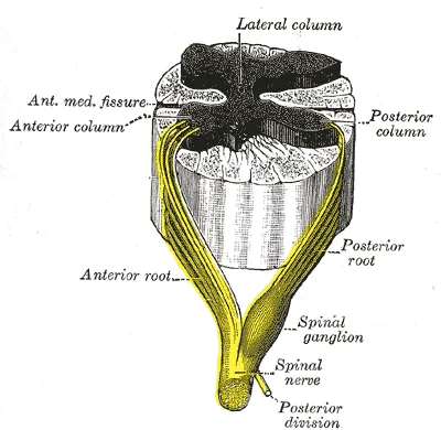
Long description
Formation of spinal nerve from roots of spinal cord

Figure 1: Formation of spinal nerve from roots of spinal cord.
The spinal cord is the primary conduit for neural information, extending from the brainstem down through the vertebral column. Its length is divided into regions corresponding to the vertebral column: cervical, thoracic, lumbar, and sacral. The name of a spinal cord region corresponds to the level at which spinal nerves pass through the intervertebral foramina. Crucially, the spinal cord is not the full length of the vertebral column because the spinal cord does not grow significantly longer after the first or second year, but the skeleton continues to grow. This disparity means that spinal cord segments do not align with their corresponding vertebral bones. For instance, the sacral spinal cord is at the level of the upper lumbar vertebral bones. The spinal nerves extend from their various levels to the proper level of the vertebral column. [1] [2]
Figure 2Gray661
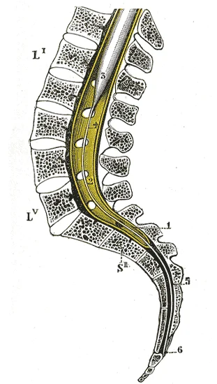
Long description
Sagittal section of vertebral canal to show the lower end of the medulla spinalis and the filum terminale. Li, Lv. First and fifth lumbar vertebræ. Sii. Second sacral vertebra. 1. Dura mater. 2. Lower part of tube of dura mater. 3. Lower extremity of medulla spinalis. 4. Intradural, and 5, Extradura

Figure 2: Sagittal section of vertebral canal to show the lower end of the medulla spinalis and the filum terminale (li, lv).
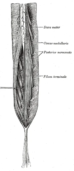
At the inferior end, the cord tapers into the conus medullaris. The nerves that emerge from the spinal cord pass through the intervertebral foramina at the respective levels. As the vertebral column grows, these nerves grow with it and result in a long bundle of nerves that resembles a horse’s tail and is named the cauda equina. This structure contains the lumbar and sacral nerve roots descending to exit at their appropriate levels. [1]
Figure 3Gray671
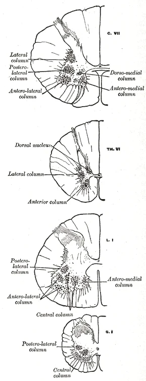
Long description
Transverse sections of the medulla spinalis at different levels to show the arrangement of the principal cell columns.

Figure 3: Transverse sections of the medulla spinalis at different levels to show the arrangement of the principal cell columns.
DefinitionSpinal Cord—lower
The inferior termination of the spinal cord, typically ending as the conus medullaris around the L1/L2 vertebral level in adults, below which lies the cauda equina.
DefinitionConus medullaris
The conical terminal end of the spinal cord, marking the transition from the solid cord to the filamentous cauda equina.
The spinal nerves are the nerves connected to the spinal cord. The arrangement of these nerves is much more regular than that of the cranial nerves. All of the spinal nerves are combined sensory and motor axons that separate into two nerve roots. The sensory axons enter the spinal cord as the dorsal nerve root. The motor fibers, both somatic and autonomic, emerge as the ventral nerve root. This separation allows for distinct pathways for afferent and efferent signals. [2]
DefinitionSpinal cord levels and
The anatomical designation of the cord based on the vertebral level of the exiting nerve roots, noting the mismatch between cord segment and vertebral body.
Stabilisation of the cord within the dural sac is provided by the denticulate ligaments. These structures are positioned between the anterior and posterior roots of the spinal nerves. Traditionally, the DLs are described as 20–21 fibrous spinal projections that stabilize the spinal cord within the spinal canal. Each DL is covered by a leptomeningeal layer that continues with th [2][3]
| Feature | Description |
|---|---|
| Origin | Lateral aspect of the spinal cord pia mater |
| Attachment | Inner aspect of the dura mater |
| Number | 20–21 projections per side |
| Function | Stabilisation and suspension of the spinal cord |
| Composition | Collagen fibers arranged in longitudinal, transverse, and oblique positions |
The lumbar and sacral plexuses arise from the lower cord segments.
The spinal cord is a single structure that forms part of the central nervous system, along with the brain. It maintains the tube structure from its embryonic development as the neural tube, unlike the brain which develops into distinct regions. [1]
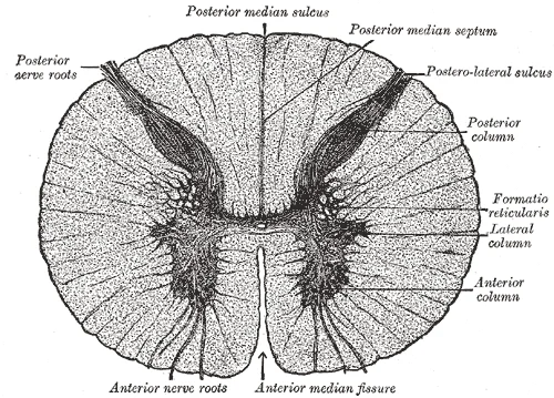
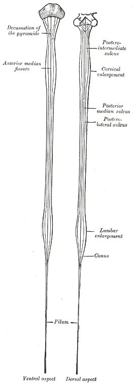
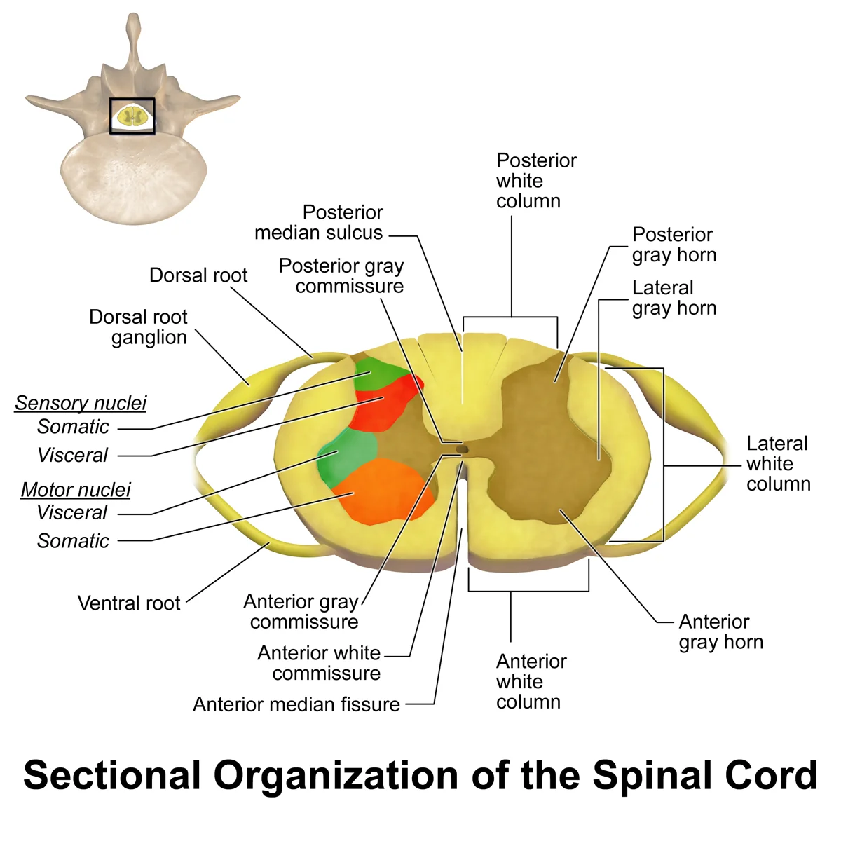
On the external surface of the spinal cord, several anatomical features are visible. The anterior midline is marked by the anterior median fissure, while the posterior midline is marked by the posterior median sulcus. Axons emerging from the anterior side do so through the ventral (anterior) nerve root. [1]
The spinal cord is divided into regions based on vertebral levels: cervical, thoracic, lumbar, and sacral. The cord is shorter than the vertebral column, with nerve roots extending downward to form the cauda equina in the lower lumbar and sacral regions. The spinal cord is surrounded by the meninges, consisting of the dura mater, arachnoid mater, and pia mater.
Figure 4Gray665
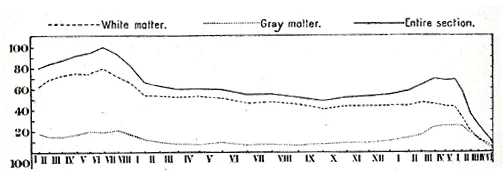
Long description
Curves showing the sectional area at different levels of the cord. The ordinates show the area in sq. mm. (Donaldson and Davis.)

Figure 4: Curves showing the sectional area at different levels of the cord.
Figure 5Gray662
Long description
Cauda equina and filum terminale seen from behind. The dura mater has been opened and spread out, and the arachnoid has been removed.

Figure 5: Cauda equina and filum terminale seen from behind.
The spinal cord has two enlargements: the cervical enlargement (around C5-T1) and the lumbar enlargement (around T12-L1), which correspond to the regions innervating the limbs. The conus medullaris is the tapered end of the spinal cord, typically at the L1-L2 level. From this point, the spinal nerves continue as the cauda equina.
Figure 6Gray664
Long description
Transverse section of the medulla spinalis in the mid-thoracic region.

Figure 6: Transverse section of the medulla spinalis in the mid-thoracic region.
The dorsal and ventral nerve roots emerge from the posterior and anterior aspects respectively, joining to form spinal nerves. The dentate ligaments extend laterally from the pia mater to attach to the dura mater, providing lateral support to the spinal cord.
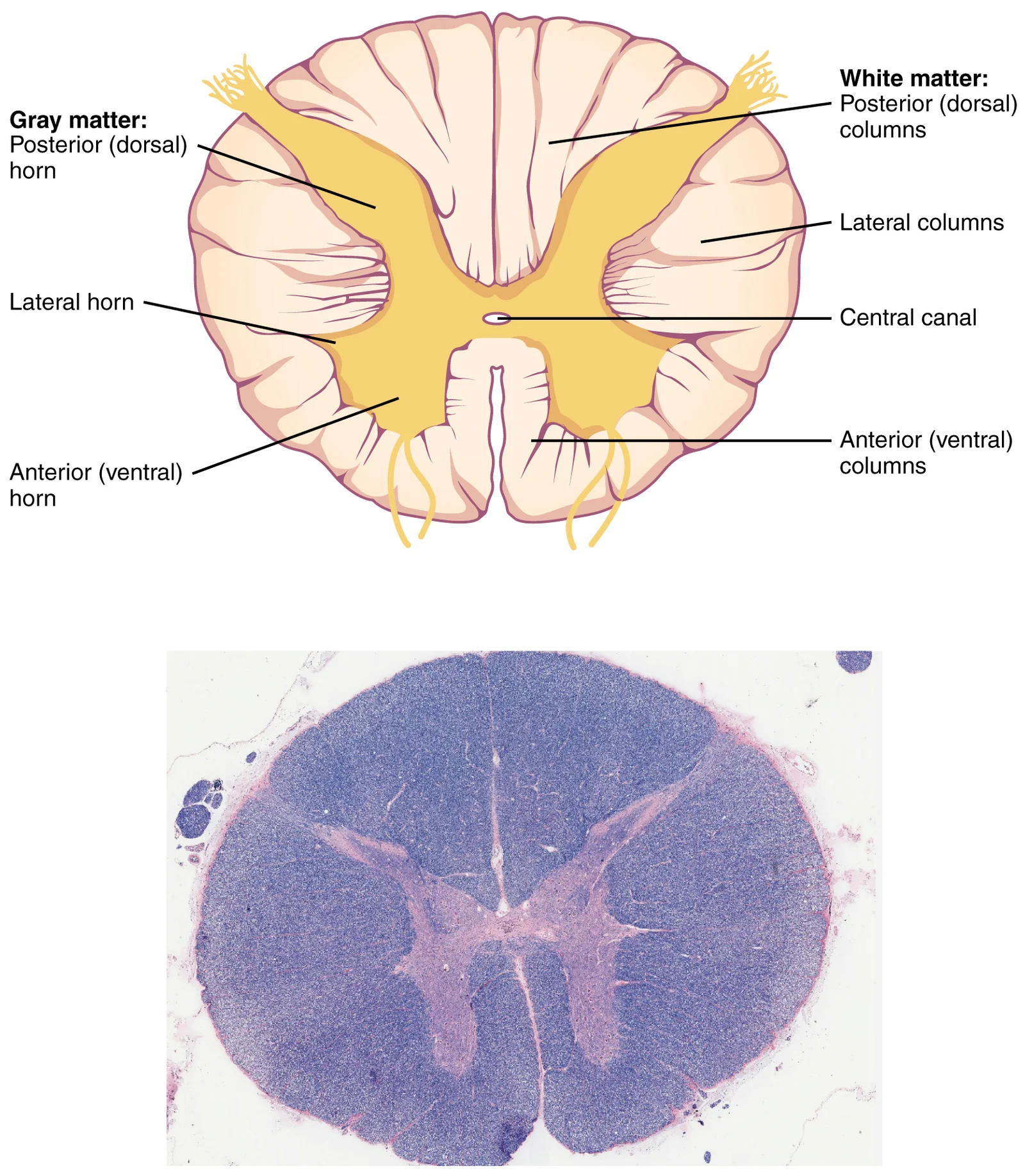
The spinal cord is organized into gray matter (central H-shaped region) and white matter (surrounding region). The gray matter is divided into anterior (ventral) and posterior (dorsal) horns, with the anterior horns containing motor neurons and the posterior horns receiving sensory input.
Spinal nerves emerge from the spinal cord and are classified as mixed nerves containing both sensory and motor fibers. Unlike cranial nerves, all spinal nerves are mixed in function. Spinal nerves reorganize through plexuses (except for thoracic spinal nerves, which directly give rise to intercostal nerves) to form the systemic nerves that innervate the body. [2]
The spinal cord is a cylindrical extension of the central nervous system that transmits neural signals between the brain and the periphery. In cross-section, it presents a distinctive 'H' or butterfly-shaped region of grey matter surrounded by white matter. This organisation allows for the segregation of sensory input, motor output, and integrative processing.
The grey matter is composed of neuronal cell bodies, dendrites, and unmyelinated axons. It is arranged into horns that project into the surrounding white matter. The anterior (ventral) horn contains somatic motor neurons that innervate skeletal muscles. These are large multipolar neurons, some of which are among the largest cells in the body, with cell bodies possibly several hundred micrometers in diameter. For instance, the motor neuron controlling the big toe resides in the sacral spinal cord, maintaining an axon that may reach a meter in length to the muscle belly. [1]
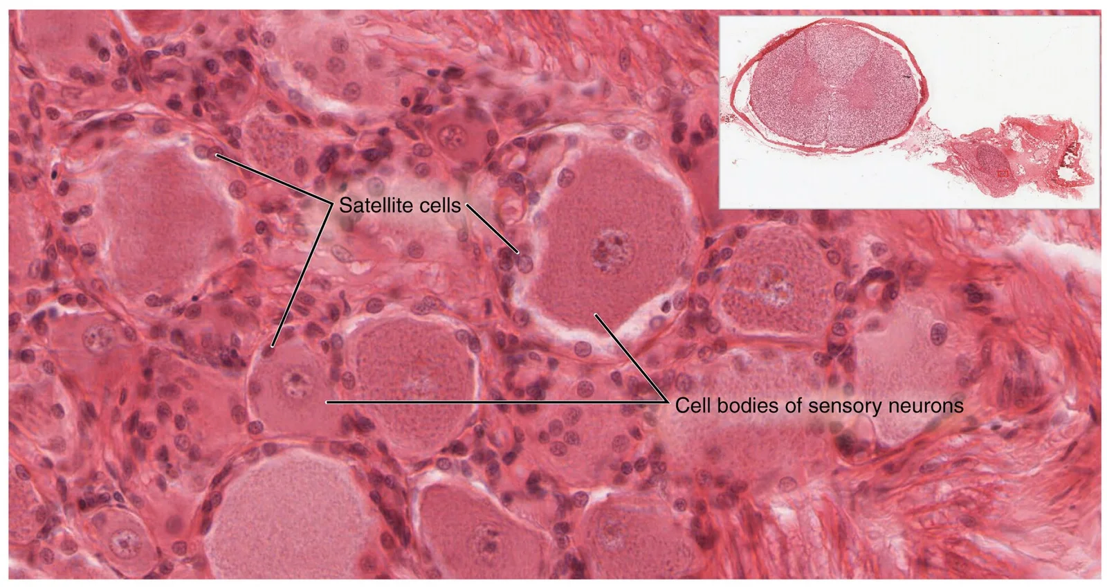
The posterior (dorsal) horn contains interneurons and sensory relay nuclei. These structures receive input from sensory neurons whose cell bodies lie in the dorsal root ganglia. The axons of these sensory neurons enter the spinal cord via the dorsal nerve root.
The grey matter is further subdivided into ten Rexed laminae, which provide a cytoarchitectonic map of neuronal types and connections. Laminae I to VI are primarily located in the posterior horn and process sensory information. Laminae VII is found in the intermediate zone and contains autonomic nuclei. Laminae VIII and IX are in the anterior horn, with Lamina IX containing the large somatic motor neurons described above. Lamina X surrounds the central canal.
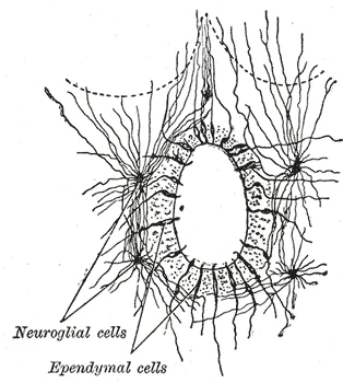
Figure 7Gray667
Long description
Section of central canal of medulla spinalis, showing ependymal and neuroglial cells. (v. Lenhossek.)

Figure 7: Section of central canal of medulla spinalis, showing ependymal and neuroglial cells (v.
The white matter consists of myelinated axons organised into columns (funiculi) and tracts. These tracts carry ascending sensory information to the brain and descending motor commands from the brain. The anterior and lateral columns are composed of many different groups of axons of both ascending and descending tracts. The descending tracts carry motor commands down from the brain to the spinal cord to control output to the periphery. [1]
The central grey matter is shaped like a butterfly, with anterior horns projecting ventrally and posterior horns projecting dorsally. The white matter surrounds the grey matter, divided into anterior, lateral, and posterior funiculi by the anterior and posterior median fissures and sulci.
DefinitionSpinal Cord Anatomy
The spinal cord consists of central grey matter containing neuronal cell bodies and peripheral white matter containing myelinated axon tracts.
| Component | Location | Primary Function |
|---|---|---|
| Anterior Horn | Grey matter, ventral | Contains somatic motor neurons |
| Posterior Horn | Grey matter, dorsal | Processes sensory input |
| White Matter | Peripheral to grey | Ascending and descending tracts |
| Central Canal | Centre of grey matter | Contains cerebrospinal fluid |
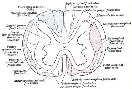
Figure 8Gray672
Long description
Diagram of the principal fasciculi of the spinal cord.

Figure 8: Diagram of the principal fasciculi of the spinal cord.
This structure is continuous with the brain stem, which comprises the midbrain, pons, and medulla. The brain stem acts as the central relay station, housing the major ascending and descending pathways between the spinal cord and the cerebrum. [1]
At the superior end, the spinal cord merges with the medulla oblongata. Superior to the medulla lies the pons, which serves as the main connection with the cerebellum. It is connected to the brain stem primarily at the pons, where it receives a copy of the descending input from the cerebrum to the spinal cord. The midbrain coordinates sensory representations of the visual, auditory, and somatosensory perceptual spaces. [1]
In terms of afferent and efferent pathways, the spinal cord processes sensory input and generates motor output. The cranial nerves connect through the brain stem and provide the brain with the sensory input and motor output associated with the head and neck.
Within the spinal cord itself, the gray matter is subdivided into regions referred to as horns. The posterior horn is responsible for sensory processing. The anterior horn sends out motor signals to the skeletal muscles. These autonomic neurons connect to ganglia outside the central nervous system. Sensory ganglia are found on the dorsal root of all spinal nerves. Autonomic ganglia include the sympathetic chain ganglia, which receive central input from the lateral horn of the thoracic and upper lumbar spinal cord. [1] [2]
Figure 91318b DRG
Long description
The slide includes both a cross-section of the lumbar spinal cord and a section of the dorsal root ganglion (see also ) (tissue source: canine). LM × 1600. (Micrograph provided by the Regents of University of Michigan Medical School © 2012)

Figure 9: The slide includes both a cross-section of the lumbar spinal cord and a section of the dorsal root ganglion (see also ) (tissue source: canine) (lm × 1600).
There are eight pairs of cervical nerves (C1 to C8), twelve thoracic nerves (T1 to T12), five pairs of lumbar nerves (L1 to L5), five pairs of sacral nerves (S1 to S5), and one pair of coccygeal nerves. The nerves emerge from the vertebral column through the intervertebral foramen at their respective levels. For instance, C1 emerges between the first cervical vertebra and the occipital bone, while C8 emerges between the seventh cervical vertebra and the first thoracic vertebra. Thoracic nerves T2 through T11 emerge as intercostal nerves between the ribs. [2]
DefinitionDorsal and Ventral
Dorsal and ventral are used interchangeably with posterior and anterior, particularly in reference to nerves and the structures of the spinal cord. [1]
| Region | Number of Pairs | Designation |
|---|---|---|
| Cervical | 8 | C1 to C8 |
| Thoracic | 12 | T1 to T12 |
| Lumbar | 5 | L1 to L5 |
| Sacral | 5 | S1 to S5 |
| Coccygeal | 1 | Coccygeal |
The spinal cord is a cylindrical structure extending from the medulla oblongata, terminating at the conus medullaris, typically at the L1 or L2 vertebral level in adults. Below this level, the subarachnoid space contains the cauda equina, a bundle of lumbar and sacral nerve roots. These segments correspond to the increased neuronal mass required for limb innervation.
Figure 10Gray663
Long description
Diagrams of the medulla spinalis. Labelled illustration from Wikimedia Commons, used under its open licence.

Figure 10: Diagrams of the medulla spinalis.
The spinal cord is protected by three meningeal layers. The arachnoid mater lies beneath, and the pia mater is the delicate inner layer that adheres directly to the cord surface. The pia mater sends lateral denticulate ligaments, fibrous strips that anchor the cord to the dura, limiting excessive movement. These ligaments are present from the foramen magnum to T12, with fibrous bands attaching to the dura in the cervical and lower thoracic regions to allow mobility.
Internally, the cord consists of grey matter arranged in an 'H' shape, surrounded by white matter. The grey matter contains cell bodies and is divided into anterior (ventral), posterior (dorsal), and lateral horns. The anterior horns contain motor neurons, while the posterior horns receive sensory input. The white matter is organized into columns (funiculi): anterior, lateral, and posterior. These columns contain ascending sensory tracts and descending motor tracts.
The blood supply is critical for cord function. It is formed by branches from the vertebral arteries. The posterior spinal arteries, usually paired, run along the posterolateral sulci, supplying the posterior columns. These are reinforced by segmental medullary arteries, which arise from the aorta and enter through the intervertebral foramina.
Ischaemia in the anterior spinal artery territory leads to anterior cord syndrome, characterised by loss of motor function and pain/temperature sensation, with preserved proprioception and vibration sense (posterior columns).
Figure 111313 Spinal Cord Cross Section
Long description
The cross-section of a thoracic spinal cord segment shows the posterior, anterior, and lateral horns of gray matter, as well as the posterior, anterior, and lateral columns of white matter. LM × 40. (Micrograph provided by the Regents of University of Michigan Medical School © 2012)

Figure 11: The cross-section of a thoracic spinal cord segment shows the posterior, anterior, and lateral horns of gray matter, as well as the posterior, anterior, and lateral columns of white matter (lm × 40).
DefinitionConus Medullaris
The tapered, lower end of the spinal cord, typically ending at L1-L2.
DefinitionCauda Equina
The collection of lumbar and sacral nerve roots descending within the dural sac below the conus medullaris.
| Region | Vertebral Level | Cord Segment | Function |
|---|---|---|---|
| Cervical | C1-C7 | C1-C8 | Upper limb innervation |
| Thoracic | T1-T12 | T1-T12 | Trunk and autonomic |
| Lumbar | L1-L2 | L1-S3 | Lower limb innervation |
| Sacral | S1-S5 | S1-Co1 | Pelvic organs |
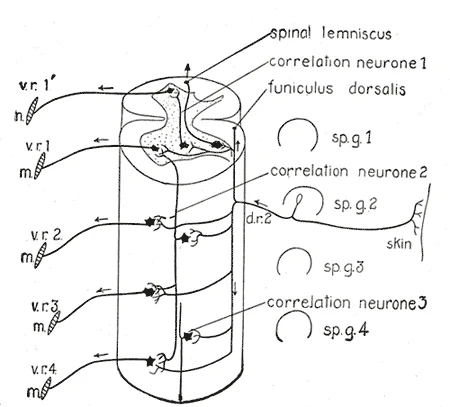
Figure 12Gray758
Long description
Diagram of the spinal cord reflex apparatus. Some of the connections of a single afferent neuron from the skin ( d.r. 2) are indicated: d.r. 2, dorsal root from second spinal ganglion; m, muscles; sp.g. 1 to sp.g. 4, spinal ganglia; v.r. 1′ to v.r. 4, ventral roots. (After Herrick.)

Figure 12: Diagram of the spinal cord reflex apparatus.
This structural arrangement ensures that the brain receives comprehensive data about the internal and external environment while issuing precise motor commands to skeletal muscles. [1]
The spinal cord extends from the medulla oblongata down to the conus medullaris, typically ending at the level of the first or second lumbar vertebra in adults. Above this level, the meninges and cerebrospinal fluid provide essential protection. The gray matter, shaped like a butterfly, contains the cell bodies of interneurons and motor neurons.
Motor control originates in the primary motor cortex, located in the precentral gyrus of the frontal lobe. The upper motor neurons in this region send axons down through the brainstem and into the spinal cord, where they synapse with lower motor neurons. These lower motor neurons then exit the spinal cord via the ventral roots to directly stimulate skeletal muscle contraction. Adjacent to the primary motor cortex, the premotor area is responsible for thinking of a movement to be made, while the [1]
Sensory information from the body is processed in the parietal lobe, which handles somatosensation such as touch, pain, and temperature. Visual processing begins in the occipital lobe, specifically in areas 17 and 18, which are responsible for primary visual perception. Auditory information is processed in the temporal lobe, which also plays a crucial role in memory formation. The thalamus acts as a major relay station, directing sensory information to the appropriate cortical areas. The hypothalamus, part of the diencephalon, coordinates homeostatic functions through the autonomic and endocrine systems. [1]
Reflex arcs within the spinal cord allow for rapid, involuntary responses to stimuli, such as withdrawing a hand from a hot surface, without waiting for brain processing. This integration of sensory input and motor output ensures both protective responses and coordinated voluntary movement. The spinal cord’s ability to process information locally while communicating with higher brain centers is fundamental to maintaining posture, balance, and interaction with the environment.
Figure 13Spinal Cord Sectional Anatomy
Long description
Spinal Cord Sectional Anatomy. Animation in the reference. [ 1 ]

Figure 13: Spinal Cord Sectional Anatomy (animation in the reference).
The spinal cord is encased within three protective layers known as the spinal meninges. From superficial to deep, these are the dura mater, the arachnoid mater, and the pia mater. The space between the arachnoid and pia mater is the subarachnoid space, which contains cerebrospinal fluid. This fluid acts as a cushion, protecting the delicate neural tissue from mechanical shock. Understanding the relationship between these meningeal layers is critical when interpreting imaging studies or performing lumbar punctures.
Below this level, the vertebral canal contains the lumbar and sacral nerve roots, which descend vertically before exiting their respective intervertebral foramina. This bundle of nerve roots resembles a horse’s tail, hence the term cauda equina. Recognising this distinction is vital because lesions affecting the cord above the conus produce upper motor neuron signs, whereas compression of the cauda equina produces lower motor neuron signs.
Lesions of the spinal cord present with distinct clinical syndromes depending on the level and extent of damage. A comprehensive understanding of these patterns allows for precise localisation of the pathology. The following table summarises key lesion types and their clinical correlates.
| Syndrome | Key Features | Anatomical Basis |
|---|---|---|
| Brown-Séquard | Ipsilateral motor loss, contralateral pain/temperature loss | Hemisection of the cord |
| Anterior Cord | Motor loss, pain/temperature loss; preserved proprioception | Anterior spinal artery infarction |
| Central Cord | Upper limb weakness > lower limb weakness; cape-like sensory loss | Cervical spondylosis or trauma |
| Cauda Equina | Lower motor neuron signs, saddle anaesthesia, bowel/bladder dysfunction | Compression of nerve roots below L1/L2 |
| Nerve Root | Sensory Dermatom | Motor Myotome | Reflex |
|---|---|---|---|
| C5 | Lateral arm | Biceps | Biceps |
| C6 | Lateral forearm/thumb | Wrist extensors | Brachioradialis |
| C7 | Middle finger | Triceps | Triceps |
| L4 | Medial leg | Quadriceps | Patellar |
| S1 | Lateral foot | Plantar flexors | Achilles |
Imaging plays a pivotal role in diagnosing these conditions. Magnetic resonance imaging is the gold standard for visualising soft tissue structures, including the spinal cord, meninges, and nerve roots. It provides detailed views of disc herniations, tumours, and inflammatory changes. For instance, a herniated disc at the L4/L5 level may compress the L5 nerve root, leading to specific motor and sensory deficits. In cases where the spinal cord is compressed, such as in severe spinal stenosis, the resulting myelopathy can lead to progressive neurological decline. Early detection through appropriate imaging allows for timely surgical intervention, potentially preventing permanent disability. The cauda equina syndrome, in particular, constitutes a surgical emergency due to the risk of irreversible bladder and bowel dysfunction if decompression is delayed.
Clinical pearlThoracic/Lumbar Nerve Exit
Thoracic and lumbar nerves emerge between the vertebra of the same designation and the next one below. Sacral nerves exit via sacral foramina.
Students frequently conflate the anatomical location of sensory processing with the functional identity of the lobes. A primary error is assuming that memory is stored exclusively in the temporal lobe as a distinct entity. The temporal lobe is associated with primary auditory sensation, known as Brodmann’s areas 41 and 42 in the superior temporal lobe. However, because regions of the temporal lobe are part of the limbic system, memory is an important function associated with that lobe. The critical distinction is that structures in the temporal lobe are responsible for establishing long-term memory, but the ultimate location of those memories is usually in the region in which the sensory perception was processed. Memory is essentially a sensory function; memories are recalled sensations such as the smell of Mom’s baking or the sound of a barking dog. Even memories of movement are really the memory of sensory feedback from those movements, such as stretching muscles or the movement of the skin around a joint. [1]
Another frequent mistake involves the terminology of the basal nuclei. Students often use the term "basal ganglia" interchangeably with "basal nuclei" without recognising the potential for confusion. The basal nuclei are also referred to as the basal ganglia, although that is potentially confusing because the term ganglia is typically used for peripheral structures. It is vital to remember that the basal nuclei are responsible for cognitive processing, the most important function being that associated with planning movements. For example, while a student is sitting in a classroom listening to a lecture, the basal nuclei will keep the urge to jump up and scream from actually happening. This inhibitory control is a key functional aspect often overlooked in basic anatomical descriptions. [1]
Students also misidentify the primary somatosensory cortex. It is incorrect to place this in the frontal lobe. The main sensation associated with the parietal lobe is somatosensation, meaning the general sensations associated with the body. Posterior to the central sulcus is the postcentral gyrus, the primary somatosensory cortex, which is identified as Brodmann’s areas 1, 2, and 3. All of the tactile senses are processed in this area, including touch, pressure, tickle, pain, itch, and vibration, as well as more general senses of the body such as proprioception and kinesthesia, which are the senses of body position and movement, respectively. Confusing the precentral gyrus (motor) with the postcentral gyrus (sensory) is a common error. [1]
Exam trapSensory vs Motor
Do not confuse the precentral gyrus (motor) with the postcentral gyrus (sensory). The postcentral gyrus is the primary somatosensory cortex, identified as Brodmann’s areas 1, 2, and 3.
A further pitfall relates to the composition of the denticulate ligament. Students may assume it is muscular or neural. Elvan et al. analyzed ten histological DL specimens from C5 and ten from T6 in five cadavers (five right-sided and five left-sided specimens at each respective level). Their analysis showed that the DLs were composed of collagen fibrous bundles. There is a continuity between the collagen fibers located in the dura mater, the collagen fibers located inside the denticulate ligament, and the collagen fibers of the subpial compartment. [3]
DefinitionDenticulate Ligament
The denticulate ligament is composed of collagen fibrous bundles, providing structural continuity between the dura mater and the pia mater.
This knowledge is applied at the bedside to localise neurological deficits. When a patient presents with loss of touch, pressure, tickle, pain, itch, and vibration, the clinician must identify the postcentral gyrus as the affected area. The main sensation associated with the parietal lobe is somatosensation, meaning the general sensations associated with the body. Posterior to the central sulcus is the postcentral gyrus, the primary somatosensory cortex, which is identified as Brodmann’s areas 1, 2, and 3. Understanding that all of the tactile senses are processed in this area allows for precise localisation of parietal lobe lesions.
In the operating theatre, awareness of the brain stem is critical. The brain stem is composed of the midbrain, pons, and medulla. It controls the head and neck region of the body through the cranial nerves. There are control centers in the brain stem that regulate the cardiovascular and respiratory systems. Surgeons must respect these structures to avoid catastrophic failure of autonomic regulation. [1]
On imaging, radiologists use the location of Brodmann’s areas to interpret functional scans. The temporal lobe is associated with primary auditory sensation, known as Brodmann’s areas 41 and 42 in the superior temporal lobe. If a patient has auditory agnosia, imaging may reveal pathology in these specific areas. Furthermore, the olfactory nerve and optic nerve are responsible for the sense of smell and vision, respectively[1]. Damage to these nerves can be traced back to their specific origins and pathways. [2]
In examinations, students must accurately describe cranial nerve functions. The oculomotor nerve is responsible for eye movements by controlling four of the extraocular muscles[1]. It is also responsible for lifting the upper eyelid when the eyes point up, and for pupillary constriction[1]. The trochlear nerve and the abducens nerve are both responsible for eye movement, but do so by controlling different extraocular muscles[1]. The trigeminal nerve is responsible for cutaneous sensations of the face and controlling the muscles of mastication[1]. The facial nerve is responsible for the muscles involved in facial expressions, as well as part of the sense of taste and the production of saliva[1]. The vestibulocochlear nerve is responsible for the senses of hearing and balance[1]. [2]
Cranial Nerve Localisation
- Patient presents with eye movement deficit...
- Question: Is pupillary constriction affected?
Yes
- Oculomotor nerve (CN III) lesion
No
- Question: Is it vertical or horizontal?
Vertical
- Trochlear nerve (CN IV) lesion
Horizontal
- Abducens nerve (CN VI) lesion
The basal nuclei play a role in behavioural control observed in clinical settings. The basal nuclei are responsible for cognitive processing, the most important function being that associated with planning movements. The basal forebrain contains nuclei that are important in learning and memory. The limbic cortex is the region of the cerebral cortex that is part of the limbic system, a collection of structures involved in emotion, memory, and behavior. Understanding these functions helps in assessing patients with cognitive or emotional disturbances. [1]
| Nerve | Function |
|---|---|
| Oculomotor | Eye movements, eyelid lifting, pupillary constriction |
| Trochlear | Eye movement (vertical) |
| Abducens | Eye movement (horizontal) |
| Trigeminal | Facial sensation, mastication |
| Facial | Facial expression, taste, saliva |
| Vestibulocochlear | Hearing, balance |
The cerebrum comprises the outer gray matter cortex and deep nuclei belonging to three functional groups. [1] The basal nuclei are responsible for cognitive processing, primarily planning movements. The temporal lobe is associated with primary auditory sensation in Brodmann’s areas 41 and 42. Memory is a sensory function; ultimate memory location is where sensory perception was processed. The postcentral gyrus is the primary somatosensory cortex, identified as Brodmann’s areas 1, 2, and 3. The brain stem controls the head and neck via cranial nerves and regulates cardiovascular/respiratory systems. The denticulate ligament is composed of collagen fibrous bundles continuous with dura and pia. The oculomotor nerve controls four extraocular muscles, eyelid lifting, and pupillary constriction[1].
Answer each one in your head first, then open the answer.
Which Brodmann’s areas constitute the primary somatosensory cortex?
Show answerHide answer
Areas 1, 2, and 3What is the primary function of the basal nuclei?
Show answerHide answer
Planning movementsWhich nerve is responsible for pupillary constriction?
Show answerHide answer
Oculomotor nerveWhat tissue composes the denticulate ligament?
Show answerHide answer
Collagen fibrous bundlesWhich lobe is associated with primary auditory sensation?
Show answerHide answer
Temporal lobe
- Betts JG, Young KA, Wise JA, et al. The Central Nervous System. OpenStax, Anatomy and Physiology 2e (CC BY-NC-SA 4.0). Accessed .
- Betts JG, Young KA, Wise JA, et al. The Peripheral Nervous System. OpenStax, Anatomy and Physiology 2e (CC BY-NC-SA 4.0). Accessed .
- Casteel AT, Dumont LM, Samrid R, et al. A comprehensive review of the denticulate ligaments: application to surgery of the craniocervical junction and spine. Anatomy & cell biology 2026;59(2):230-237. doi:10.5115/acb.25.376. Accessed .
Figure credits
- Figure 1: Henry Vandyke Carter, via Wikimedia Commons, Public domain, Public domain.
- Figure 2: Henry Vandyke Carter, via Wikimedia Commons, Public domain, Public domain.
- Figure 3: Henry Vandyke Carter, via Wikimedia Commons, Public domain, Public domain.
- Figure 4: Henry Vandyke Carter, via Wikimedia Commons, Public domain, Public domain.
- Figure 5: Henry Vandyke Carter, via Wikimedia Commons, Public domain, Public domain.
- Figure 6: Henry Vandyke Carter, via Wikimedia Commons, Public domain, Public domain.
- Figure 7: Henry Vandyke Carter, via Wikimedia Commons, Public domain, Public domain.
- Figure 8: Henry Vandyke Carter, via Wikimedia Commons, Public domain, Public domain.
- Figure 9: OpenStax, via Wikimedia Commons, CC BY 4.0, CC BY 4.0. resized.
- Figure 10: Henry Vandyke Carter, via Wikimedia Commons, Public domain, Public domain.
- Figure 11: OpenStax, via Wikimedia Commons, CC BY 4.0, CC BY 4.0. resized.
- Figure 12: Henry Vandyke Carter, via Wikimedia Commons, Public domain, Public domain.
- Figure 13: BruceBlaus, via Wikimedia Commons, CC BY-SA 4.0, CC BY-SA 4.0.
Educational content for medical students and doctors. It is not a substitute for clinical judgement or local guidelines.