Ascending spinal tracts
Ascending spinal tracts carry somatosensory signals from the body to the brain via distinct pathways.
Ascending pathways transmit peripheral sensations to the brain, with each sensory modality following a specific route through the central nervous system[1]. The two primary systems are the dorsal column–medial lemniscus pathway and the spinothalamic tract, both composed of three successive neurons[1]. The dorsal column system handles fine touch, vibration, and proprioception, while the spinothalamic tract carries pain and temperature information[1]. [2]
This pathway begins with axons from dorsal root ganglion neurons entering the dorsal root and joining the dorsal column white matter[1]. Within the dorsal column, axons from lower body levels position themselves medially, while those from upper levels position themselves laterally[1]. The tract is divided into two components: the fasciculus gracilis, which carries signals from the legs and lower body, and the fasciculus cuneatus, which carries signals from the upper body and arms[1]. The second neuron in this pathway decussates in the brainstem, specifically in the medulla, before ascending to the thalamus[1].
The spinothalamic tract also originates from dorsal root ganglion neurons, which synapse in the dorsal horn of the spinal cord[1]. Axons from these second neurons decussate within the spinal cord at the same level where the information entered, then ascend to the thalamus[1]. This pathway is primarily responsible for pain and temperature sensations[1].
While the dorsal column and spinothalamic tracts are the major conscious sensory pathways, the spinocerebellar tracts carry unconscious proprioceptive information to the cerebellum for motor coordination. The spinoreticular and spinotectal tracts convey pain and arousal signals to the reticular formation and tectum, respectively, contributing to the emotional and autonomic aspects of pain. These pathways are less emphasized in basic sensory testing but are integral to the full sensory experience. [1] [2]
Figure 1Gray763
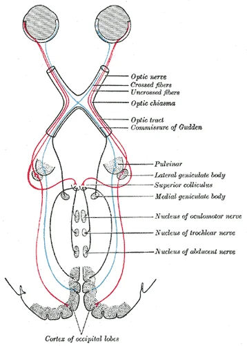
Long description
Scheme showing central connections of the optic nerves and optic tracts.

Figure 1: Scheme showing central connections of the optic nerves and optic tracts.
The trigeminal pathway carries somatosensory information from the face, head, mouth, and nasal cavity[1]. Like other sensory pathways, it involves three successive neurons[1]. Axons from the trigeminal ganglion enter the brainstem at the pons and project to three locations[1]. The spinal trigeminal nucleus in the medulla receives pain and temperature information, similar to the spinothalamic tract[1]. The chief sensory nucleus in the pons and the mesencephalic nuclei in the midbrain receive touch, pressure, vibration, and proprioception, similar to the dorsal column system[1]. Axons from the second neuron decussate and ascend to the thalamus along the trigeminothalamic tract[1].
Sensory tests assess the function of these major ascending tracts[1]. The dorsal column pathway conveys fine touch, vibration, and proprioceptive information, while the spinothalamic pathway conveys pain and temperature[1]. Within the spinal cord, these systems are segregated, with dorsal column information ascending ipsilaterally and decussating in the medulla, whereas spinothalamic information decussates at the level of entry and ascends contralaterally[1]. Failure to recognize two points may indicate a dorsal column pathway deficit[1]. [2]
The somatic nervous system relies on precise relay stations to transmit sensory information from the periphery to the cerebral cortex. This transmission follows a consistent three-neuron chain, ensuring that data regarding touch, pain, temperature, and proprioception reaches conscious awareness. Understanding the origin, course, and termination of these neurons is fundamental to localising neurological lesions. [1]
Figure 21417 Ascending Pathways of Spinal Cord
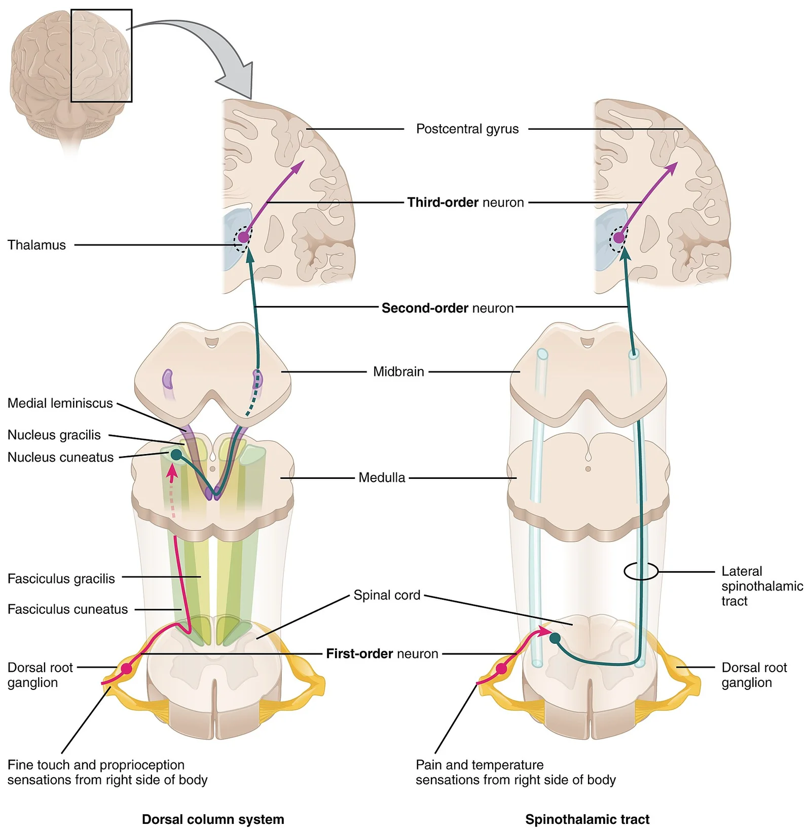
Long description
The dorsal column system and spinothalamic tract are the major ascending pathways that connect the periphery with the brain.

Figure 2: The dorsal column system and spinothalamic tract are the major ascending pathways that connect the periphery with the brain.
The third-order neuron resides in the thalamus, projecting to the primary somatosensory cortex.
The dorsal column-medial lemniscus pathway carries fine touch, vibration, and proprioception. The first-order neuron enters the spinal cord and ascends ipsilaterally in the dorsal columns. Axons from the lower body travel in the fasciculus gracilis, while those from the upper body travel in the fasciculus cuneatus. These axons synapse on second-order neurons in the nucleus gracilis or nucleus cuneatus within the medulla. The axons of these second-order neurons decussate as internal arcuate fibres and ascend as the medial lemniscus. This pathway maintains a topographical arrangement, preserving the relationship between body regions. The third-order neurons are located in the ventral posterolateral nucleus of the thalamus, which relays information to the cerebral cortex. [1]
Figure 3Gray758
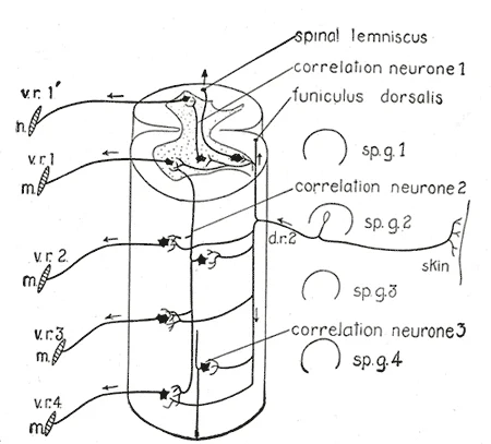
Long description
Diagram of the spinal cord reflex apparatus. Some of the connections of a single afferent neuron from the skin ( d.r. 2) are indicated: d.r. 2, dorsal root from second spinal ganglion; m, muscles; sp.g. 1 to sp.g. 4, spinal ganglia; v.r. 1′ to v.r. 4, ventral roots. (After Herrick.)

Figure 3: Diagram of the spinal cord reflex apparatus.
Figure 4Gray676
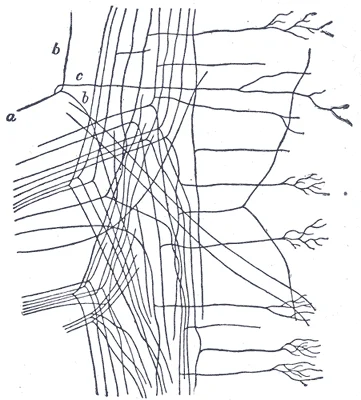
Long description
Posterior roots entering medulla spinalis and dividing into ascending and descending branches. (Van Gehuchten.) a. Stem fiber. b, b. Ascending and descending limbs of bifurcation. c. Collateral arising from stem fiber.

Figure 4: Posterior roots entering medulla spinalis and dividing into ascending and descending branches (Van Gehuchten.) a.
Figure 5Gray669
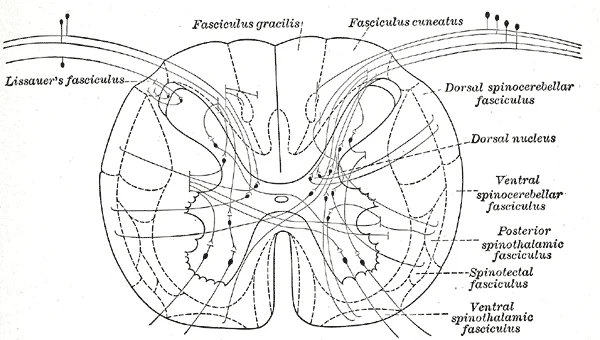
Long description
Diagram showing a few of the connections of afferent (sensory) fibers of the posterior root with the efferent fibers from the ventral column and with the various long ascending fasciculi.

Figure 5: Diagram showing a few of the connections of afferent (sensory) fibers of the posterior root with the efferent fibers from the ventral column and with the various long ascending fasciculi.
In contrast, the spinothalamic tract carries pain and temperature sensation. The first-order neuron synapses in the dorsal horn of the spinal cord. The second-order neuron decussates immediately within the spinal cord via the anterior white commissure and ascends in the lateral spinothalamic tract. The third-order neuron is again located in the thalamus.
Lesions at different levels produce distinct sensory deficits. A lesion of the dorsal column above the decussation in the medulla causes loss of proprioception and fine touch on the ipsilateral side of the body below the lesion. Conversely, a lesion of the spinothalamic tract in the spinal cord causes loss of pain and temperature on the contralateral side, as these fibres have already crossed.
The spinocerebellar tracts convey unconscious proprioception to the cerebellum. These pathways do not reach the thalamus for conscious perception but instead aid in motor coordination.
The dorsal columns are located posteriorly, containing the fasciculus gracilis medially and fasciculus cuneatus laterally. The spinothalamic tract is situated in the anterolateral quadrant. This spatial arrangement is critical for interpreting imaging and clinical findings.
DefinitionDecussation
The crossing over of nerve fibres from one side of the central nervous system to the other, typically at the level of the medulla or spinal cord.
| Tract | Sensation | Decussation Level | First-Order Synapse |
|---|---|---|---|
| Dorsal Column | Fine touch, vibration, proprioception | Medulla (internal arcuate fibres) | Nucleus gracilis/cuneatus |
| Spinothalamic | Pain, temperature | Spinal cord (anterior white commissure) | Dorsal horn |
| Spinocerebellar | Unconscious proprioception | Varies (mostly cerebellum) | Spinal cord/cerebellum |
The ascending spinal tracts carry sensory information from the periphery to the brain through a complex pathway. The diagram shows the spinal cord, brainstem, thalamus, and cerebral cortex, with the ascending pathways traveling upward through these structures.
The journey begins in the periphery where sensory receptors detect stimuli. For somatosensory input from the body, the information enters through the spinal cord. [1] [2]
From the spinal cord, the sensory pathways ascend through the brainstem. In the brainstem, additional nuclei process the information. Similarly, the vestibular system enters the brainstem and influences activity in the cerebellum, spinal cord, and cerebral cortex. [1]
All sensory pathways, except for the olfactory system, reach the thalamus in the diencephalon. The thalamus is a collection of several nuclei categorized into three anatomical groups: an anterior nucleus, a medial nucleus, and a lateral group of nuclei. The special and somatic senses connect to the lateral nuclei, where their information is relayed to the appropriate sensory cortex of the cerebrum. [1]
Finally, the sensory information reaches the cerebral cortex. In the somatosensory cortex, there is a topographical map of the human body known as the homunculus. The external genitals, feet, and lower legs are represented on the medial face of the gyrus within the longitudinal fissure. As the gyrus curves out of the fissure, the body map continues through the thighs, hips, trunk, shoulders, arms, and hands, with the head and face just lateral to the fingers. [1]
Course of Ascending Spinal Tracts
- Peripheral receptorsDetect sensory stimuli
- Spinal cordSomatic sensory input enters here
- BrainstemAdditional processing occurs in nuclei
- Question: Which sensory system?
Vestibular
- Vestibular nucleiMost axons terminate here
- CerebellumSome axons project directly
Other senses
- ThalamusRelay station for sensory information
- Cerebral cortexFinal processing in sensory areas
Ascending spinal tracts carry sensory information from the periphery to the brain, enabling us to perceive our environment and our own bodies. These tracts transmit different modalities of sensation, including touch, proprioception, vibration, temperature, and pain. Each tract has specific functions and carries particular types of sensory information.
The dorsal column-medial lemniscus pathway is responsible for fine touch, vibration, and proprioception. This pathway begins with sensory receptors in the skin, muscles, and joints. The first-order neurons enter the spinal cord and ascend ipsilaterally in the dorsal columns (fasciculus gracilis and fasciculus cuneatus) to the medulla. In the medulla, these neurons synapse with second-order neurons that decussate and ascend as the medial lemniscus to the thalamus. From the thalamus, third-order neurons project to the primary somatosensory cortex.
First-order neurons enter the spinal cord and synapse with second-order neurons in the dorsal horn. These second-order neurons decussate immediately and ascend contralaterally in the spinothalamic tract to the thalamus. From the thalamus, third-order neurons project to the primary somatosensory cortex. [2]
The spinocerebellar tracts carry proprioceptive information to the cerebellum. [2]
The dorsal columns carry fine touch, vibration, and proprioception ipsilaterally. The spinothalamic tract carries pain, temperature, and crude touch contralaterally. The spinocerebellar tracts carry proprioceptive information to the cerebellum, with the dorsal tract remaining ipsilateral and the ventral tract decussating twice.
DefinitionSensory modalities
Ascending spinal tracts carry different sensory modalities: fine touch, vibration, proprioception, pain, temperature, and crude touch.
To understand the clinical consequences of spinal tract damage, one must first appreciate the precise anatomical course of each pathway. Ascending tracts carry sensory information from the periphery to the brain. When a lesion interrupts these pathways, the resulting deficit depends on the specific tract involved, the level of the spinal cord, and whether the lesion is unilateral or bilateral.
The dorsal columns are located posteriorly, adjacent to the posterior median septum. The spinothalamic tract lies anterolaterally, within the anterior and lateral funiculi. The corticospinal tract occupies the lateral funiculus, lateral to the spinothalamic tract. Understanding this spatial arrangement allows for localising lesions based on the pattern of sensory loss. For instance, a lesion affecting only the dorsal columns will spare pain and temperature sensation, which travel in the spinothalamic tract.
The dorsal columns (fasciculus gracilis and cuneatus) carry fine touch, vibration, and proprioception. These fibres ascend ipsilaterally in the spinal cord. A unilateral lesion of the dorsal columns results in ipsilateral loss of these modalities below the level of the lesion. The fasciculus gracilis carries information from the lower limbs and lower trunk, while the fasciculus cuneatus carries information from the upper limbs and upper trunk. The fasciculus cuneatus is present only above the T6 level. Therefore, a lesion at T4 will affect both fasciculi, whereas a lesion at T8 will spare the fasciculus cuneatus.
The spinothalamic tract carries pain, temperature, and crude touch. These fibres decussate within the spinal cord, typically within one or two segments of entry, before ascending contralaterally. Consequently, a unilateral lesion of the spinothalamic tract results in contralateral loss of pain and temperature sensation, starting one or two segments below the level of the lesion. This is because the fibres entering at the level of the lesion have not yet crossed.
The spinocerebellar tracts carry unconscious proprioception to the cerebellum. These tracts ascend ipsilaterally. Lesions here result in ataxia and incoordination on the ipsilateral side, but this is often masked by other motor deficits in acute settings.
| Tract | Modalities | Decussation | Lesion Deficit |
|---|---|---|---|
| Dorsal Columns | Fine touch, vibration, proprioception | Medulla | Ipsilateral loss below lesion |
| Spinothalamic | Pain, temperature, crude touch | Spinal Cord | Contralateral loss below lesion |
| Spinocerebellar | Unconscious proprioception | None | Ipsilateral ataxia |
Localising lesions requires correlating the sensory deficit with the anatomical level. For example, a patient with loss of vibration sense in the left leg but preserved pain and temperature sensation suggests a lesion of the left dorsal column at the corresponding thoracic level. If pain and temperature are also lost on the right side, this suggests a hemisection of the cord (Brown-Séquard syndrome), where the dorsal column is damaged ipsilaterally and the spinothalamic tract is damaged contralaterally.
Clinical pearlClinical Pearl
Remember that the spinothalamic tract decussates shortly after entry, so a spinal cord lesion causes contralateral pain and temperature loss starting one or two segments below the injury.
Exam trapCommon Trap
Do not confuse the dorsal columns with the spinothalamic tract. Dorsal column lesions cause ipsilateral deficits, while spinothalamic lesions cause contralateral deficits.
The assessment of ascending spinal tracts relies on specific clinical signs that localise lesions within the central nervous system.
Clinical pearlChiasmal Lesion
Pituitary masses compressing the optic chiasm cause bitemporal hemianopia by affecting decussating fibres, sparing ipsilateral fibres.
Somatosensory testing evaluates the integrity of the spinothalamic and dorsal column-medial lemniscus pathways using simple bedside tools. The perception of pain can be tested using the broken end of the cotton-tipped applicator. When the tuning fork is still, the metal against the skin can be perceived as a cold stimulus. Using the cotton tip of the applicator, or even just a fingertip, the perception of tactile movement can be assessed as the stimulus is drawn across the skin for approximately 2–3 cm. The patient would be asked in what direction the stimulus is moving. All of these tests are repeated in distal and proximal locations and for different dermatomes to assess the spatial specificity of perception. [2]
DefinitionDorsal Column Function
The dorsal columns mediate vibration, proprioception, and fine touch, tested via tuning forks and two-point discrimination.
DefinitionSpinothalamic Function
The spinothalamic tract mediates pain and temperature sensation, tested via pinprick and cold discrimination.
To differentiate between these tracts, clinicians must understand their distinct modalities. The dorsal columns carry large, myelinated fibres that ascend ipsilaterally before decussating in the medulla. Damage here results in loss of vibration and proprioception below the level of the lesion. In contrast, the spinothalamic tract carries small, unmyelinated or lightly myelinated fibres that decussate shortly after entering the spinal cord. Lesions here cause contralateral loss of pain and temperature sensation.
| Modality | Tract | Test Tool |
|---|---|---|
| Vibration | Dorsal Column | Tuning fork |
| Pain | Spinothalamic | Broken applicator |
| Light Touch | Both | Cotton tip |
| Temperature | Spinothalamic | Cold metal |
Exam trapDermatomal Mapping
Always test distal and proximal sites to map the level of sensory loss accurately across dermatomes.
The spatial specificity of perception is crucial for localising lesions. By repeating tests in distal and proximal locations, clinicians can determine if the deficit is segmental or long-tract in nature. For instance, a complete transection of the spinal cord will result in a sensory level, with loss of all modalities below that segment. Partial syndromes, such as Brown-Séquard syndrome, demonstrate the lateralisation of these tracts: ipsilateral loss of vibration and proprioception with contralateral loss of pain and temperature.
Sensory Assessment
- StartAssess patient sensation
- Question: Test Vibration?
Yes
- Use tuning fork on ulna
No
- Proceed to pain test
- Question: Test Pain?
Yes
- Use broken applicator
No
- Assess temperature
Understanding these clinical tests allows for precise anatomical localisation. Always correlate clinical findings with neuroimaging to confirm the anatomical site of pathology.
The dorsal column-medial lemniscus pathway carries fine touch, vibration, and proprioception. First-order neurons ascend ipsilaterally in the spinal cord, terminating in the nucleus gracilis (lower limb) or nucleus cuneatus (upper limb) in the medulla. Second-order neurons decussate in the medulla and ascend as the medial lemniscus to the thalamus. Third-order neurons project to the postcentral gyrus for conscious perception. A lesion in the medulla causes contralateral loss of fine touch and proprioception, while a spinal cord lesion causes ipsilateral loss below the lesion level. [1]
The medial geniculate nucleus projects to the auditory cortex in the temporal lobe.
The superior colliculus is another target of the optic tract. Visual fields are processed contralaterally: the right visual field is processed in the left visual cortex, and the left visual field in the right visual cortex. [1]
Two main streams process this information: the ventral stream (temporal lobe) for object identification and memory, and the dorsal stream (parietal lobe) for spatial location and movement.
Two-point discrimination assesses the density of sensory endings and receptive field size in the skin. Sensitivity is highest in the fingertips. The test involves touching the skin with two points (e.g., forceps or calipers) while the patient’s eyes are closed, asking them to distinguish one versus two stimuli. [2]
Exam trapMedial lemniscus decussation
The decussation of the second neuron occurs in the medulla, not the spinal cord. Therefore, a medullary lesion causes contralateral sensory loss, whereas a spinal cord lesion causes ipsilateral loss below the level of injury.
Clinical pearlVisual field crossing
The optic chiasm sorts visual information so that each hemisphere processes the contralateral visual field. The right visual field is processed in the left occipital lobe, and the left visual field in the right occipital lobe.
| Pathway | First-order termination | Second-order projection | Third-order termination |
|---|---|---|---|
| Dorsal column | Nucleus gracilis/cuneatus (medulla) | Medial lemniscus (decussates in medulla) | Postcentral gyrus |
| Auditory | Inferior colliculus (midbrain) | Medial geniculate nucleus (thalamus) | Auditory cortex (temporal lobe) |
| Visual | Lateral geniculate nucleus (thalamus) | — | Primary visual cortex (occipital lobe) |
Dorsal column-medial lemniscus pathway
- First-order neuron ascends in spinal cord
- Terminates in nucleus gracilis or cuneatus in medulla
- Second-order neuron decussates in medulla
- Ascends as medial lemniscus to thalamus
- Third-order neuron projects to postcentral gyrus
Students frequently confuse the anatomical course of ascending tracts with their functional outcomes, leading to errors in localising lesions. A primary error is assuming that all sensory information crosses immediately at the level of entry. In reality, the point of decussation varies significantly by tract. For instance, the dorsal column-medial lemniscus pathway crosses in the medulla, whereas the spinothalamic tract crosses in the spinal cord. Misidentifying this level leads to incorrect localisation of spinal cord injuries.
Another frequent mistake involves the somatotopic organisation of the cortex. Students often assume a uniform representation of the body. However, the sensory homunculus demonstrates that areas with high receptor density, such as the hands and face, occupy disproportionately large cortical areas. Failing to appreciate this distortion can lead to misunderstandings regarding the extent of cortical damage required to produce specific sensory deficits.
Visual processing presents another common trap. Learners often forget that the visual field is inverted and reversed upon entering the visual cortex. Light from the superior visual field falls on the inferior retina, and this topography is maintained such that the superior region of the visual cortex processes the inferior visual field. Confusing the visual field with the retinal image can result in errors when interpreting visual field defects on imaging. [1]
Gustatory pathways are also frequently misremembered. It is incorrect to assume a single cranial nerve mediates all taste. The sensory pathway for gustation travels along the facial and glossopharyngeal cranial nerves. These nerves synapse with neurons of the solitary nucleus in the brain stem. Axons from the solitary nucleus then project to the ventral posterior nucleus of the thalamus. Finally, axons from the ventral posterior nucleus project to the gustatory cortex of the cerebral cortex. Overlooking the involvement of multiple cranial nerves can lead to incomplete clinical assessments of taste disorders. [1]
The role of the thalamus is often underestimated. It is not merely a passive relay but a critical integration point. In the somatic nervous system, the thalamus is an important relay for communication between the cerebrum and the rest of the nervous system. Students may overlook its role in filtering and directing sensory information to appropriate cortical areas. [1]
Integration of sensory modalities is another area of confusion. Students might treat vision, touch, and taste as isolated experiences. However, the brain integrates these distinct modalities so that we experience the world as a continuous whole. Our experience of these senses is not divided. Instead, we experience what can be referred to as a seamless percept. Failing to recognise this integration can hinder understanding of complex neurological syndromes where multiple senses are affected. [1]
Exam trapVisual Inversion
Remember that the visual field is inverted and reversed as it enters the visual cortex—up is down, and left is right. The cortex processes this information so that the final conscious perception is correct.
DefinitionSensory Homunculus
The somatosensory cortex maps the locations of somatosensory receptors in the body onto the cortex. This mapping is often depicted using a sensory homunculus. [1]
At the bedside, knowledge of ascending tracts is essential for neurological examination. When a patient presents with sensory loss, the clinician must determine if the lesion is peripheral, spinal, or central. Understanding the decussation points allows the clinician to predict whether sensory loss will be ipsilateral or contralateral. For example, a lesion above the medulla will affect the contralateral side of the body for dorsal column modalities, but the ipsilateral side for spinothalamic modalities if the lesion is below the decussation. This distinction is crucial for localising the lesion.
In the operating theatre, anaesthetists and surgeons use this knowledge to monitor sensory function during procedures involving the spinal cord or brain. Intraoperative monitoring of somatosensory evoked potentials relies on the integrity of these ascending pathways. If the signal is lost, it indicates potential damage to the dorsal columns or medial lemniscus. Similarly, visual evoked potentials assess the integrity of the visual pathways, ensuring that the optic tracts and visual cortex are functioning correctly.
On imaging, radiologists interpret scans based on the anatomical organisation of these tracts. A lesion in the thalamus, for instance, can disrupt multiple sensory modalities because the thalamus is an important relay for communication between the cerebrum and the rest of the nervous system. The diencephalon is beneath the cerebrum and includes the thalamus and hypothalamus. Identifying a thalamic stroke requires understanding that it may affect sensory, motor, and even autonomic functions due to the hypothalamus's role. The hypothalamus has both somatic and autonomic functions.
In examinations, students are tested on their ability to trace the pathway of specific sensations. Questions often require identifying the nucleus, decussation point, and cortical termination of a sensory pathway. For gustation, students must know that taste information travels via the facial and glossopharyngeal nerves to the solitary nucleus. From there, it projects to the ventral posterior nucleus of the thalamus. Finally, it reaches the gustatory cortex. Memorising this sequence is vital for answering questions on cranial nerve functions and cortical processing.
Visual field defects are another common examination topic. Students must understand that the left field of view of each eye is processed on the right side of the brain. Conversely, the right field of view of each eye is processed on the left side of the brain. This knowledge allows for the localisation of lesions in the optic chiasm, optic tract, or visual cortex. For example, a lesion in the right optic tract will cause a left homonymous hemianopia. [1]
Sensory Pathway Localisation
- Patient presents with sensory deficitIdentify modality: touch, pain, temperature, taste, or vision
- Question: Is the deficit visual?
Yes
- Check visual fieldsLeft field processed on right brain; Right field processed on left brain
- Localise lesionBased on field defect pattern
No
- Question: Is the deficit gustatory?
Yes
- Check cranial nervesFacial and glossopharyngeal nerves
- Trace pathwaySolitary nucleus -> Ventral posterior thalamus -> Gustatory cortex
No
- Assess somatosensoryCheck dorsal columns vs spinothalamic tract
- Localise lesionBased on decussation level
- The dorsal column–medial lemniscus pathway transmits fine touch, vibration, and proprioception, with second-order neurons decussating in the medulla.
- The spinothalamic tract carries pain, temperature, and crude touch, and its second-order neurons decussate within the spinal cord.
- Spinocerebellar tracts convey unconscious proprioceptive information to the cerebellum to assist with motor coordination.
- The trigeminal pathway handles somatosensory information from the face and head, utilizing the spinal trigeminal nucleus for pain and temperature.
- A unilateral lesion of the dorsal columns in the spinal cord results in the ipsilateral loss of fine touch, vibration, and proprioception below the level of the lesion.
- A unilateral lesion of the spinothalamic tract in the spinal cord causes a contralateral loss of pain and temperature sensation starting one or two segments below the injury.
- Except for the olfactory system, all sensory pathways reach the thalamus, which serves as a relay station before information is sent to the cerebral cortex.
- The somatosensory cortex contains a topographical map of the body called the homunculus, where areas with high receptor density occupy disproportionately large cortical areas.
Answer each one in your head first, then open the answer.
Why does a spinal cord lesion cause different lateralization for vibration versus pain sensation?
Show answerHide answer
Vibration travels in the dorsal columns and ascends ipsilaterally before decussating in the medulla, whereas pain travels in the spinothalamic tract and decussates shortly after entering the spinal cord.What sensory deficits are characteristic of Brown-Séquard syndrome?
Show answerHide answer
This syndrome involves a hemisection of the cord, resulting in ipsilateral loss of vibration and proprioception and contralateral loss of pain and temperature.Why would a spinal lesion at the T8 level spare the fasciculus cuneatus?
Show answerHide answer
The fasciculus cuneatus carries information from the upper body and is only present above the T6 level.How is the visual field processed in the occipital lobe?
Show answerHide answer
Visual fields are processed contralaterally, meaning the right visual field is processed in the left visual cortex and the left visual field in the right visual cortex.Which structures are involved in the transmission of taste from the periphery to the cortex?
Show answerHide answer
Taste travels via the facial and glossopharyngeal nerves to the solitary nucleus, then to the ventral posterior nucleus of the thalamus, and finally to the gustatory cortex.
- Betts JG, Young KA, Wise JA, et al. Central Processing. OpenStax, Anatomy and Physiology 2e (CC BY-NC-SA 4.0). Accessed .
- Betts JG, Young KA, Wise JA, et al. The Sensory and Motor Exams. OpenStax, Anatomy and Physiology 2e (CC BY-NC-SA 4.0). Accessed .
Figure credits
- Figure 1: Henry Vandyke Carter, via Wikimedia Commons, Public domain, Public domain.
- Figure 2: OpenStax College, via Wikimedia Commons, CC BY 3.0, CC BY 3.0. resized.
- Figure 3: Henry Vandyke Carter, via Wikimedia Commons, Public domain, Public domain.
- Figure 4: Henry Vandyke Carter, via Wikimedia Commons, Public domain, Public domain.
- Figure 5: Henry Vandyke Carter, via Wikimedia Commons, Public domain, Public domain.
Educational content for medical students and doctors. It is not a substitute for clinical judgement or local guidelines.