Descending spinal tracts
Overview of descending motor pathways and somatic reflexes in the spinal cord.
ALIASE: Motor tracts, Pyramidal and extrapyramidal systems, Spinal reflex arcs
The motor output from the cortex descends into the brain stem and to the spinal cord to control the musculature through motor neurons. Neurons located in the primary motor cortex, named Betz cells, are large cortical neurons that synapse with lower motor neurons in the brain stem or in the spinal cord. The two descending pathways travelled by the axons of Betz cells are the corticobulbar tract and the corticospinal tract, respectively. Both tracts are named for their origin in the cortex and their targets—either the brain stem (the term “bulbar” refers to the brain stem as the bulb, or enlargement, at the top of the spinal cord) or the spinal cord. [1]
These two descending pathways are responsible for the conscious or voluntary movements of skeletal muscles. Any motor command from the primary motor cortex is sent down the axons of the Betz cells to activate lower motor neurons in either the cranial motor nuclei or in the ventral horn of the spinal cord. Conversely, the axons of the corticospinal tract are largely contralateral, meaning that they cross the midline of the brain stem or spinal cord and synapse on the opposite side of the body. Therefore, the right motor cortex of the cerebrum controls muscles on the left side of the body, and vice versa. [1]
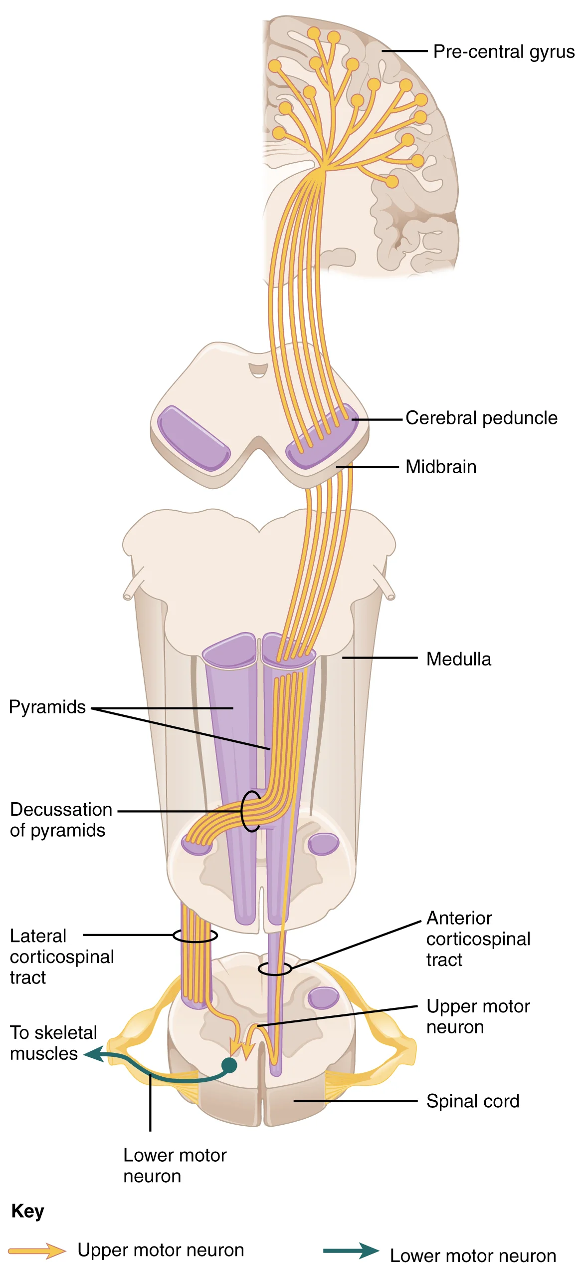
The corticospinal tract descends from the cortex through the deep white matter of the cerebrum. It then passes between the caudate nucleus and putamen of the basal nuclei as a bundle called the internal capsule. The tract then passes through the midbrain as the cerebral peduncles, after which it burrows through the pons. Upon entering the medulla, the tracts make up the large white matter tract referred to as the pyramids. The defining landmark of the medullary-spinal border is the pyramidal dec [1]
Because movements of the body trunk involve both sides of the body, the anterior corticospinal tract is not entirely contralateral. Some collateral branches of the tract will project into the ipsilateral ventral horn to control synergistic muscles on that side of the body, or to inhibit antagonistic muscles through interneurons within the ventral horn. Through the influence of both sides of the body, the anterior corticospinal tract can coordinate postural muscles in broad movements of the body. These coordinating axons in the anterior corticospinal tract are often considered bilateral, as they are both ipsilateral and contralateral. [1]
The tectospinal tract projects from the midbrain to the spinal cord and is important for postural movements that are driven by the superior colliculus. The name of the tract comes from an alternate name for the superior colliculus, which is the tectum. The reticulospinal tract connects the reticular system, a diffuse region of gray matter in the brain stem, with the spinal cord. This tract influences trunk and proximal limb muscles related to posture and locomotion. The reticulospinal tract also contributes to muscle tone and influences autonomic functions. The vestibulospinal tract connects the brain stem nuclei of the vestibular system with the spinal cord. This allows posture, movement, and balance to be modulated on the basis of equilibrium information provided by the vestibular system. [1]
DefinitionReflex Arc
A simple somatic reflex does not include the higher centers discussed for conscious or voluntary aspects of movement. Reflexes can be spinal or cranial, depending on the nerves and central components that are involved. [1]
This chapter began by introducing reflexes as an example of the basic elements of the somatic nervous system. Simple somatic reflexes do not include the higher centers discussed for conscious or voluntary aspects of movement. Reflexes can be spinal or cranial, depending on the nerves and central components that are involved. The example described at the beginning of the chapter involved heat and pain sensations from a hot stove causing withdrawal of the arm through a connection in the spinal cord that leads to contraction of the biceps brachii. The description of this withdrawal reflex was simplified, for the sake of the introduction, to emphasize the parts of the somatic nervous system. But to consider reflexes fully, more attention needs to be given to this example. [1]
As you withdraw your hand from the stove, you do not want to slow that reflex down. As the biceps brachii contracts, the antagonistic triceps brachii needs to relax. Because the neuromuscular junction is strictly excitatory, the biceps will contract when the motor nerve is active. Skeletal muscles do not actively relax. Instead the motor neuron needs to “quiet down,” or be inhibited. In the hot-stove withdrawal reflex, this occurs through an interneuron in the spinal cord. The interneuron receives a synapse from the axon of the sensory neuron that detects that the hand is being burned. In response to this stimulation from the sensory neuron, the interneuron then inhibits the motor neuron that controls the triceps brachii. [1]
The reflex helps to maintain muscles at a constant length. A common example of this reflex is the knee jerk that is elicited by a rubber hammer struck against the patellar ligament in a physical exam. [1]
NoteClinical Note
The acute effect of damage to descending pathways on neural function within the lumbosacral spinal cord has been studied using a full thoracic transection, providing evidence that 5-HT release maintains a brake on neural activity within the dorsal horn that counters the development of over-excitation. [2]
Figure 11426 Corticospinal Pathway
Long description
The major descending tract that controls skeletal muscle movements is the corticospinal tract. It is composed of two neurons, the upper motor neuron and the lower motor neuron. The upper motor neuron has its cell body in the primary motor cortex of the frontal lobe and synapses on the lower motor ne

Figure 1: The major descending tract that controls skeletal muscle movements is the corticospinal tract.
The upper motor neurons are located in the cortex and brainstem, projecting to lower motor neurons in the ventral horn. The lower motor neurons then exit the spinal cord to innervate skeletal muscle.
The primary motor cortex is located in the precentral gyrus of the frontal lobe. This area receives input from several regions that aid in planning movement, and its principal output stimulates spinal cord neurons to stimulate skeletal muscle contraction. The cortex is arranged in a topographical map known as the motor homunculus. Neurons responsible for musculature in the feet and lower legs are in the medial wall of the precentral gyrus, with the thighs, trunk, and shoulder at the crest of the longitudinal fissure. The hand and face are in the lateral face of the gyrus. The relative space allotted for different regions is exaggerated in muscles that have greater innervation. The greatest amount of cortical space is given to muscles that perform fine, agile movements, such as the muscles of the fingers and the lower face. The “power muscles” that perform coarser movements, such as the buttock and back muscles, occupy much less space on the motor cortex. [1]
The motor components of the somatic nervous system begin with the frontal lobe of the brain, where the prefrontal cortex is responsible for higher functions such as working memory. The integrative and associate functions of the prefrontal lobe feed into the secondary motor areas, which help plan movements. The premotor cortex and supplemental motor area then feed into the primary motor cortex that initiates movements. Large Betz cells project through the corticobulbar and corticospinal tracts to synapse on lower motor neurons in the brain stem and ventral horn of the spinal cord, respectively. These connections are responsible for generating movements of skeletal muscles. [1]
Figure 2Gray670
Long description
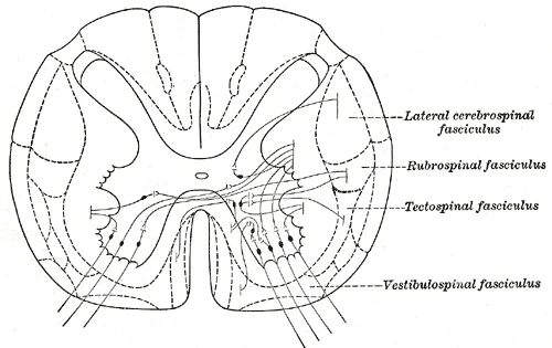
Diagram showing possible connection of long descending fibers from higher centers with the motor cells of the ventral column through association fibers.

Figure 2: Diagram showing possible connection of long descending fibers from higher centers with the motor cells of the ventral column through association fibers.
This influence over the appendicular muscles means that the lateral corticospinal tract is responsible for moving the muscles of the arms and legs. The ventral horn in both the lower cervical spinal cord and the lumbar spinal cord both have wider ventral horns, representing the greater number of muscles controlled by these motor neurons. The cervical enlargement is particularly large because there is greater control over the fine musculature of the upper limbs, particularly of the fingers. [1]
The anterior corticospinal tract is responsible for controlling the muscles of the body trunk. These axons do not decussate in the medulla. Instead, they remain in an anterior position as they descend the brain stem and enter the spinal cord. These axons then travel to the spinal cord level at which they synapse with a lower motor neuron. Upon reaching the appropriate level, the axons decussate, entering the ventral horn on the opposite side of the spinal cord from which they entered. In the ventral horn, these axons synapse with their corresponding lower motor neurons. The lower motor neurons are located in the medial regions of the ventral horn, because they control the axial muscles of the trunk. [1]
All of these motor pathways project to the spinal cord to synapse with motor neurons in the ventral horn of the spinal cord. These lower motor neurons are the cells that connect to skeletal muscle and cause contractions. These neurons project through the spinal nerves to connect to the muscles at neuromuscular junctions. One motor neuron connects to multiple muscle fibers within a target muscle. The number of fibers that are innervated by a single motor neuron varies on the basis of the precision necessary for that muscle and the amount of force necessary for that motor unit. The quadriceps, for example, have many fibers controlled by single motor neurons for powerful contractions that do not need to be precise. The extraocular muscles have only a small number of fibers controlled by each motor neuron because moving the eyes does not require much force, but needs to be very precise. [1]
The axons will also branch to innervate multiple muscle fibers. Together, the motor neuron and all the muscle fibers that it controls make up a motor unit. Motor units vary in size. Some may contain up to 1000 muscle fibers, such as in the quadriceps, or they may only have 10 fibers, such as in an extraocular muscle. The number of muscle fibers that are part of a motor unit corresponds to the precision of control of that muscle. Also, muscles that have finer motor control have more motor units connecting to them, and this requires a larger topographical field in the primary motor cortex. [1]
Course of Descending Spinal Tracts
- OriginDescending tracts originate from multiple brain regions including motor cortices, basal nuclei, cerebellum, brain stem nuclei (superior colliculus, red nucleus, vestibular nuclei), and reticular formation
- PathwayTracts descend through the CNS, with serotonergic fibers specifically traveling through the dorsolateral fasciculus (DLF)
- Question: Target Region?
Spinal Cord
- Termination in Dorsal HornFibers terminate primarily in laminae I and II of the dorsal horn
- Termination in Ventral HornMotoneurons in ventral horn (lamina IX) receive input from descending serotonergic fibers from raphe obscurus and raphe pallidus
- Sympathetic RegulationSympathetic neurons in thoracic cord receive inputs from ventrolateral medulla
Descending input from the secondary motor cortices, basal nuclei, and cerebellum connects to the origins of these tracts in the brainstem. [1]
This termination pattern allows for the modulation of sensory input and the dampening of neural excitation through serotonin (5-HT). [2]
This innervation is critical for motor control and the maintenance of muscle tone. This pathway is essential for autonomic regulation, including cardiovascular control. [2]
The spinal cord tissue itself is surrounded by cerebrospinal fluid and protected by bony structures in the spinal canal. It is soft in nature and passes up and down to mediate signal connections between the brain and different segments of the spinal cord. This loss can contribute to pathophysiology after spinal cord injury (SCI). For instance, sympathetic dysfunction, such as autonomic dysreflexia, often occurs as a result of the loss of descending sympathetic fibers above the T6 region. This condition is characterized by acute bouts of hypertension and bradycardia induced by stimuli below the injury. Understanding the precise course and termination of these tracts is vital for localizing lesions and predicting clinical outcomes in neuroanatomy. [3] [2]
These pathways drive voluntary movement and regulate the capacity for neural plasticity within the spinal cord. The integrity of these tracts is fundamental to the recovery of function, as recovery depends on the remodeling and integrity of neural circuits. Damage to these pathways, such as the breakage of neuronal axons and the death of neurons, causes dysfunction of neural circuits. [2] [3]
A subset of these descending fibres release the neurotransmitter serotonin (5-HT), which affects spinal cord function in alternative ways depending upon the region innervated and the receptor type engaged. This inhibitory tone is crucial for preventing over-excitation. [2]
Loss of descending 5-HT fibers promotes the development of maladaptive plasticity by enabling a state of over-excitation. This over-excitation can fuel cell death and foster the development of spasticity, pain, and autonomic dysreflexia. The transformation in brain-derived neurotrophic factor (BDNF) function is also linked to this mechanism. [2]
This suggests that nociceptive signals during medical procedures under anesthesia may adversely affect spinal function. Understanding the specific modalities carried by these tracts allows clinicians to predict deficits and target rehabilitation strategies, such as neuromodulation technology, to improve limb function. [2]
Descending spinal tracts are the primary motor pathways that convey voluntary movement commands from the cerebral cortex and brainstem to the anterior horn cells of the spinal cord. Understanding their anatomy requires a systematic approach: origin, course, relations, and termination. The major tracts are the corticospinal, rubrospinal, vestibulospinal, reticulospinal, and tectospinal tracts.
The anterior corticospinal tract is located in the anterior funiculus.
DefinitionCorticospinal Tract
The largest descending motor pathway, originating in the cerebral cortex, decussating in the medulla, and terminating in the spinal cord to control voluntary movement.
The corticicospinal tract originates in the primary motor cortex, premotor areas, and somatosensory cortex. Approximately 85-90% of fibres cross at the pyramidal decussation in the medulla to form the lateral corticospinal tract, which descends in the lateral funiculus. The remaining 10-15% descend ipsilaterally as the anterior corticospinal tract in the anterior funiculus, crossing near their termination level. Lesions above the decussation cause contralateral spastic paralysis. Lesions below the decussation cause ipsilateral spastic paralysis. [2]
Exam trapDecussation Site
Confusing the level of decussation leads to incorrect localisation of lesions. Remember: most fibres cross in the medulla, not the spinal cord.
The rubrospinal tract originates in the red nucleus of the midbrain. It crosses immediately in the ventral tegmental decussation and descends in the lateral funiculus, just anterior to the lateral corticospinal tract. It terminates in the cervical and upper thoracic spinal cord, influencing flexor muscle tone. Lesions are rare in isolation but contribute to upper limb motor control.
Lesions result in loss of postural tone and difficulty standing.
These tracts modulate muscle tone and autonomic functions. Lesions can cause abnormal muscle tone and autonomic instability.
It terminates in the cervical spinal cord, coordinating head and eye movements in response to visual and auditory stimuli. Lesions are uncommon but may affect reflexive head turning.
| Tract | Origin | Course | Primary Function |
|---|---|---|---|
| Lateral Corticospinal | Cerebral Cortex | Decussates in Medulla; Lateral Funiculus | Voluntary fine motor control |
| Anterior Corticospinal | Cerebral Cortex | Descends Ipsilateral; Crosses at Level | Axial muscle control |
| Rubrospinal | Red Nucleus | Crosses in Midbrain; Lateral Funiculus | Flexor tone (upper limb) |
| Lateral Vestibulospinal | Lateral Vestibular Nucleus | Ipsilateral; Anterior Funiculus | Extensor tone, posture |
| Pontine Reticulospinal | Pontine Reticular Formation | Ipsilateral; Anterior Funiculus | Extensor facilitation |
| Tectospinal | Superior Colliculus | Crosses in Midbrain; Anterior Funiculus | Head/eye coordination |
Clinical assessment of descending tracts involves testing muscle strength, tone, and reflexes. Spasticity and hyperreflexia indicate upper motor neuron lesions. Flaccidity suggests lower motor neuron or acute shock phase lesions. Understanding the specific tract involvement helps localise the lesion level and aetiology.
Reflexes represent the simplest functional circuits within the somatic nervous system, relying on minimal neuronal connections to produce rapid, involuntary responses to stimuli. For instance, a withdrawal reflex from a painful stimulus requires only the sensory fiber entering the spinal cord and the motor neuron projecting to the relevant muscle. While antagonist and postural muscles can be coordinated with this withdrawal, making the circuitry more complex, the core mechanism remains direct. [1]
DefinitionReflex Arc
The simplest circuit in the somatic nervous system, typically involving a sensory neuron and a motor neuron.
Stretch reflexes serve a critical homeostatic function by maintaining a constant length of muscles. They achieve this by causing a contraction of a muscle to compensate for a stretch that is sensed by a specialized receptor known as a muscle spindle. This mechanism ensures postural stability and rapid correction of limb position against unexpected loads. [1]
Clinical pearlCorneal Reflex
A specialized protective reflex involving the trigeminal and facial nerves to blink the eyelid.
A specialized reflex to protect the surface of the eye is the corneal reflex, also referred to as the eye blink reflex. When the cornea is stimulated by a tactile stimulus, or even by bright light in a related reflex, blinking is initiated. The sensory component of this reflex travels through the trigeminal nerve, which carries somatosensory information from the face, or through the optic nerve if the stimulus is bright light. The motor response travels through the facial nerve and innervates the orbicularis oculi bilaterally. This reflex is commonly tested during a physical exam using an air puff or a gentle touch of a cotton-tipped applicator. [1]
| Reflex | Sensory Nerve | Motor Nerve | Effector Muscle | Stimulus |
|---|---|---|---|---|
| Corneal | Trigeminal (V) | Facial (VII) | Orbicularis oculi | Tactile touch or air puff |
| Withdrawal | Spinal afferents | Spinal efferents | Flexor muscles | Painful stimulus |
| Stretch | Muscle spindle | Alpha motor neuron | Same muscle | Muscle stretch |
Understanding these reflex arcs is essential for localizing lesions within the nervous system. Damage to the sensory limb of the corneal reflex results in an absent blink response to ipsilateral stimulation, while damage to the facial nerve prevents the motor response regardless of the side stimulated. Similarly, the integrity of the stretch reflex arc depends on the continuity of the muscle spindle, the afferent pathway, the spinal cord synapse, and the efferent motor neuron. Any disruption in this chain alters the clinical presentation, providing vital diagnostic information regarding the level and nature of neurological injury.
Exam trapSerotonin Loss
Do not confuse the loss of serotonergic fibres with general motor loss; it specifically drives spasticity and autonomic dysregulation. [2]
DefinitionExtrapyramidal System
This system includes descending connections outside the corticospinal pathway, which features the pyramids in the medulla. [1]
NoteMicroglial Peaks
Microglial activation can have two peaks: one at 7 days post-injury and another at 60 days post-injury. [3]
DefinitionInstrumental Conditioning
This learning involves maintaining a limb in a flexed position to minimize exposure to noxious stimulation. [2]
DefinitionPrimary Injury
Primary injury causes irreversible mechanical damage, leading to axonal disruption, degeneration, and neuronal death.
Exam trapChronic Pain
Sensitization of dorsal horn neurons interferes with adaptive learning and promotes cell loss when the spinal cord is contused. [2]
NoteAutonomic Function
Loss of serotonergic fibres is linked to sustained motor activity (spasticity) and dysregulation of autonomic function. [2]
Students frequently confuse the functional roles of descending pathways, particularly regarding pain modulation and motor control. A primary error is assuming that all descending serotonergic (5-HT) fibres are uniformly antinociceptive. In reality, the effect depends on the receptor subtype engaged. 5-HT1A/B/D and 5-HT7 receptors are primarily antinociceptive, whereas 5-HT2 and 5-HT3 receptors are pronociceptive.
A significant pitfall lies in understanding the mechanism of spasticity following spinal cord injury (SCI). It is incorrect to attribute spasticity solely to upper motor neuron disinhibition without considering spinal motoneuron changes. Weeks after disconnection from supraspinal input, spinal motoneurons compensate for the loss of 5-HT by transitioning into an excitable state. This state makes them easily responsive to excitatory transmitters such as glutamate. This leads to the activation of 5-HT receptors that facilitate sustained firing of voltage-gated persistent Ca2+ and Na+ currents, also called persistent inward currents (PICs), causing muscle contractions. Murray et al. showed that this effect is due to the activity of 5-HT2 receptors. PICs and spasms are easily triggered by innocuous stimuli such as touch or muscle stretching. [2]
Another frequent error concerns the developmental shift in GABAergic function. Students may assume GABA is always inhibitory. However, early in development, the inward flow of Cl− is augmented by NKCC1, maintaining a high intracellular concentration of the anion. Under these conditions, engaging the GABA-A receptor allows Cl− to flow out of the cell, which has a depolarizing (excitatory) effect. Later in development, increased expression of KCC2 lowers the intracellular concentration of Cl−, restoring inhibitory function. After injury, the downregulation of KCC2 can revert GABA to a depolarizing state. How these pathways affect KCC2 depends upon PLC: if PLC is absent, KCC2 is upregulated; if PLC is engaged, KCC2 is downregulated. [2]
Finally, students often overlook the supraspinal contribution to secondary injury. It is a mistake to think that local spinal mechanisms alone drive all aspects of post-injury pathology. A T2 transection also blocked the activation of pro-inflammatory cytokines, and signals indicative of cell death, at the site of injury. Furthermore, a pharmacological transection at T2 blocked the adverse effect nociceptive stimulation has on long-term recovery. Because a rostral transection is sufficient to downregulate KCC2 in the caudal tissue, these findings imply that the shift in GABA function does not, by itself, enable nociception-induced hemorrhage after injury. The implication is that an additional, brain-dependent, process is engaged that plays an essential role in driving pain-induced tissue loss after injury. [2]
Exam trapReceptor Specificity
Do not generalise 5-HT effects. 5-HT1A/B/D and 5-HT7 are primarily antinociceptive while 5-HT2 and 5-HT3 are pronociceptive. [2]
DefinitionPersistent Inward Currents
PICs are sustained firing of voltage-gated persistent Ca2+ and Na+ currents facilitated by 5-HT receptors, leading to muscle contractions.
Knowledge of descending spinal tracts is applied at the bedside to interpret neurological deficits and manage complications of spinal cord injury. Clinicians use this understanding to explain spasticity to patients. Spasticity after SCI is a product of overactive, unregulated motor neurons within the spinal cord that create muscle spasms. Recognising that these spasms are driven by PICs and 5-HT2 receptor activity helps guide pharmacological interventions, such as using agents that modulate these specific receptors. It also explains why innocuous stimuli, such as touch or muscle stretching, can trigger severe spasms. [2]
In the operating theatre and during acute management, understanding the supraspinal drive to inflammation is crucial. The discovery that rostral transection blocks nociception-induced hemorrhage and pro-inflammatory cytokine activation suggests that brain-dependent processes drive secondary tissue loss. This knowledge informs strategies to mitigate secondary injury, potentially by targeting the communication pathways between the brain and the spinal cord during the acute phase of trauma. It highlights that pain management is not just about comfort but may influence structural outcomes by reducing brain-dependent inflammatory cascades. [2]
On imaging, particularly MRI, students must correlate structural damage with functional loss. The defining characteristic of the somatic nervous system is that it controls skeletal muscles. Somatic senses inform the nervous system about the external environment, but the response to that is through voluntary muscle movement. Damage to specific tracts visible on imaging must be interpreted in the context of these voluntary and involuntary controls. For instance, damage to dorsal region fibres is linked to antinociception rather than motor function. Therefore, a lesion in the dorsal columns might present with sensory deficits without immediate motor paralysis, whereas ventral damage affects the motor output. [1] [2]
In examinations, students are tested on the precise anatomical and physiological principles. The loss of mature neurons is the main cause of functional defects after SCI, and this is directly involved in the induction of programmed cell death (PCD) in neural cells, including apoptosis, necroptosis, autophagy, and ferroptosis. It was reported that apoptosis in neurons could be detected at 4 h and peaked at 8 h after SCI. The total number of axons at the injured site decreased immediately, and the downward trend continued during the subacute phase and reached a minimum during the chronic phase. The results showed that cytoplasmic Nissl material was decreased in neurons within a few minutes after mild SCI, and the lesion area expanded and cavitated within the next 7 days. Oxidative stress is an important cause for ongoing neuronal damage long after initial trauma. Examiners expect candidates to distinguish between primary mechanical damage and these secondary biochemical cascades. [3]
Descending Modulation Pathways
- StartDescending Input
- Question: Receptor Type?
5-HT1A/B/D/7
- Antinociceptive Effect
5-HT2/3
- Pronociceptive Effect
- Question: Post-Injury Context?
Yes
- BDNF Quietens Excitation
No
- BDNF Promotes Sensitization
The somatic nervous system controls skeletal muscles, with responses typically voluntary but sometimes unconscious. 5-HT1A/B/D and 5-HT7 receptors are primarily antinociceptive, while 5-HT2 and 5-HT3 are pronociceptive. Spasticity after SCI involves spinal motoneurons transitioning to an excitable state responsive to glutamate. Persistent inward currents (PICs) are facilitated by 5-HT receptors and cause muscle contractions. Early in development, GABA is excitatory due to high intracellular Cl− maintained by NKCC1. Later in development, KCC2 expression lowers intracellular Cl−, making GABA inhibitory. After injury, downregulation of KCC2 can revert GABA to a depolarizing, excitatory state. Brain-dependent processes drive pain-induced tissue loss, as rostral transection blocks secondary injury markers.
Answer each one in your head first, then open the answer.
Which 5-HT receptors are primarily antinociceptive?
Show answerHide answer
5-HT1A/B/D and 5-HT7What currents facilitate sustained firing of voltage-gated channels in spasticity?
Show answerHide answer
Persistent inward currents (PICs)Which transporter maintains high intracellular Cl− early in development?
Show answerHide answer
NKCC1What happens to BDNF function after injury when PLC is present?
Show answerHide answer
BDNF promotes neural excitationAt what time point does neuronal apoptosis peak after SCI?
Show answerHide answer
8 h
- Betts JG, Young KA, Wise JA, et al. Motor Responses. OpenStax, Anatomy and Physiology 2e (CC BY-NC-SA 4.0). Accessed .
- Fauss GNK, Hudson KE, Grau JW. Role of Descending Serotonergic Fibers in the Development of Pathophysiology after Spinal Cord Injury (SCI): Contribution to Chronic Pain, Spasticity, and Autonomic Dysreflexia. Biology 2022;11(2):234. doi:10.3390/biology11020234. Accessed .
- Hu X, Xu W, Ren Y, et al. Spinal cord injury: molecular mechanisms and therapeutic interventions. Signal transduction and targeted therapy 2023;8(1):245. doi:10.1038/s41392-023-01477-6. Accessed .
Figure credits
Educational content for medical students and doctors. It is not a substitute for clinical judgement or local guidelines.