Brainstem
The brainstem connects the cerebrum to the spinal cord, housing vital autonomic and motor pathways.
It is traditionally divided into three main regions: the midbrain, the pons, and the medulla oblongata. The spinothalamic tracts, which carry pain and temperature sensations, terminate in the thalamus, which lies immediately rostral to the upper brainstem. The brainstem contains critical structures for generating and experiencing emotion, particularly within the tegmental regions. While much research has focused on cortical mechanisms, the brainstem’s interconnected networks serve as output centers for motor and autonomic regulatory systems. [2]
From a ventral perspective, the brainstem displays the prominent pyramids of the medulla, which contain the corticospinal tracts. These large, myelinated axons transmit motor impulses from the cerebral cortex to the anterior horn cells of the spinal cord. In the dorsal view, with the cerebellum removed, the fourth ventricle is visible, separating the pons and medulla from the cerebellum. [2]
Figure 1Gray690
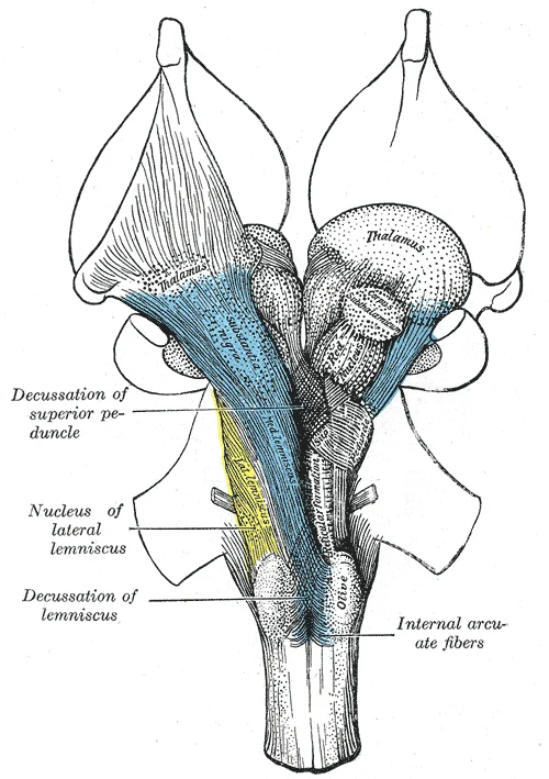
Long description
Deep dissection of brain-stem. Ventral view.

Figure 1: Deep dissection of brain-stem (ventral view).
Figure 2Gray689
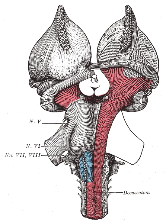
Long description
Superficial dissection of brain-stem. Ventral view.

Figure 2: Superficial dissection of brain-stem (ventral view).
Representations of the body of varying degrees of complexity that exist at multiple levels in the Ascending network, including the nucleus of the tractus solitarius. It [2]
The dorsal motor nucleus of the vagus is another critical component of the dorsal vagal complex. It is located in the medulla, adjacent to the nucleus of the tractus solitarius. This nucleus contains the cell bodies of parasympathetic motor neurons that innervate the heart, lungs, and gastrointestinal tract. The nucleus ambiguus, which contains motor neurons for the pharynx and larynx, is also located in this region and shows similar marker expression.
The pedunculopontine nucleus and the laterodorsal tegmental nucleus are the main cholinergic cell groups in the human brainstem. They provide the major cholinergic innervations of the thalamic relay nuclei and the reticular nucleus of the thalamus. In contrast, cortical structures receive cholinergic innervations from cell groups outside the brainstem, specifically in the basal forebrain. These cholinergic pathways are essential for arousal and attention, linking the brainstem to higher cortical functions. [2]
Basic respiratory rhythm is generated in the respiratory neuron network of the lower brainstem. A region critically important for inspiratory burst generation is the pre-Bötzinger complex, located within the ventral respiratory group. This complex is bilaterally located in the reticular formation of the ventrolateral medulla. It occupies a limited portion within the ventral respiratory group, situated just ventral to the nucleus ambiguus. The pre-Bötzinger complex is thought to play a significant role in the generation of respiratory rhythm in adults. [3]
DefinitionDorsal Vagal Complex
The dorsal vagal complex consists of the nucleus of the solitary tract, the dorsal motor nucleus of the vagus, and the area postrema, all located in the medulla.
| Nucleus | Location | Primary Function |
|---|---|---|
| Nucleus Tractus Solitarius | Medulla | Visceral sensory processing, emotion |
| Dorsal Motor Nucleus | Medulla | Parasympathetic motor output |
| Pre-Bötzinger Complex | Ventrolateral Medulla | Respiratory rhythm generation |
| Pedunculopontine Nucleus | Pons/Midbrain | Cholinergic innervation of thalamus |
Brainstem Respiratory Control
- Respiratory Rhythm GenerationPre-Bötzinger complex in ventrolateral medulla
- Question: Inspiration Phase?
Yes
- Inspiratory BurstPre-I neurons fire prior to inspiration
No
- Expiration PhasePassive recoil or active expiratory muscles
The brainstem serves as the critical conduit between the forebrain and the spinal cord, integrating complex motor, sensory, and autonomic functions. While traditionally viewed as a simple relay station, contemporary neuroanatomy recognises it as a site of sophisticated processing, particularly regarding posture, gait, and emotional regulation. The structure is divided into three main segments: the midbrain (mesencephalon), the pons, and the medulla oblongata.
Figure 3Gray719
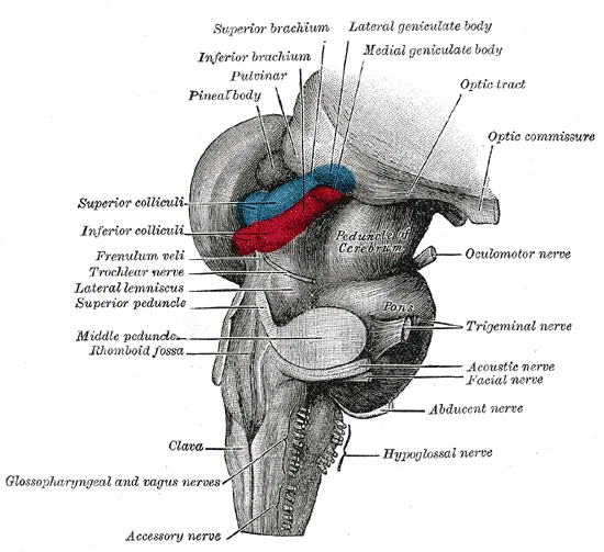
Long description
Hind- and mid-brains; postero-lateral view.

Figure 3: Hind- and mid-brains; postero-lateral view.
The midbrain is identified by the cerebral peduncles anteriorly and the tectum posteriorly. The pons is characterised by its broad, bulging anterior surface, formed by transverse pontine fibres and the basilar artery running in the pontomedullary sulcus. The medulla oblongata tapers inferiorly, continuous with the spinal cord, and is marked by the pyramids anteriorly and the olives laterally. Understanding this gross anatomy is essential for localising lesions, as the brainstem’s compact structure means that small pathological changes can affect multiple cranial nerve nuclei and long tracts simultaneously.
Figure 4Gray685
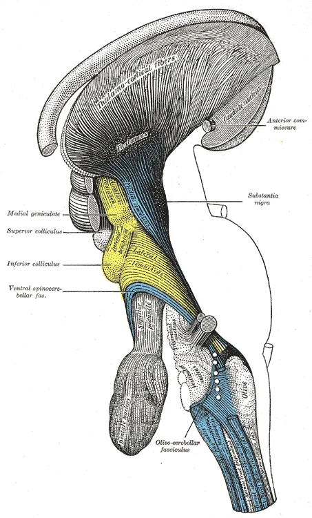
Long description
Deep dissection of brain-stem. Lateral view.

Figure 4: Deep dissection of brain-stem (lateral view).
Figure 5Gray684
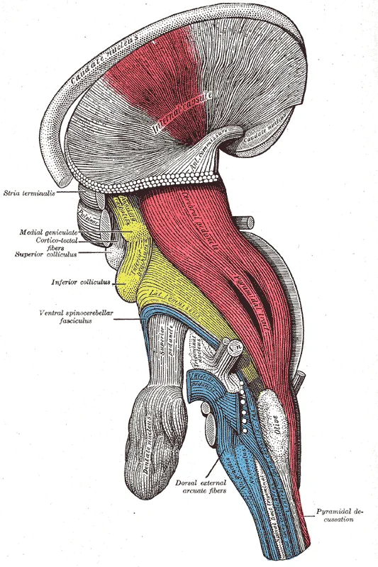
Long description
Deep dissection of brain-stem. Lateral view.

Figure 5: Deep dissection of brain-stem (lateral view).
Figure 6Gray683
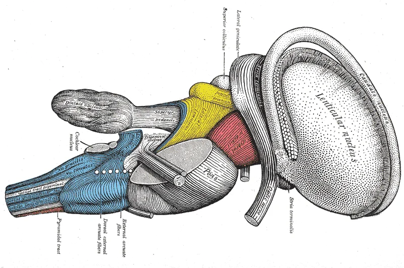
Long description
Dissection of brain-stem. Lateral view. Labelled illustration from Wikimedia Commons, used under its open licence.

Figure 6: Dissection of brain-stem (lateral view).
Figure 7Gray682
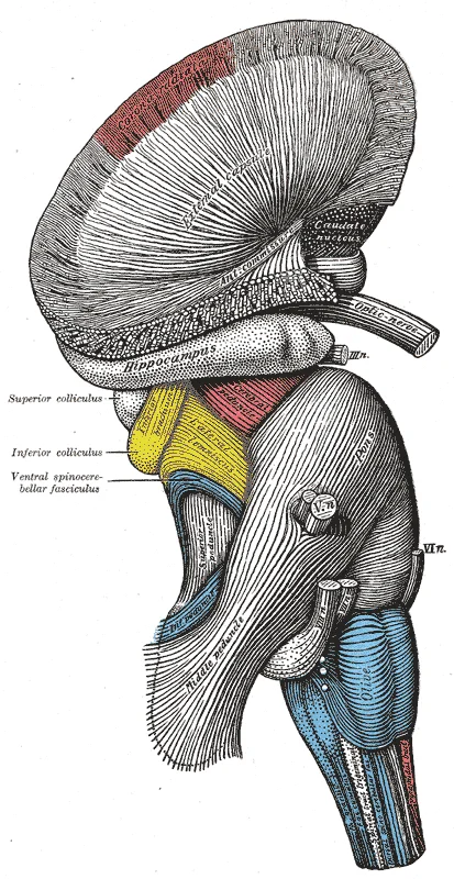
Long description
Superficial dissection of brain-stem. Lateral view.

Figure 7: Superficial dissection of brain-stem (lateral view).
This highlights the clinical relevance of the midbrain’s role in motor control. The brainstem’s function is far more complicated than we have presumed, involving multifaceted roles of the reticulospinal system. [1]
DefinitionBrainstem Networks
The “emotional brainstem” is comprised of three major networks – Ascending, Descending and Modulatory. [2]
The Descending motor network is subdivided into medial projections from the reticular formation that modulate the gain of inputs impacting emotional salience, and lateral projections from the periaqueductal gray, hypothalamus and amygdala that activate characteristic emotional behaviors. [2]
| Network | Primary Components | Function |
|---|---|---|
| Ascending | Spinothalamic tracts | Transmit sensory info to rostral structures |
| Descending | Reticular formation, PAG | Modulate motor gain and emotional behaviors |
| Modulatory | Raphe nuclei, VTA, LC | Coordinate interactions between networks |
Finally, the brainstem is home to a group of modulatory neurotransmitter pathways, such as those arising from the raphe nuclei (serotonergic), ventral tegmental area (dopaminergic) and locus coeruleus (noradrenergic), which form a Modulatory network that coordinates interactions between the Ascending and Descending networks. Integration of signaling within these three networks occurs at all levels of the brainstem, with progressively more complex forms of integration occurring in the hypothalamus and thalamus. [2]
Medullary respiratory rhythm generators pFRG and preBötC are described in both anatomy and physiology. The primary rhythms are modulated by various regions in the brainstem including the pons. This anatomical arrangement ensures that basic life- [3]
Figure 8Gray792
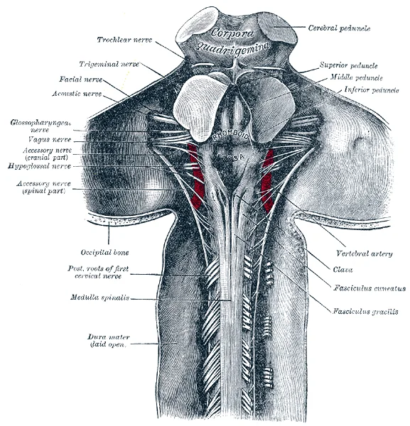
Long description
Upper part of medulla spinalis and hind- and mid-brains; posterior aspect, exposed in situ .

Figure 8: Upper part of medulla spinalis and hind- and mid-brains; posterior aspect, exposed in situ.
The brainstem is divided into three distinct levels: the midbrain, pons, and medulla oblongata. Each level contains specific nuclei, tracts, and laminae that correspond to cranial nerve functions and vital autonomic control. Understanding the internal architecture requires examining cross-sections at each level to identify the spatial relationship between motor, sensory, and reticular structures.
The midbrain is the most superior part of the brainstem. In cross-section, it is characterised by the cerebral peduncles anteriorly and the tectum posteriorly. The cerebral peduncles contain descending motor tracts, including the corticospinal and corticobulbar fibres. Posterior to the cerebral aqueduct lies the periaqueductal grey, a key component of the emotional motor system involved in pain modulation and autonomic control. The substantia nigra and red nucleus are prominent motor nuclei located within the tegmentum. Cranial nerve nuclei present in the midbrain include the oculomotor (CN III) and trochlear (CN IV) nuclei, which control extraocular movements. The superior colliculus, located in the dorsal aspect, is involved in visual processing and orientation.
The pons serves as a bridge between the midbrain and medulla. It is distinguished by the large basilar pons anteriorly, which contains transverse and longitudinal fibres connecting the cerebral cortex to the cerebellum. The pontine nuclei relay information from the cortex to the cerebellum via the middle cerebellar peduncles. The tegmentum contains the reticular formation, which regulates vigilance and motor coordination. Cranial nerve nuclei in the pons include the abducens (CN VI), facial (CN
The caudal third of the locus coeruleus () and the caudal raphe nuclei both send projections downward to the spinal cord, as depicted in Figure , and are responsible for descending pain modulation. [2]
The cranial nerve nuclei are arranged in columns, with motor nuclei medial and sensory nuclei lateral.
The brainstem serves as the critical nexus for afferent and efferent pathways, integrating sensory input with motor output to regulate vital functions and emotional responses. Understanding these connections requires tracing the flow of information from peripheral sources to central processing centres and finally to effector organs.
Sensory information reaches the brainstem via cranial nerves and ascending spinal tracts. The nociceptive and visceral sensory pathways are particularly significant. The nociceptive fibers in the anterolateral pathway give off collaterals at every level that converge with projections from visceral sensory neurons in the brainstem. From lamina I of the spinal cord, ascending projections arise and travel to the caudal brainstem. These projections specifically target the nucleus of the tractus solitariu [2]
The emotional motor system’s lateral part consists of projections primarily from the periaqueductal gray, as well as more rostral structures such as the amygdala and hypothalamus, to the lateral tegmentum in the caudal pons and medulla. [2]
DefinitionNucleus of the Tractus Solitarius
A key relay station in the medulla that receives visceral and taste sensations via cranial nerves and nociceptive/visceral projections from the spinal cord.
Motor output from the brainstem controls respiration, arousal, and locomotion. Respiratory rhythm generators in the medulla produce inspiratory and expiratory activities. The output occurs as motoneuron activities through efferent networks in the brainstem and spinal cord. In the newborn, the medulla alone can generate a three- or four-phase respiratory pattern. However, in adults, pontine structures are necessary for the generation of the normal three-phase respiratory pattern. [3]
Arousal and emotional modulation involve cholinergic pathways. Hyperpolarization of GABAergic neurons in the reticular nucleus of the thalamus by cholinergic projections from the brainstem results in disinhibition of thalamic nuclei. This influences the level of arousal by gating connections between thalamic relay nuclei and cortical regions. Ascending cholinergic projections from the laterodorsal tegmental nucleus to the forebrain form a mesolimbic pathway related to aversive emotional states. These poorly studied cholinergic pathways are part of the ascending reticular activating system and influence emotion primarily through modulation of dopaminergic signaling. [2]
| Nucleus/Region | Primary Function | Key Connections |
|---|---|---|
| Nucleus of Tractus Solitarius | Visceral and taste sensation | Receives input from vagus, glossopharyngeal, facial nerves and spinal cord [2] |
| PreBötC | Respiratory rhythm generation | Projects to contralateral preBötC and brainstem-spinal cord networks |
| Laterodorsal Tegmental Nucleus | Arousal and aversive states | Ascending cholinergic projections to forebrain and diencephalon |
| Mesopontine Tegmentum | Locomotor and emotional control | Convergence point for appetitive, defensive, and exploratory systems |
The brainstem receives its vascular supply from a complex network of arteries arising from both the vertebrobasilar system and the internal carotid arteries. Understanding this supply is critical for localising lesions, as different territories supply distinct functional regions.
They unite at the pontomedullary junction to form the single midline basilar artery. This system supplies the majority of the brainstem. The basilar artery runs along the ventral surface of the pons, giving off paramedian, short circumferential, and long circumferential branches. These vessels penetrate the brainstem to supply the medial and lateral structures respectively. Occlusion of these vessels leads to specific syndromes depending on the level of the brainstem affected. [1]
The PICA is typically the largest branch of the vertebral artery. It supplies the lateral medulla and the inferior cerebellum. A classic lesion here causes Lateral Medullary Syndrome (Wallenberg syndrome), characterised by ipsilateral ataxia, Horner’s syndrome, and loss of pain and temperature sensation on the ipsilateral face and contralateral body.
AICA arises from the lower part of the basilar artery. It supplies the lateral pons and the middle cerebellar peduncle. Infarction here results in Lateral Pontine Syndrome, involving ipsilateral facial paralysis, deafness, and ataxia, along with contralateral loss of pain and temperature sensation.
Lesions here may cause ipsilateral ataxia and contralateral sensory loss, often with oculomotor nerve palsies due to proximity to the cerebral peduncles.
These small branches arise directly from the basilar and vertebral arteries. They supply the medial structures of the brainstem, including the corticospinal tracts, medial lemnisci, and cranial nerve nuclei such as the hypoglossal (XII) and abducens (VI) nuclei. Medial syndromes typically present with contralateral hemiparesis and loss of proprioception, sparing pain and temperature sensation.
The hierarchical organisation of these vessels means that small vessel disease can affect specific nuclei. For instance, the dorsal columns of the spinal cord continue as the medial lemnisci, carrying discriminatory sensation, deep touch and proprioception[1]. These pathways are supplied by paramedian branches. Disruption here leads to loss of fine touch and vibration sense. Conversely, the anterolateral pathway, composed of the spinothalamic tracts, carries nociceptive and temperature-related signals[1]. These are supplied by lateral branches. Recognising the vascular territory helps distinguish between medial and lateral brainstem lesions in clinical practice. [2]
The brainstem serves as the critical conduit and processing hub for motor control, posture, and vital autonomic rhythms. It integrates supraspinal commands with spinal output to coordinate complex behaviours.
Locomotion is not merely a spinal reflex but a centrally generated pattern. The locomotor network in the spinal cord is functionally organized by interneurons that generate locomotor rhythm (i.e., the CPG) and locomotor pattern in addition to motoneurons. This central pattern generator may consist of interneurons that mediate flexion reflexes. Premotor interneurons, such as Ia interneurons, Ib interneurons and Renshaw cells, may contribute to locomotor pattern formation. Signals from the pattern formation interneurons may be transmitted to the target motoneurons. Supraspinal signals from the cerebral cortex and the brainstem act on these interneurons and motoneurons so that locomotion is initiated and modulated. [1]
Postural muscle tone is regulated by a counterbalance between the excitatory and inhibitory systems. Such interactions may occur at the levels of the brainstem and spinal cord as follows. Reciprocal connection exists between the cholinergic and monoaminergic nuclei. The mesopontine cholinergic neurons receive direct serotonergic projection from the DR and noradrenergic projection from the LC. These monoaminergic projections may inhibit cholinergic neurons. Conversely, cholinergic input to DR inhibits serotonergic neurons by the mediation of GABA neurons. The medial PRF (NRPo) receives monoaminergic projections from the DR/LC and cholinergic projections from the PPN/LDT. [1]
Cortical control of posture involves abundant cortico-fugal projections to the brainstem reticular formation from the premotor cortices. The projection from the primary motor cortex (M1) to the PPN had a gloss somatotopical representation: orofacial, forelimb, and hindlimb representations tended to be arranged in orderly manner from medial to lateral. Inputs from the M1, SMA and PM largely overlapped. The PPN received partially separate but essentially convergent cortical inputs not only from multiple motor-related areas representing the same body part, but also from multiple regions representing diverse body parts.
There are three distinct functions in its rhythmic burst generation. The first is burst formation. Its “persistence” is essential to burst generation. The neurons use their N-shaped I–V relationship where, within the negative slope domain, the cell would recursively auto-depolarize into a burst. The burst termination is the second function that is achieved by cumulative high-voltage-sensitive inactivation of gNaP at every action potential through the burst until spike generation is no longer possible. The third is its rhythm determination. [3]
This gNaP could be blocked with low concentrations of its blockers [riluzole (5 µM) or tetrodotoxin (5 nM)] when they were precisely microinfused into preBötC in slice preparations. Such blockade would cause the network to become incapable of generating rhythmic inspiration (as well as gasping ). [3]
The HCRG is distinct from the UCIN because it is located in the ventral portion of the ventral horn at the level of the spinomedullary junction with the C2 segment. The HCRG consists of interneurons and motoneurons, which are responsive to both respiratory and metabolic acidosis. [3]
Interoception, which is the sense of the internal condition of the body, and emotional feeling, may share a common route through the brainstem to the anterior insular cortex. The interoceptive system, represented in the cortex by the insula and adjacent regions of the frontal operculum, is particularly important for the internal simulation of observed emotion in humans and for the experience of complex social emotions. The other body map in the somatosensory cortex, which is built from dorsal column inputs and segments of the anterolateral pathway, contributes to affective understanding by simulation of facial expressions. [2]
Serotonin from the raphe nuclei appears to perform different functions in anxiety, stress, depression, and social behavior. This diversity arises because it acts through a diverse set of receptors. Its role may vary with the level of arousal. [2]
The pons lies centrally, characterized by the basilar pontine fibres and the pontine nuclei. The medulla oblongata is inferior, continuous with the spinal cord. Key structures include the reticular formation, which spans the entire length, and the cranial nerve nuclei embedded within the parenchyma.
Figure 9Blausen 0115 BrainStructures
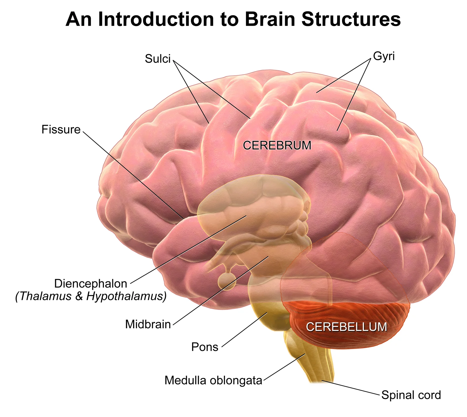
Long description
Introduction to Brain Structures. See a full animation of this medical topic and other brain structures.

Figure 9: Introduction to Brain Structures.
Localising brainstem lesions requires the integration of cranial nerve deficits with long tract signs, a method known as crossed signs. A lesion in the brainstem typically affects the cranial nerve nuclei at that level and the descending or ascending tracts before they decussate. Understanding this anatomy is critical for distinguishing brainstem pathology from cerebral or spinal cord lesions.
Above this level, in the pons and midbrain, the corticospinal tracts are ipsilateral to the cranial nerve nuclei they pass near. The viewer should note how a lesion affecting the left side of the midbrain would damage the left oculomotor nerve nucleus (causing left ptosis and a down-and-out eye) while sparing the corticospinal tract until it decussates lower down, potentially causing right-sided hemiparesis if the lesion is large enough to affect the tract before decussation, though typically midbrain lesions cause ipsilateral CN III palsy and contralateral hemiparesis if the cerebral peduncle is involved.
| Syndrome | Level | Cranial Nerve Deficit | Long Tract Deficit |
|---|---|---|---|
| Weber's | Midbrain | Ipsilateral CN III palsy | Contralateral hemiparesis |
| Millard-Gubler | Pons | Ipsilateral CN VI/VII palsy | Contralateral hemiparesis |
| Wallenberg | Medulla | Ipsilateral Horner's, ataxia | Contralateral pain/temp loss |
The table above summarises classic localising signs. In Weber’s syndrome, a lesion in the midbrain affects the cerebral peduncle and the oculomotor nerve. In Millard-Gubler syndrome, a ventral pontine lesion affects the facial and abducens nuclei or fascicles, causing ipsilateral facial and abducens palsies, along with contralateral hemiparesis. Wallenberg syndrome, or lateral medullary syndrome, involves the lateral medulla. It presents with ipsilateral Horner’s syndrome, ataxia, and loss of pain and temperature sensation on the face, but contralateral loss of pain and temperature in the body. These patterns rely on the precise anatomical arrangement of nuclei and tracts within the confined space of the brainstem.
Clinical pearlCrossed Signs
Ipsilateral cranial nerve deficit with contralateral body deficit localises the lesion to the brainstem.
DefinitionDecussation
The crossing over of nerve fibres from one side of the nervous system to the other, typically in the medulla for the corticospinal tract.
The brainstem is the critical conduit for motor, sensory, and autonomic pathways, linking the forebrain and cerebellum to the spinal cord. Understanding its organisation requires distinguishing between the functional nuclei of the cranial nerves and the integrative centres within the reticular formation.
The reticular formation modulates muscle tone and locomotion through specific nuclei. The pedunculopontine nucleus (PPN) plays a key role in these processes. Optimal sites for evoking locomotion are located within the cuneiform nucleus (CNF) and rostral to this nucleus. [1]
Medullary reticulospinal neurons (RSNs) are classified by their firing patterns during hypertonus and atonia states. Hypertonus-related RSNs are defined as having a firing frequency greater than 10 Hz during hypertonus. These neurons are located in the ventromedial part of the medullary reticular formation (MRF). In contrast, atonia-related RSNs are located in the dorsomedial part of the MRF. [1]
RSNs are further grouped by their inputs from the mesencephalic locomotor region (MLR) and the pontine reticular nucleus oralis (NRPo).
| Group | Stimulation Response | Conduction Velocity (m/s) |
|---|---|---|
| Group 1 | Excited by MLR only | 85.6 ± 12.6 |
| Group 2 | Excited by MLR, Inhibited by NRPo | 85.4 ± 16.3 |
| Group 3 | Excited by MLR and NRPo | 94.3 ± 15.5 |
| Group 4 | Excited by NRPo only | 95.2 ± 15.7 |
Brainstem dopaminergic neurons are located in the midbrain and are divided into three contiguous groups: the retrorubral field, the substantia nigra pars compacta, and the ventral tegmental area. Two major ascending pathways arise from these clusters. The nigrostriatal pathway originates from the substantia nigra pars compacta. The mesocorticolimbic pathway originates from the ventral tegmental area. [2]
The preBötzinger complex (preBötC) is a key respiratory centre. Neurons in the preBötC send axonal projections to various respiratory-related regions, including the Bötzinger complex, ventral respiratory group, and parafacial respiratory group. They also project to the hypoglossal motor nuclei and nuclei tractus solitarius. [3]
The brainstem modulates affective states through domains of valence and arousal. Valence correlates with pleasantness ratings, heart rate, and facial muscle activity. Arousal correlates with skin conductance, interest ratings, and viewing time for stimuli. These domains influence attention allocation and long-term memory formation. [2]
Students frequently confuse the functional roles of distinct brainstem nuclei, particularly regarding muscle tone and posture. A primary error is assuming all reticular formation neurons promote tone. In reality, the majority of reticulospinal neurons (RSNs) activated by the mesencephalic locomotor region (MLR) are distributed in the ventral part of the medullary reticular formation, corresponding to the nucleus reticularis magnocellularis (NRMc). These specific neurons contribute to increasing the majority of RSNs in Groups 3 and 4 are located in the dorsomedial medullary reticular formation, corresponding to the NRGc [1]
Another significant pitfall involves the neural substrate for anticipatory postural adjustments. Students may attribute these adjustments solely to the cerebellum. However, probabilistic diffusion tractography in primates demonstrates that the supplementary motor area (SMA) and the dorsal premotor cortex have strong connections with the lateral and medial pedunculopontine nuclei (PPN), respectively. The SMA specifically contributes to anticipatory postural adjustment for step initiation. This process is notably impaired in patients with Parkinson’s disease, highlighting the clinical relevance of these brainstem-cortical loops. Furthermore, dysfunction in the projection from the cerebral cortex to the vermis may underlie some forms of dystonia, rather than being a purely peripheral issue. [1]
Misinterpretation of respiratory rhythm generation is also common. Some may assume that long-lasting spinal respiratory activity is the primary driver of breathing. However, transection experiments revealed that this activity was exclusively recorded from cervical ventral roots and never observed in the cranial or phrenic nerves. Only non-respiratory motoneurons exhibited rhythmic depolarization in phase with these bursts, making it unlikely that this activity is involved in respiratory function. The actual respiratory rhythm generator is located in the medulla, with spinal contributions being secondary or non-respiratory in nature. [3]
Finally, students often misunderstand the mechanism of the rostrocaudal gradient in inspiratory motor activity. It is incorrect to assume this gradient is organized entirely by bulbospinal neurons and motoneurons. Evidence suggests that inhibitory inspiratory interneurons are involved in generating this gradient. These interneurons in the rostral thoracic segments may project their axons to the caudal thoracic segments, forming a gradient that is the inverse of the inhibitory synaptic inputs to the motoneurons. This complexity means that simple excitatory models are insufficient to explain respiratory motor patterns. [3]
Exam trapTone Confusion
Do not assume all reticular formation neurons increase tone; the NRGc is associated with atonia, while the NRMc increases tone.
This knowledge is applied at the bedside to understand movement disorders. In Parkinson’s disease, the impairment of anticipatory postural adjustment for step initiation is a key clinical feature. Recognizing the role of the SMA and its connections to the PPN helps explain why these patients struggle with initiating movement and maintaining balance. Clinicians use this understanding to tailor rehabilitation strategies that focus on cueing and external triggers to bypass impaired internal postural adjustments.
In the operating theatre and anaesthetic practice, understanding brainstem control of tone is vital. The distinction between hypertonus-related and atonia-related pathways allows anaesthetists to manage muscle tone during surgery. Knowledge of the NRMc and NRGc distributions helps in predicting responses to anaesthetic agents that target the reticular formation. For instance, agents that enhance GABAergic transmission may affect the NRGc, promoting relaxation, while those affecting serotonergic pathways might influence tone differently.
On imaging, radiologists and neurologists use these anatomical distinctions to localise lesions. If a patient presents with hypertonus, imaging may focus on the ventral medullary reticular formation (NRMc) to identify lesions affecting tone-increasing pathways. Conversely, atonia might prompt a search for lesions in the dorsomedial MRF (NRGc). Understanding the connections between the SMA, dorsal PM, and PPN also aids in interpreting functional imaging studies in movement disorders, linking cortical dysfunction to brainstem output.
In examinations, students must accurately describe the origins and functions of these nuclei. Questions often test the specific roles of the NRMc and NRGc in muscle tone control. Students must recall that the majority of MLR-activated RSNs are in the ventral MRF (NRMc) and contribute to increasing muscle tone. They must also know that the NRGc is associated with atonia. Additionally, understanding the role of inhibitory interneurons in the rostrocaudal gradient of inspiratory activity is crucial for respiratory physiology questions. Students should be able to explain that inhibitory interneurons in rostral segments project caudally to shape the inspiratory motor pattern.
Clinical pearlParkinson’s Link
Impaired anticipatory postural adjustment in Parkinson’s disease links cortical SMA function to brainstem PPN connections.
Postural Control Pathways
- StartAssess postural control
- Question: Is step initiation impaired?
Yes
- Check SMA-PPN pathwayLook for Parkinson’s features
No
- Question: Is tone abnormal?
Yes
- Check NRMc/NRGcVentral vs Dorsomedial MRF
No
- NormalNo specific pathway deficit
The NRMc in the ventral MRF contains RSNs that increase muscle tone. The NRGc in the dorsomedial MRF contains RSNs associated with atonia. The SMA connects to the PPN and aids anticipatory postural adjustment. Parkinson’s disease impairs anticipatory postural adjustment for step initiation. Long-lasting spinal respiratory activity is not primarily respiratory in function. Inhibitory interneurons help generate the rostrocaudal gradient of inspiratory activity. Serotonin projections can enhance anxiety or contribute to depression depending on the target. Fear-induced analgesia involves the dorsal/lateral periaqueductal gray via non-opioid mechanisms.
Answer each one in your head first, then open the answer.
Which nucleus in the ventral MRF is associated with increasing muscle tone?
Show answerHide answer
Nucleus reticularis magnocellularis (NRMc)What is the primary role of the SMA in postural control?
Show answerHide answer
Anticipatory postural adjustment for step initiationWhich brainstem region is linked to atonia-related RSNs?
Show answerHide answer
Dorsomedial MRF (NRGc)What type of interneurons are involved in the rostrocaudal gradient of inspiratory activity?
Show answerHide answer
Inhibitory inspiratory interneuronsWhich periaqueductal gray region produces non-opioid analgesia in fear?
Show answerHide answer
Dorsal/lateral periaqueductal gray
- Takakusaki K, Chiba R, Nozu T, Okumura T. Brainstem control of locomotion and muscle tone with special reference to the role of the mesopontine tegmentum and medullary reticulospinal systems. Journal of neural transmission (Vienna, Austria : 1996) 2016;123(7):695-729. doi:10.1007/s00702-015-1475-4. Accessed .
- Venkatraman A, Edlow BL, Immordino-Yang MH. The Brainstem in Emotion: A Review. Frontiers in neuroanatomy 2017;11:15. doi:10.3389/fnana.2017.00015. Accessed .
- Ikeda K, Kawakami K, Onimaru H, et al. The respiratory control mechanisms in the brainstem and spinal cord: integrative views of the neuroanatomy and neurophysiology. The journal of physiological sciences : JPS 2017;67(1):45-62. doi:10.1007/s12576-016-0475-y. Accessed .
Figure credits
- Figure 1: Henry Vandyke Carter, via Wikimedia Commons, Public domain, Public domain.
- Figure 2: Henry Vandyke Carter, via Wikimedia Commons, Public domain, Public domain.
- Figure 3: Henry Vandyke Carter, via Wikimedia Commons, Public domain, Public domain.
- Figure 4: Henry Vandyke Carter, via Wikimedia Commons, Public domain, Public domain.
- Figure 5: Henry Vandyke Carter, via Wikimedia Commons, Public domain, Public domain.
- Figure 6: Henry Vandyke Carter, via Wikimedia Commons, Public domain, Public domain.
- Figure 7: Henry Vandyke Carter, via Wikimedia Commons, Public domain, Public domain.
- Figure 8: Henry Vandyke Carter, via Wikimedia Commons, Public domain, Public domain.
- Figure 9: BruceBlaus. When using this image in external sources it can be cited as: Blausen.com staff (2014). "Medical gallery of Blausen Medical 2014". WikiJournal of Medicine 1 (2). DOI:10.15347/wjm/2014.010. ISSN 2002-4436., via Wikimedia Commons, CC BY 3.0, CC BY 3.0.
Educational content for medical students and doctors. It is not a substitute for clinical judgement or local guidelines.